Diagnostic Imaging W12 - Imaging the Lower Extremity
1/36
There's no tags or description
Looks like no tags are added yet.
Name | Mastery | Learn | Test | Matching | Spaced | Call with Kai |
|---|
No study sessions yet.
37 Terms
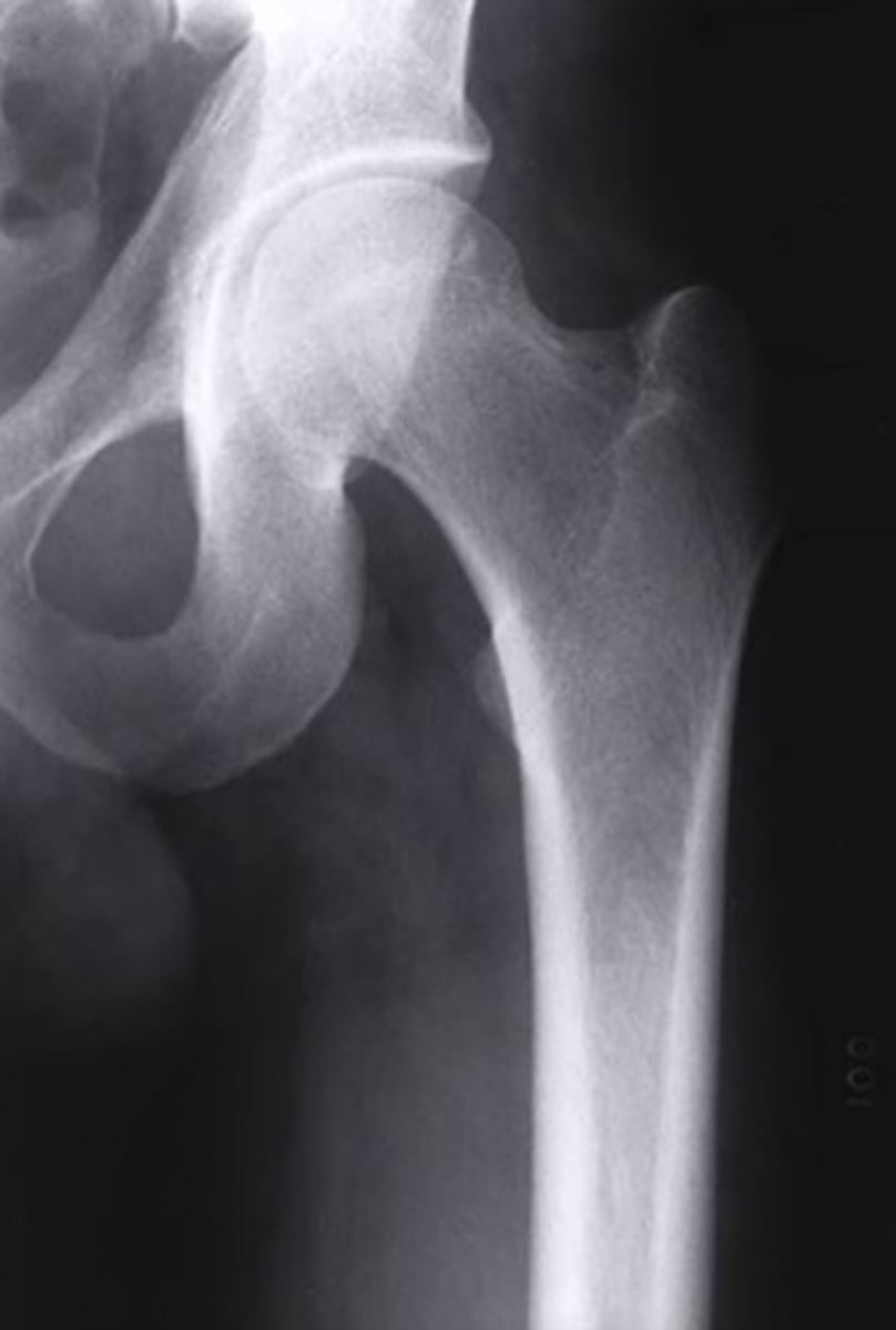
Which radiographic view of the hip allows for assessment of the femoral neck angle?
anterior-posterior
3 multiple choice options
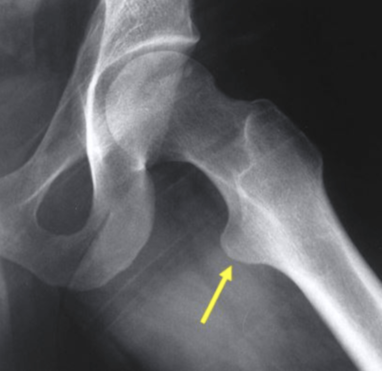
Which structure is highlighted by the yellow arrow in this frog leg lateral view of the hip?
lesser trochanter
3 multiple choice options
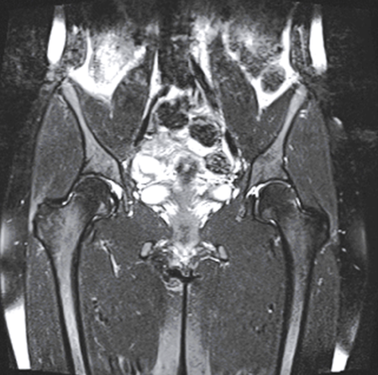
What type of image is shown here of the pelvis/hips?
coronal MRI
3 multiple choice options
Which of the following is a criterion of the Ottawa Knee Rules for radiographic imaging?
tenderness of the fibular head
3 multiple choice options
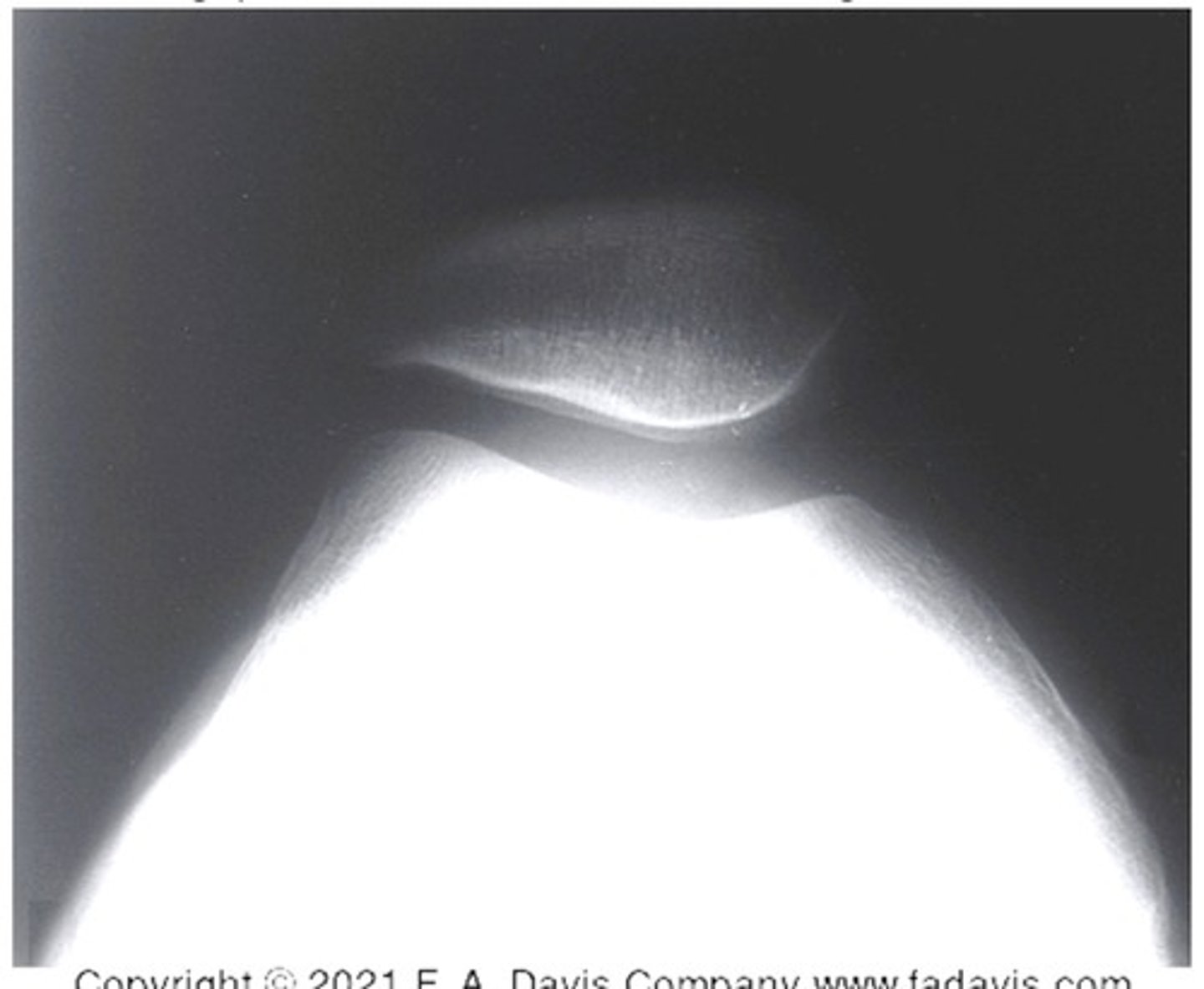
Which radiographic view of the knee is shown in the image?
tangential view of the patellofemoral joint
3 multiple choice options
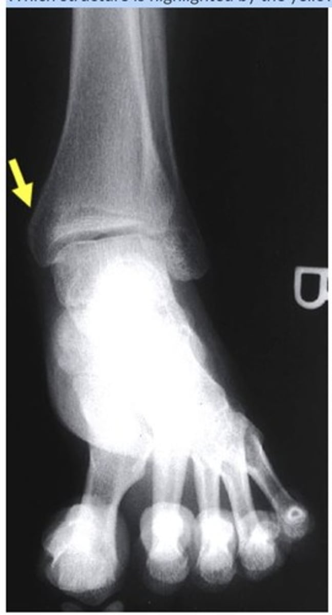
Which structure is highlighted by the yellow arrow?
medial malleolus
3 multiple choice options
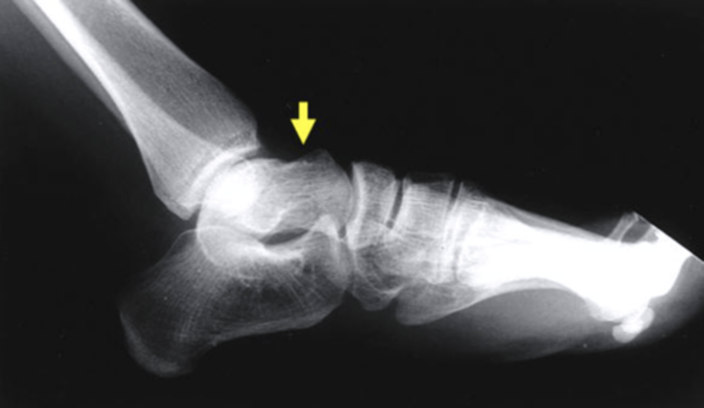
Which structure is highlighted by the yellow arrow?
talus
3 multiple choice options
your patient is 20 years old with a right lateral ankle sprain 10 days ago. he injured his ankle while running down the stairs and missing a step, landing with the ankle in a plantar flexed and inverted position. after performing the subjective examination, which of the following tests would you perform to rule out anterior talofibular ligament (ATFL) pathology?
anterior drawer test
3 multiple choice options
you learn that your patient was able to bear weight immediately after their ankle injury. however, you would like to use the Ottawa Ankle Rules to help rule out a possible ankle fracture and referral requirement. which of the following is a component of the Ottawa Ankle Rules?
bony tenderness to posterior edge of fibular/tip of lateral malleolus
3 multiple choice options
the examination for an ankle fracture with Ottawa Ankle Rules was negative. however, palpation of the anterior process of the lateral calcaneus and heel tap reproduced that patient's pain. due to the findings, should the clinician's refer the patient to an appropriate health care provider for imaging?
yes
1 multiple choice option
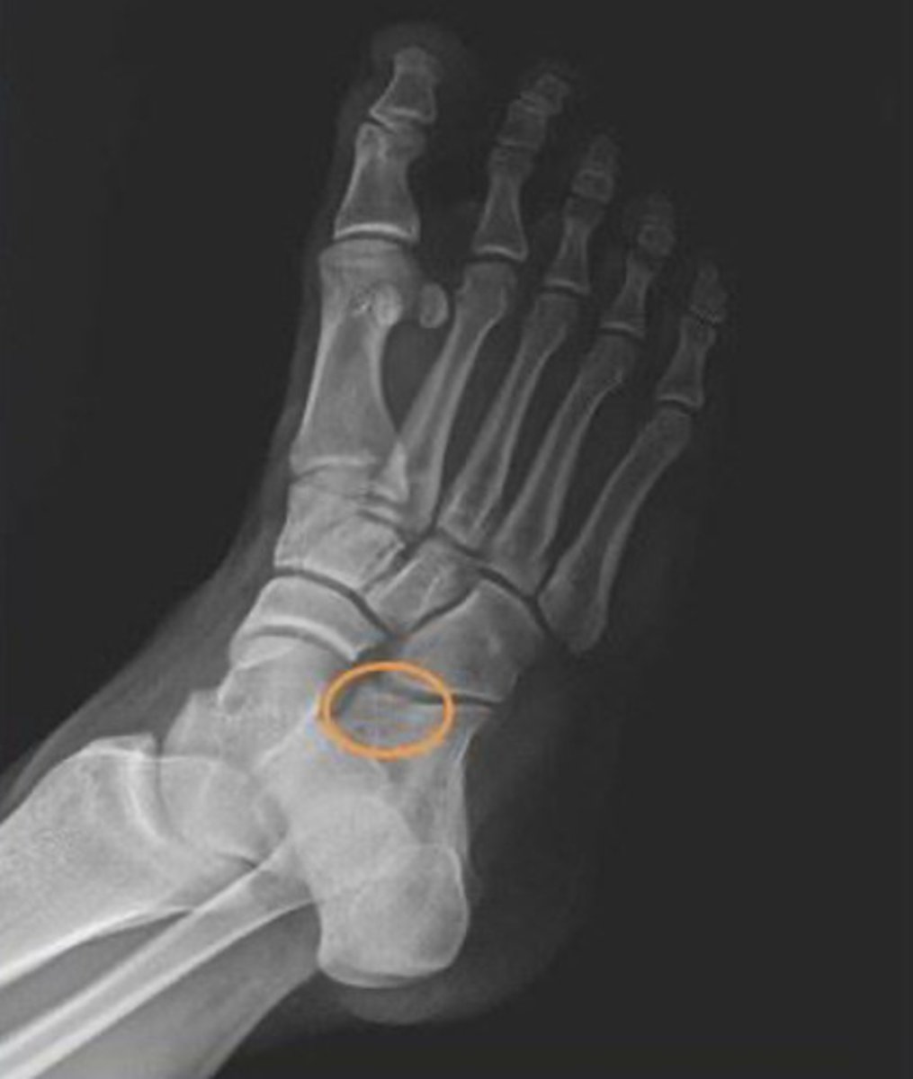
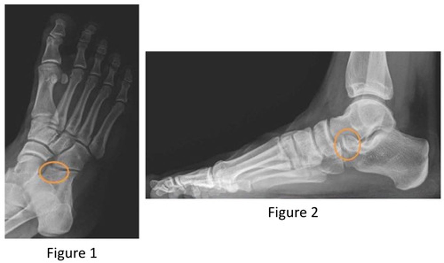
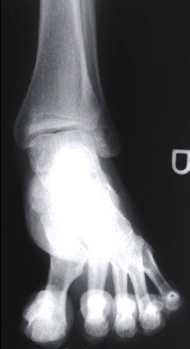
what radiograph view is this?
oblique
3 multiple choice options
what radiograph view is shown here?
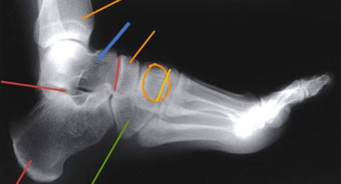
lateral
3 multiple choice options
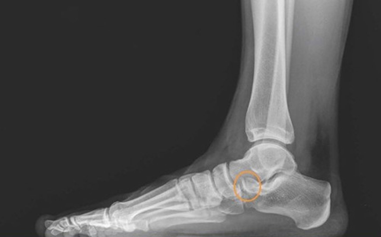
what do the orange circles in these radiographs indicate about the patient's injury to the anterior process of the calcaneus?
avulsion fracture
2 multiple choice options
after revealing a non-displaced fracture of the anterior process of the calcaneus, which of the following options will allow for the structure to heal?
controlled ankle movement (CAM) walking boot
3 multiple choice options
your 24 year old patient injured their knee playing basketball 1 day ago. this was a non-contact injury, and they describe it happening with their knee "locked out and bent sideways" (varus injury), but they deny feeling or hearing a 'pop' and was able to bear weight and walk with pain immediately after. there is large joint effusion at the right knee and their pain is 5/10 at rest and 8/10 with movement and ROM beyond 90° flexion. they present with a positive Lachman's, positive Mcmurray, and negative varus/valgus stress tests. based on these results, what types of injuries are most likely present?
ACL and meniscus
3 multiple choice options
after performing a subjective and objective exam, you suspect a fracture and want to use the Ottawa Knee Rules to determine if a radiograph referral is required. which of the following is a component of the Ottawa Knee Rules?
inability to flex the knee to 90°
3 multiple choice options
which of the following is not included in the Ottawa Knee Rules?
age >65 years
3 multiple choice options
your patient presents with a positive Lachman's, positive Mcmurray, and negative varus/valgus stress tests. what should your next steps be?
provide the pt with crutches and refer them to physician for imaging
3 multiple choice options
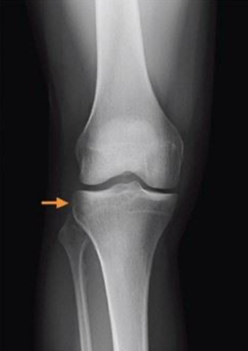
what radiograph view is this image in?
anteroposterior
3 multiple choice options

what is indicated by the orange arrow in this image?
segond fracture
3 multiple choice options
what structure is indicated by the orange arrow?
anterior cruciate ligament
3 multiple choice options
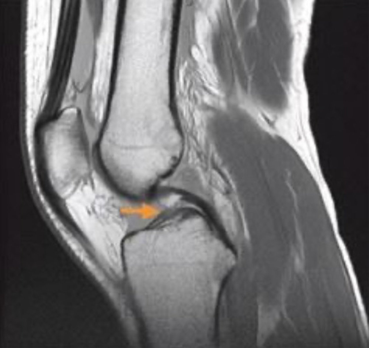
what grade would you classify the ligamentous tear in this MRI?
full-thickness
1 multiple choice option

identify the view of this magnetic resonance image (MRI)
sagittal view
2 multiple choice options

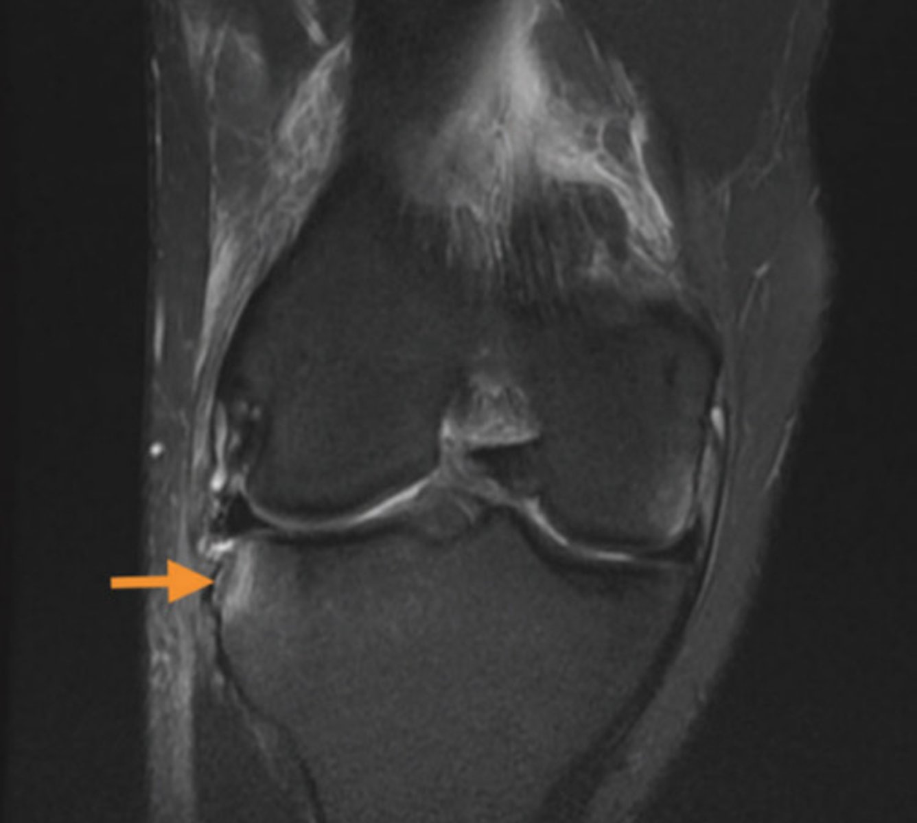
what structure is indicated by the orange arrow?
lateral meniscus, posterior horn
1 multiple choice option

identify the view of this MRI
coronal view
2 multiple choice options
your 33 year old patient has left hip pain that will "lock and give out" and has been worsening over the last 6 months. they recall no traumatic injury or specific episode that brought on the pain. they have typical ROM but painful at end ranges of flexion, IR, and ER. MMTs are all 3+/5 on the left hip. the FABER and Scour tests are both positive. based on the subjective and objective findings, what is the best next step?
refer pt to appropriate health care provider for imaging
2 multiple choice options
what view is this radiograph?
anteroposterior
3 multiple choice options
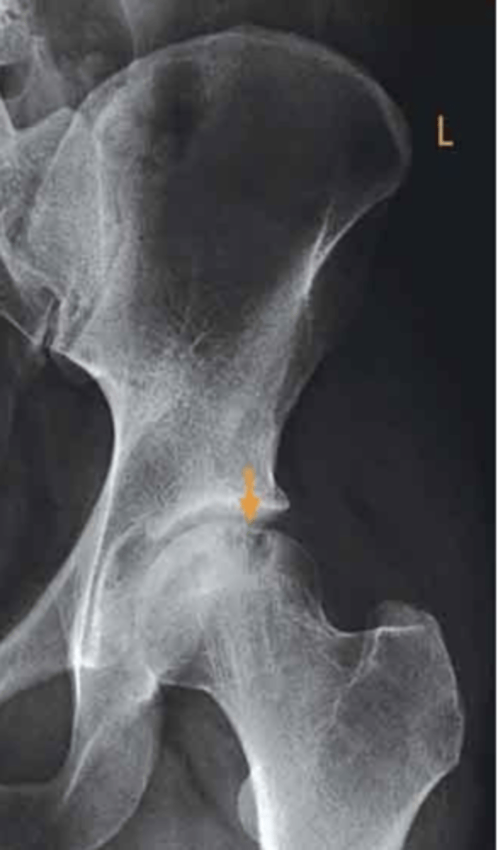
what structure is indicated by the orange arrow?
osteochondral fracture of the left femoral head
3 multiple choice options

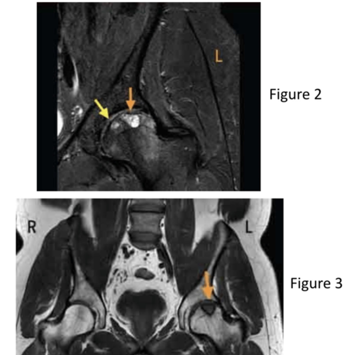
what are the MRI views of figure 2 and 3, respectively?
coronal and coronal
3 multiple choice options
after revealing an osteochondral fracture of the left femoral head, what should the next steps be for the patient?
discuss with a surgeon for possible surgical options
2 multiple choice options
which type of imaging typically evaluates for osteochondral lesion of the foot/ankle?
MRI
3 multiple choice options
which is not included in the Ottawa Ankle Rules?
tenderness at the cuboid
3 multiple choice options
which structure of the foot is highlighted by the blue line?
talus
3 multiple choice options
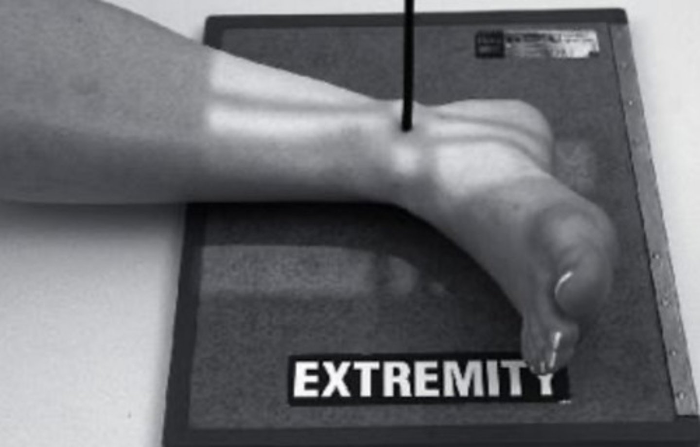
which view will result from the patient positioning as shown in this image?
lateral ankle
3 multiple choice options
what is this view?
AP
3 multiple choice options
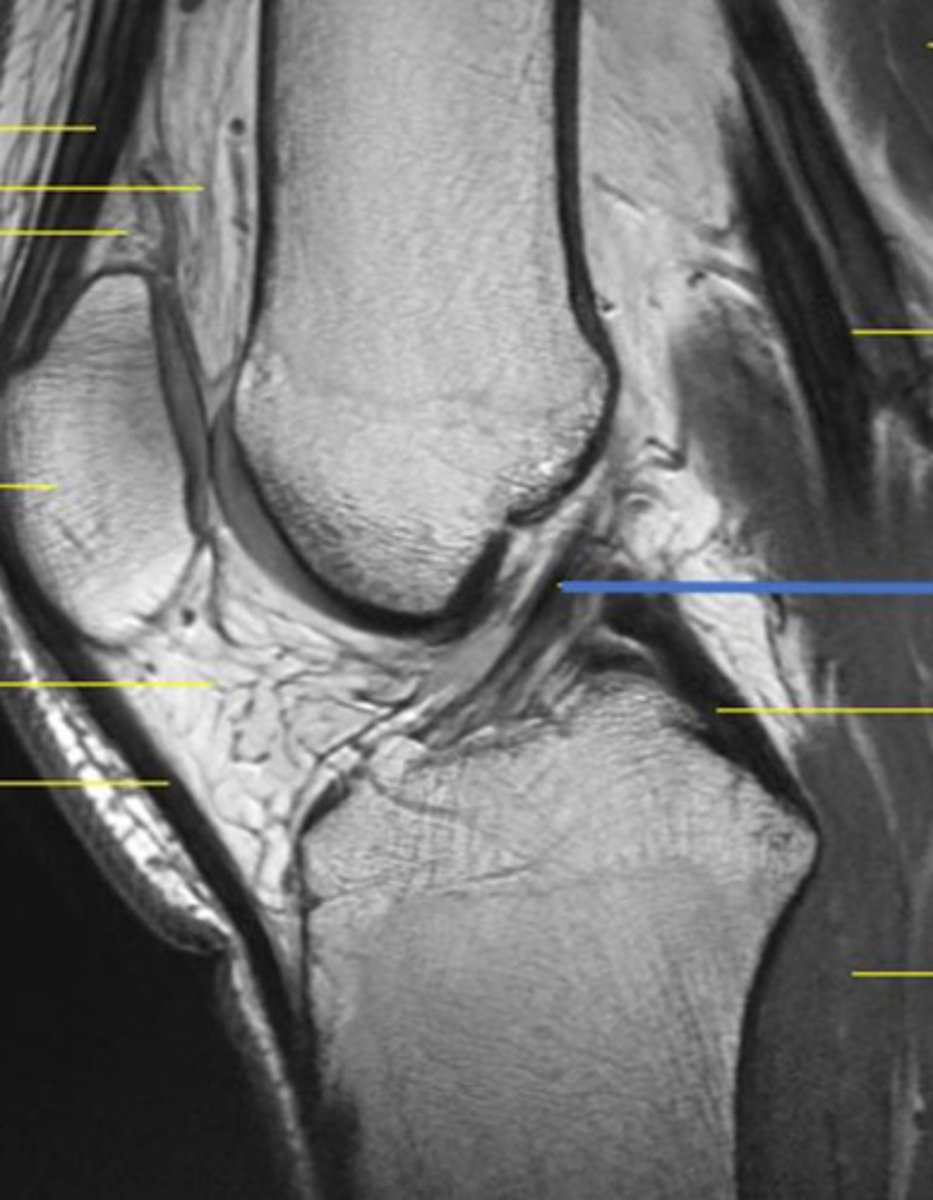
what structure of the knee is highlighted by the blue line?
ACL
3 multiple choice options
what is the name of this view of the hip?
AP
3 multiple choice options