U2 Cell Structure - Microscopy
1/55
Earn XP
Name | Mastery | Learn | Test | Matching | Spaced | Call with Kai |
|---|
No analytics yet
Send a link to your students to track their progress
56 Terms
magnification formula
magnification = image size/actual size
or I/AM
benefits of light microscope
- easily available
- cheap
- can be used out in the field
- can observe both living and dead specimens (although sometimes we use a stain which kills cells)
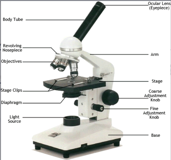
draw and label a microscope
benefits of having both objective and eyepiece lens
allows much higher magnification
reduces chromatic aberration
what adjustments would need to be made for an opaque specimen
illumination from above, in some microscopes
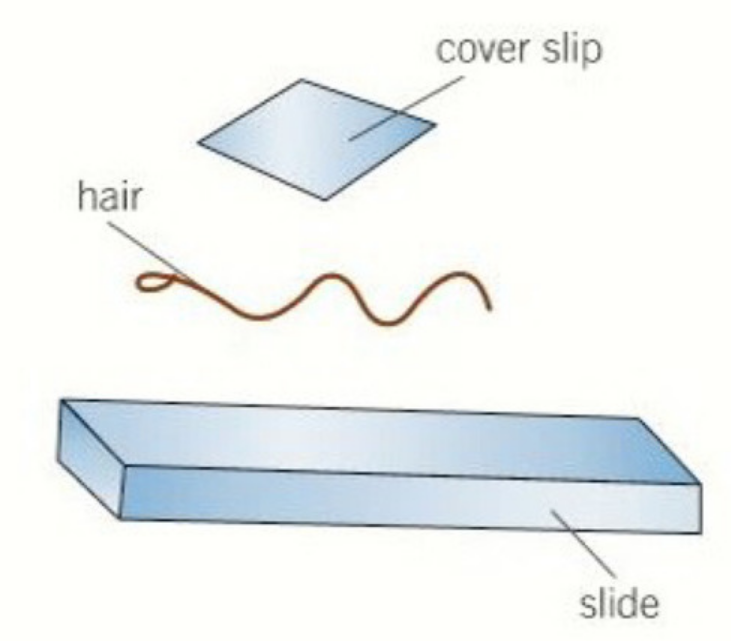
describe the dry mount preparation
solid specimens viewed whole / in thin slices (sectioning)
specimen on centre of slide with cover slip
examples include hair, pollen, muscle tissue, plants
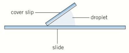
describe the slide preparation that could be used for a small water flea (2)
wet mount preparation
suspend specimen in liquid such as water or an immersion oil
place cover slip at an angle
for example aquatic samples and living organisms
describe the slide preparation for soft samples/root tips
squash sample (?)
wet mount is prepared
press on cover slip with lens tissue
depending on the material, can avoid damage to cover slip by squashing sample between two slides
for example, root tips or soft samples.

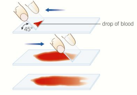
describe smear slides prep
edge of slide is used to smear the sample creating a thin even coating on another slide
then place cover slip on
for example is blood
describe the difference between brightfield microscopy and the other type of microscopy (2)
BRIGHTFIELD: objects are dark and the field is light; can be used to observe unstained microorganisms
WIDE FIELD: whole sample illuminated at once
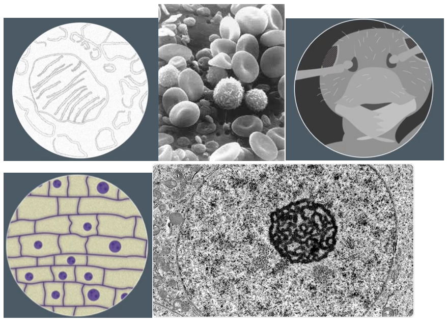
identify if there images were made by a SEM, TEM or optical microscope
1- TEM
2- SEM
3- SEM
4- optical
5- TEM
describe how an image with a light microscope would look
low contrast as most cells do not absorb a lot of light
resolution is limiting factor by wavelength and diffraction of light passing through the sample
why do we use (differential) stains
to see transparent structures (cytosol etc)
stains increase contrast as different components take up stains to different degrees, enhancing visibility
highlighting specific structures
SPEC: to identify different organelles and cell types
how to prepare a slide BEFORE staining
(i.e. how do you properly stain a slide)
Place on slide and allow to dry
It is then heat fixed by passing through a flame
the specimen will then adhere to the microscope slide and will then take up stains
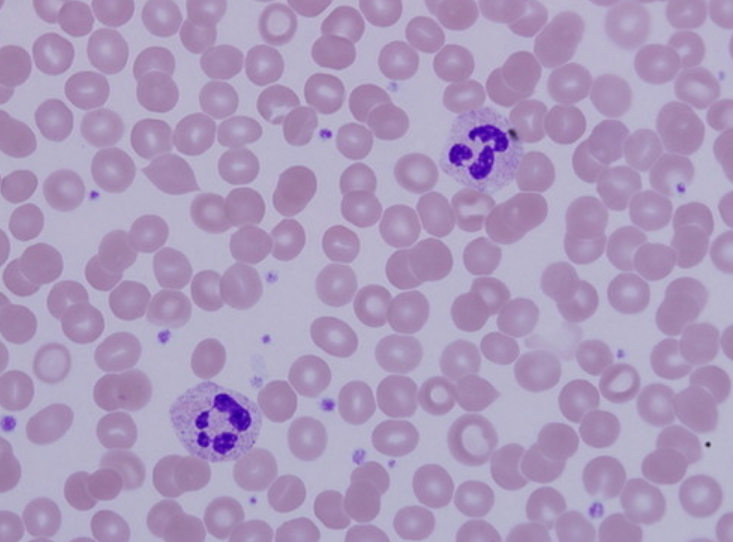
identify the key structures on the pic
pink circles: RBC (erythrocytes)
purple: white blood cells (leucocytes)
surrounding grey: plasma
exmaples of positively and negatvively charged dyes
+VE: crystal violet, methylene blue
-VE: nigrosin, congo red

what is the difference between positive and negatively charged dyes
positively charged dyes stain cell components.
negatively charged dyes stay outside cells and leave cells unstained
define differential staining
and give two examples
using two or more dyes to distinguish between two types of organism, or different organelles within a tissue sample
gram staining technique and acid-fast technique are both differential staining methods.
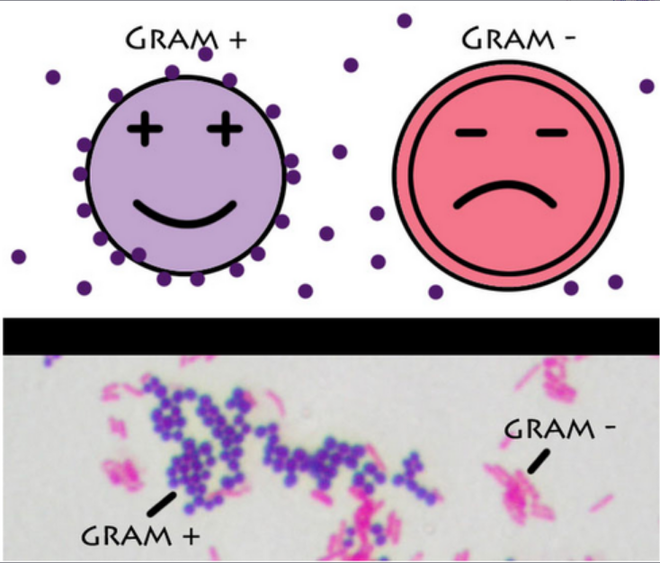
suggest and explain a method for staining a slide to differentiate thin-walled and thick-walled bacteria
gram staining technique
separates bacteria into gram+ and gram-
apply crystal violet to specimen, then iodine
- wash slide with alcohol
gram+ will retain the stain and stay blue/purple
gram- have thinner cell walls so lose the stain
stain with a counterstain (safranin dye)
the bacteria appear red
gram+ are susceptible to penicillin which inhibits cell wall formation
gram- have thinner cell walls so are not susceptible
why is iodine applied in the gram-stain technique?
why is alcohol added?
why is safranin used?
Iodine - it fixes the dye
alcohol - removes the dye from thin cell walls
safranin - stains bacteria pink but not in the presence of crystal violet
acid fast technique
- differentiates species of mycobacterium from other bacteria
carbol fuchsin dye into cells
- wash cells with dilute acid-alcohol solution
- mycobacterium aren't affected by solution and retain stain, which is bright red
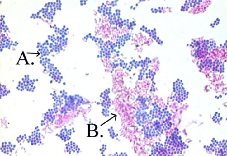
which technique?
acid-fast
which technique
gram stain (duh)
define fixing a slide
using chemicals like formaldehyde to preserve specimens in as near-neutral state as possible
(preparing for staining and killing the specimen and keeping structures)
what is sectioning
specimens dehydrated with alcohols,
then placed in a mould of wax/resin
then sliced with a microtome knife
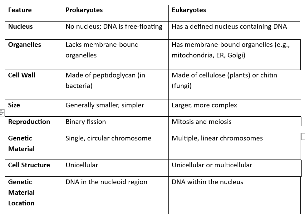
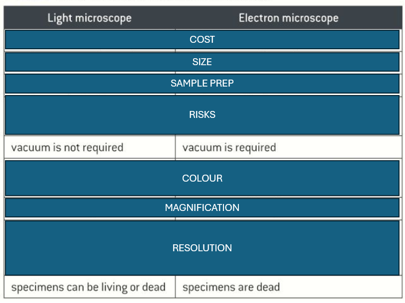
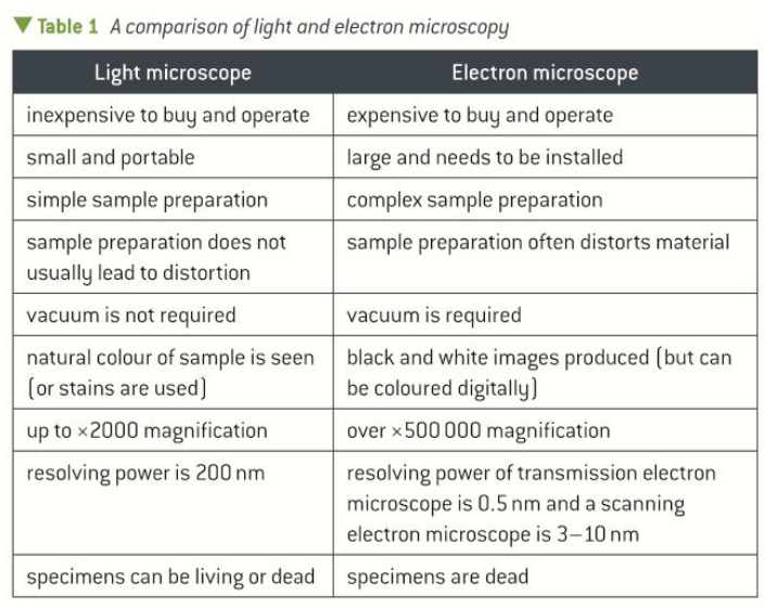
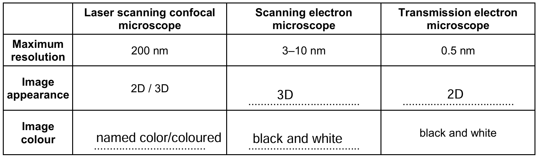
fill this table
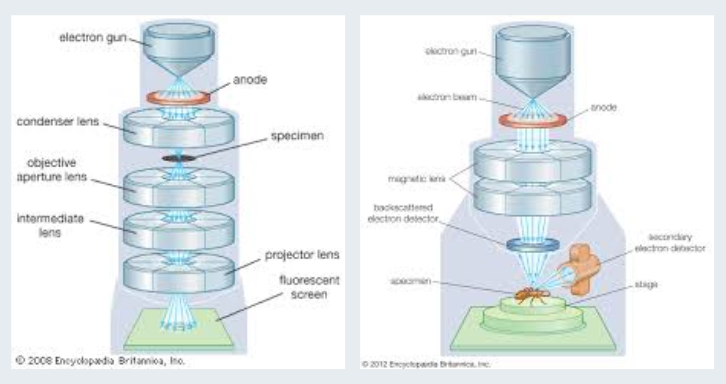
difference between transmission electron microscope and scanning electron microscope - how they work
SEM - beam of electrons sent across the surface of a specimen and the reflected electrons are collected. creates image by collecting and deflecting reflected electrons
TEM uses electrons passing through the sample and focused to create an image
compare the results from a SEM and TEM
SEM has lower resolution + magnification, but TEM has twoD images
SEM? OR TEM?
1) TEM
2) SEM
define magnification
how many times larger an image is than the actual size of the object being viewed
define resolution
the minimum distance between 2 objects where they can still be seen as 2 different objects
THE ABILITY TO SEE MORE DETAIL / SEPARATE TWO OBJECTS accPMT
why is resolution limited in light microscopes
light waves diffract when they pass near physical structures
the structures present in the specimens are close and the light reflected from individual structures can overlap due to diffraction
how do electron microscopes solve this problem
electron beams are still diffracted, but the shorter wavelength means individual beams can be closer before they overlap
problems with electron microscope
specimens can be damaged by electron beam
artefacts (structures produced due to the preparation process)
why is there a vaccuym inside an electron microscope
to ensure electron beams travel in straight lines
specimen preparation for an electron microscope
fixing using chemicals/freezing
heavy metal staining
dehydration with solvents
criteria for graphs
S ize of points - 50%
P oints plotted correctly
L ine of best fit
A xes with labels and units
T itle that’s descriptive
N o fluffy lines, extrapolation or tthick crosses
define artefact
give an example
visible structural detail caused by processing the specimen which is not a feature of the specimen
e.g. bubbles under the coverslip
give an artefact example for electron microscopes
loss of continuity of membranes, distortion of organelles, empty spaces in the cytoplasm of cells
how does a laser scanning confocal microscope work
Specimen is stained with a dye,
a laser is moved across the specimen, causing the dye to fluoresce /give off light.
This light is passed through a narrow hole through a detector which generates an image
what is LSCM used for now and later
NOW: eye disease diagnosis, drugs, endoscopy
LATER: biopsyyy
what is a eyepiece graticule?
what is a stage micrometer?
disc with a scale of 1-100 that represents different measurements with each magnification
microscope slide with a scale in micrometres, each division usually 10 micrometers. it calibrates an eyepiece graticule
how to calibrate a lens
put the stage micrometer in place and eyepiece graticule in… well, the eyepiece
get the scale on the micrometre in focus
align the scales and take a reading
remove the stage micrometer

what are the units of the EG and the SM
EG- none
SM- micrometres… duhh
how many times do you calibrate the graticule scale
3x. for each objective lens separately.
what is the length of a usual SM division
1 division = 10 micrometers
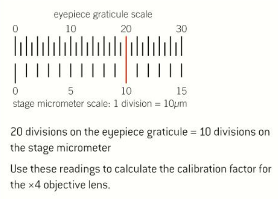
20 EG = 10 SM
20 EG = 100 MICROMETRES
1 EG = 5 MICROMETERS
The magnification factor is 5. this is also the number of micrometers per division, or µm/division on the EG
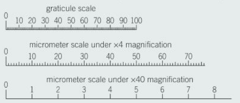
magnification factor for the x40 lens
using the same thing as before, that one division of the micrometer = 10microm
x40=0.5
using this value, find the diameter (roughly) of this atom
1 div = 0.5µm
100 div = 50µm
how to use a microscope - longhand
stage in lowest position
lowest objective lens
put slide
stage up but doesn’t touch slide (w/ coarse)
stage down until you can see specimen (w/ coarse)
fine focus up
for more detail objective lens up and fine focus up
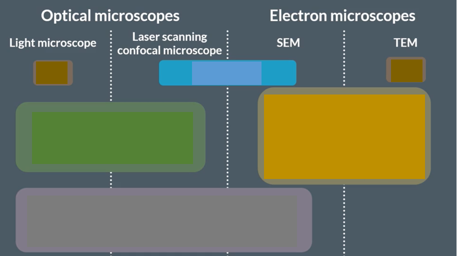
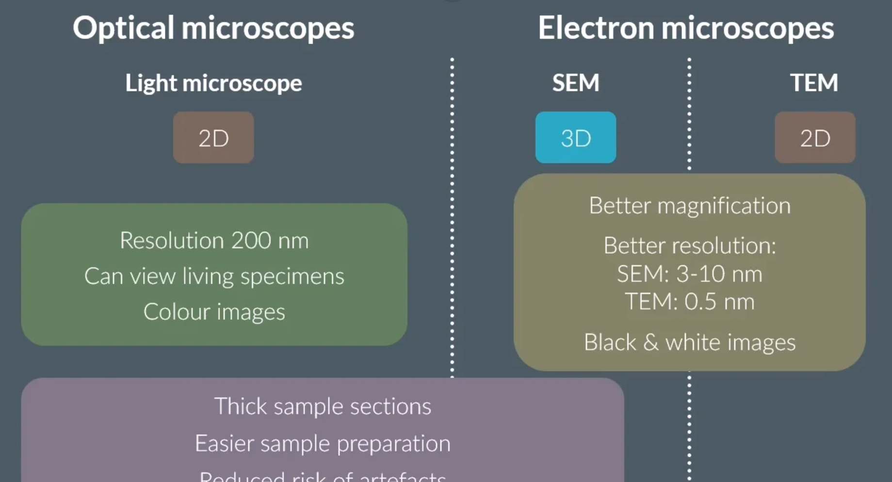
OCR accepted figures for the magnification and resolution of
light microscope
SEM
TEM
MAG
LIGHT: 1000
SEM: 100,000-500,000
TEM: 500,000 - 2mil
RES
LIGHT: 200nm
SEM: 3-10nm
TEM: 0.2-0.5nm
TEM - INSIDE THE CELL because SEM electrons bounce off it. sem only shows surface

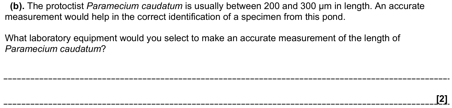
light microscope (1)
graticule (1)
compare and contrast SEMs and TEMs (5)
similarities
both use electrons
both use vacuums / dead tissue only
both have higher magnification and resolution than light microscopes
differences
electrons are transmitted through the specimen in TEM (internal structure visible); in SEM they are reflected off the surface (surface)
SEM 3D; TEM 2D
TEM gives higher resolution and maximum magnification
SEM can be used on thicker specimens than TEM
diff between prokaryotes and eukaryotes