15 Most Tested Radiograph Errors (DANB RHS)
1/18
There's no tags or description
Looks like no tags are added yet.
Name | Mastery | Learn | Test | Matching | Spaced | Call with Kai |
|---|
No analytics yet
Send a link to your students to track their progress
19 Terms
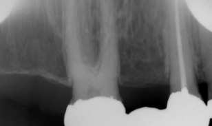
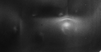
Cone Cut
What it looks like:
A clear white semicircle on the radiograph.
Cause:
The X-ray beam was not centered on the receptor.
Fix:
Center the PID properly.
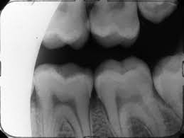
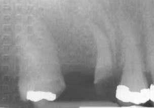
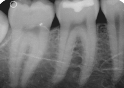
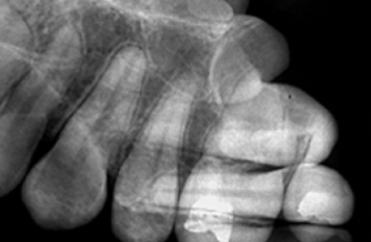
Overlapping Contacts
What it looks like:
Teeth appear stacked on top of each other and you can’t see between them.
Cause:
Incorrect horizontal angulation.
Fix:
Direct the beam through the contact points.
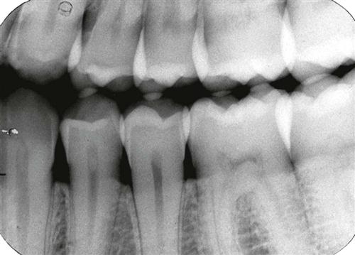
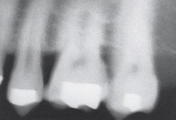
Foreshortening
What it looks like:
Teeth appear short and stubby.
Cause:
Excessive vertical angulation.
Fix:
Reduce vertical angulation.
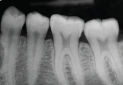
Elongation
What it looks like:
Teeth appear long and stretched.
Cause:
Insufficient vertical angulation.
Fix:
Increase vertical angulation.
Herringbone Pattern
What it looks like:
A tire-track or fishbone pattern across the image.
Cause:
Film placed backwards.
Fix:
Place the white side toward the X-ray beam.
Blurred Image
What it looks like:
The image appears fuzzy or unclear.
Cause:
Patient movement during exposure.
Fix:
Instruct patient to stay still.
Dark Radiograph
What it looks like:
Image is too dark.
Cause:
Overexposure or overdevelopment.
Fix:
Reduce exposure time.
Light Radiograph
What it looks like:
Image is too light.
Cause:
Underexposure or underdevelopment.
Fix:
Increase exposure time.
Film Bending
What it looks like:
Distorted or warped image.
Cause:
Receptor bent inside the mouth.
Fix:
Use proper film holders.
Apices Cut Off
What it looks like:
Roots of the teeth are missing from the image.
Cause:
Receptor not placed far enough into the mouth.
Fix:
Position receptor deeper.
Crowns Cut Off
What it looks like:
Top of teeth missing.
Cause:
Incorrect receptor placement.
Slanted Occlusal Plane
What it looks like:
Teeth appear tilted.
Cause:
Receptor not placed straight.
Double Image
What it looks like:
Two images overlap.
Cause:
Film exposed twice.
Partial Image
What it looks like:
Only part of the teeth appear.
Cause:
Beam not centered on receptor.
No Image
What it looks like:
Blank film.
Cause: Film not exposed.
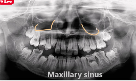
Maxillary sinus
dark area above upper molars
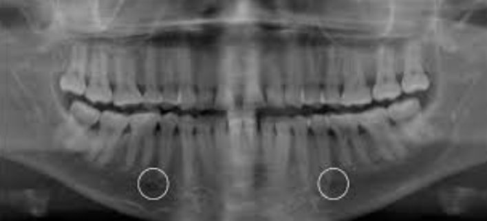
Mental foramen
dark circle near lower premolars
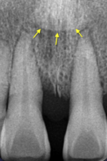
Incisive foramen
dark circle between upper central incisors
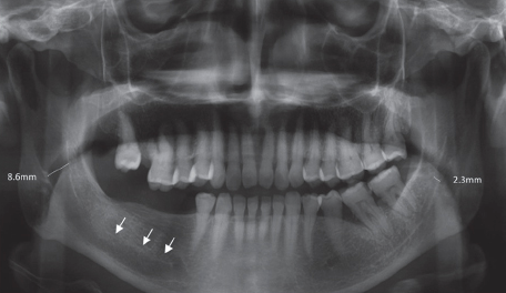
Mandibular canal
dark horizontal line in mandible