Anatomy: Final Review Topics
0.0(0)
Studied by 20 peopleCard Sorting
1/70
Earn XP
Description and Tags
Last updated 10:34 PM on 12/6/22
Name | Mastery | Learn | Test | Matching | Spaced | Call with Kai |
|---|
No analytics yet
Send a link to your students to track their progress
71 Terms
1
New cards
Absorption
Active uptake of molecules
2
New cards
Secretion
Active release of molecules
3
New cards
Absorbtion and secretion...
Usually involve microvilli= extensions to increase surface area
4
New cards
Diffusion
Molecules move down the concentration gradient
Passive (no energy used by cell)
Passive (no energy used by cell)
5
New cards
Filtration
Plasma (fluid component of blood) leaks across capillary walls
Passive (no energy used by cell)
Passive (no energy used by cell)
6
New cards
Propulsion
Cilia drive fluid along surface of epithelium
7
New cards
Functions of epithelium
Absorbtion
Secretion
Diffusion
Filtration
Propulsion
Sliding
Protection
Sensory reception
Secretion
Diffusion
Filtration
Propulsion
Sliding
Protection
Sensory reception
8
New cards
Protection
Stratified= multiple cell layers
9
New cards
Sensory reception (not nerves)
Epithelial cell generates sensory signal
Ear and tongue have special epithelial tissue
Ear and tongue have special epithelial tissue
10
New cards
Classification by layering
Simple vs stratified
11
New cards
Classification by shape
Squamous vs cubodial vs columnar
Named for the shape of the apical layer
Named for the shape of the apical layer
12
New cards
Simple squamous
Thinnest kind
No surface projections (cilia,microvilli)
Lining air sacs in lungs (aveoli)
Glomerular capsule in kidney (where filtration occurs)
No surface projections (cilia,microvilli)
Lining air sacs in lungs (aveoli)
Glomerular capsule in kidney (where filtration occurs)
13
New cards
Mesothelium
Simple squamus
Lining of closed body cavities
Lining of closed body cavities
14
New cards
Endothelium
Simple squamous
Inner lining of heart & blood vessels
Inner lining of heart & blood vessels
15
New cards
Simple cuboidal
Kidney tubules
Most glands
Larger cells provide more space for organelles
Most glands
Larger cells provide more space for organelles
16
New cards
Simple columnar (nonciliated)
Lines most of digestive tract
Provides more room for organelles than cubodial
Ex: goblet cells=secrete mucus (digestive and respiratory tracts)
Provides more room for organelles than cubodial
Ex: goblet cells=secrete mucus (digestive and respiratory tracts)
17
New cards
Simple columnar (ciliated)
Lines small bronchi (air tubes in lungs)= sweeps away debris
Also lining of uterine tube
Also lining of uterine tube
18
New cards
Psuedostratified columnar
All cells attached to basement membrane, so not truly stratified
Undifferentiated cells don't reach apical surface
Nuclei occur at different levels so tissue looks "stratified"
Undifferentiated cells don't reach apical surface
Nuclei occur at different levels so tissue looks "stratified"
19
New cards
Simple squamous major functions
Diffusion/filtration
Sliding
Sliding
20
New cards
Simple cuboidal/columnar/pseudostratified major functions
Absorbtion/secretion
Propulsion
Propulsion
21
New cards
Stratified squamous
Thickest
Function= protection
Keratinized= skin
Nonkeratinized= ends of digestive & reproductive tracts
Function= protection
Keratinized= skin
Nonkeratinized= ends of digestive & reproductive tracts
22
New cards
Stratified cuboidal
Large ducts of glands (not common)
Function= Protection and secretion
Function= Protection and secretion
23
New cards
Stratified columnar
In urethra
Function= Protection and secretion
Function= Protection and secretion
24
New cards
Transitional
Found in urinary bladdar
Apical cells change shape when tissue stretches
The shape of epithelium changes shape through the layers
Function= stretchable and protection
Apical cells change shape when tissue stretches
The shape of epithelium changes shape through the layers
Function= stretchable and protection
25
New cards
Connective tissue proper
Relatively unspecialized- "classic" CT
Cells: fibroblasts (secrete matrix) – become fibrocytes (maintain the matrix), and other cells
Six types, varying and density and types of fibers:
Loose connective tissue: lots of ground substance, fewer fibers
Dense connective tissue: lots of collagen fibers, good at resisting tension (pulling) forces
26
New cards
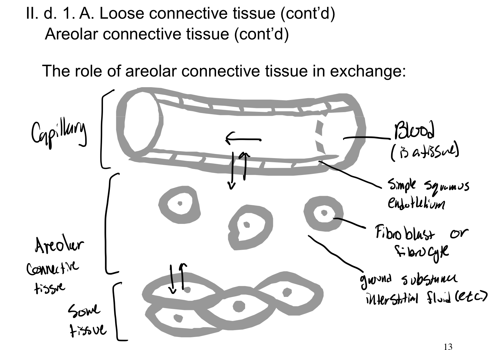
Connective tissue proper: areolar
Loose connective tissue
Most widespread, "generic" CT
Gel like matrix, lots of tissue fluid
Adjoins all epithelia
All three fiber types
Has defense cells to fight infections
Has fat cells to store energy
Most widespread, "generic" CT
Gel like matrix, lots of tissue fluid
Adjoins all epithelia
All three fiber types
Has defense cells to fight infections
Has fat cells to store energy
27
New cards
Connective tissue proper: adipose
Loose connective tissue
Matrix composition as in areolar CT
Less matrix than any other CT
Mostly fat cells
In hypodermis (superficial fascia)- deep to skin
Also "visceral fat" around internal organs
Energy storage, protection, insulation
Matrix composition as in areolar CT
Less matrix than any other CT
Mostly fat cells
In hypodermis (superficial fascia)- deep to skin
Also "visceral fat" around internal organs
Energy storage, protection, insulation
28
New cards
Connective tissue proper: Reticular CT
Loose connective tissue
The only fibers are reticular fibers
Exclusively in lymphoid structures (parts of the immune system, e.g. spleen, lymph nodes, red bone marrow)
Labyrinth used by defense cells
The only fibers are reticular fibers
Exclusively in lymphoid structures (parts of the immune system, e.g. spleen, lymph nodes, red bone marrow)
Labyrinth used by defense cells
29
New cards
Connective tissue proper: Dense regular CT
Dense connective tissue
All collagen fibers run in one direction (axis)
Resist tension in one direction (axis)
Tendons and ligaments
Fascia (deep fascia)
All collagen fibers run in one direction (axis)
Resist tension in one direction (axis)
Tendons and ligaments
Fascia (deep fascia)
30
New cards
Connective tissue proper: Dense irregular CT
Dense connective tissue
Collagen fibers oriented in various directions
Resists tension in various directions
In dermis, and fibrous capsules of organs/joints
Collagen fibers oriented in various directions
Resists tension in various directions
In dermis, and fibrous capsules of organs/joints
31
New cards
Connective tissue proper: elastic CT
Dense connective tissue
Most fibers are elastic fibers (also has collagen fibers)
Able to recoil after stretching
Bronchial tubes in lungs; artery walls; some intervertebral ligaments
Most fibers are elastic fibers (also has collagen fibers)
Able to recoil after stretching
Bronchial tubes in lungs; artery walls; some intervertebral ligaments
32
New cards
Body cavities
Open body cavities (continuous with the outside world), E.g., respiratory, digestive, reproductive, urinary tracts
Closed body cavities (not continuous with the outside world)
Closed body cavities (not continuous with the outside world)
33
New cards
Dorsal body cavity
Cranial cavity (brain)
Vertebral cavity (spinal cord)
Vertebral cavity (spinal cord)
34
New cards
Ventral body cavity
Visceral organs (viscera)
Thoratic cavity (heart, lungs)
Abdominopelvic cavity (mainly organs of digestive, urinary, reproductive systems)
Thoratic cavity (heart, lungs)
Abdominopelvic cavity (mainly organs of digestive, urinary, reproductive systems)
35
New cards
Membrane
Membrane= it's thin, pliable layer that covers or separates, E.g. plasma membrane, basement membrane
Concerned with multi cellular membranes that line the bodies cavities and surfaces
Membrane composed of epithelium layer + connective tissue layer
Concerned with multi cellular membranes that line the bodies cavities and surfaces
Membrane composed of epithelium layer + connective tissue layer
36
New cards
Membrane Functions (SPESS)
Protection
Exchange
Support
Sliding
Sensation
etc.
Exchange
Support
Sliding
Sensation
etc.
37
New cards
Mucous membrane (mucosa)
Produces mucus, enzymes, etc., by secretion (Active process)
Lines the lumen (internal space) of open body cavities, e.g., respiratory, digestive, reproductive, and urinary tracts
Lines the lumen (internal space) of open body cavities, e.g., respiratory, digestive, reproductive, and urinary tracts
38
New cards
Tissue layers (deep to superficial): Mucous membrane
Lumen
Epithelium (mucous membrane)
Lamina propria (connective tissue) (mucous membrane)
Muscle
Epithelium (mucous membrane)
Lamina propria (connective tissue) (mucous membrane)
Muscle
39
New cards
Serous Membrane
Lines the spaces between and around the organs in the ventral body cavity (a closed body cavity)
Lines outer surface of visceral organs
Lines inner surface of body wall
Produces watery fluid, mainly by filtration (passive process)- for lubrication
Mesothelium- simple squamous
Lines outer surface of visceral organs
Lines inner surface of body wall
Produces watery fluid, mainly by filtration (passive process)- for lubrication
Mesothelium- simple squamous
40
New cards
Tissue layers (deep to superficial): Serous membrane
Muscle
Connective tissue (visceral serosa)
Mesothelium (visceral serosa)
Serous cavity (fluid filled)
Mesothelium (parietal serosa)
Connective tissue (parietal serosa)
Muscle
Connective tissue (visceral serosa)
Mesothelium (visceral serosa)
Serous cavity (fluid filled)
Mesothelium (parietal serosa)
Connective tissue (parietal serosa)
Muscle
41
New cards
Serous membrane continued
Organs can have mucus membrane, serous membrane, both, or neither
Ventral body cavity (an internal body region) contains: visceral organs; serous cavity (a slit like space)
Ventral body cavity (an internal body region) contains: visceral organs; serous cavity (a slit like space)
42
New cards
Cutaneous membrane= the skin
Directly exposed to outside world
Has glands that secrete (active process)
Has glands that secrete (active process)
43
New cards
Tissue layers (deep to superficial): Cutaneous membrane
Muscle
Dermis (connective tissue) (cutaneous membrane)
Epidermis (epithelium) (cutaneous membrane)
Dermis (connective tissue) (cutaneous membrane)
Epidermis (epithelium) (cutaneous membrane)
44
New cards
Internal and external lining of G.I. Tract
Membrane= two layers (epithelium lines space; connective tissue nourishes epithelium)
mucosa (mucous membrane)
Serosa (serous membrane)
mucosa (mucous membrane)
Serosa (serous membrane)
45
New cards
mucosa (mucous membrane)
Lines lumen (interior space) of the G.I. tract
Has simple columnar epithelium throughout most of its length
Connective tissue layer called lamina propria
Has simple columnar epithelium throughout most of its length
Connective tissue layer called lamina propria
46
New cards
Serosa (serous membrane)
Called the peritoneum in abdominopelvic cavity
lines the peritoneal cavity (space between organs)
Epithelium (mesothelium) is simple squamous
CT layer
Deep (inner) lining of cavity= Visceral peritoneum= outer layer of organ wall
Superficial (outer) lining of cavity= Parietal peritoneum= inner layer of body wall
lines the peritoneal cavity (space between organs)
Epithelium (mesothelium) is simple squamous
CT layer
Deep (inner) lining of cavity= Visceral peritoneum= outer layer of organ wall
Superficial (outer) lining of cavity= Parietal peritoneum= inner layer of body wall
47
New cards
Serosa (serous membrane) (Continued)
Mesentery= double layer of peritoneum connecting parietal and visceral peritoneum
Supports G.I. tract, carries vessels and nerves, stores fat
During development, some organs in abdominopelvic cavity become buried in body wall= Secondarily retroperitoneal (e.g. pancreas)
Still has peritoneum on one side, has adventitia on the other side
Organs that keep their mesentery are called intraperitoneal (e.g. stomach)
48
New cards
Respiration function
Respiration= exchange of gases (oxygen and carbon dioxide) with the environment
Consists of 4 processes:
1. Ventilation= move air in/out of lungs
2. External respiration= gas diffuses between lungs and blood
3. Gas transport via blood
4. Internal respiration= gas diffuses between blood and cells
Steps 1 & 2 are functions of respiratory system
Steps 3 & 4 are functions of cardiovascular system
49
New cards
Zones
Conducting zone (passageways for air; no diffusion) (process=ventilation)
-external nose through most tubes in lungs
Respiratory zone= the anatomical zone where oxygen diffuses into blood (process= External respiration)
-pulmonary alveoli
-respiratory bronchioles
-external nose through most tubes in lungs
Respiratory zone= the anatomical zone where oxygen diffuses into blood (process= External respiration)
-pulmonary alveoli
-respiratory bronchioles
50
New cards
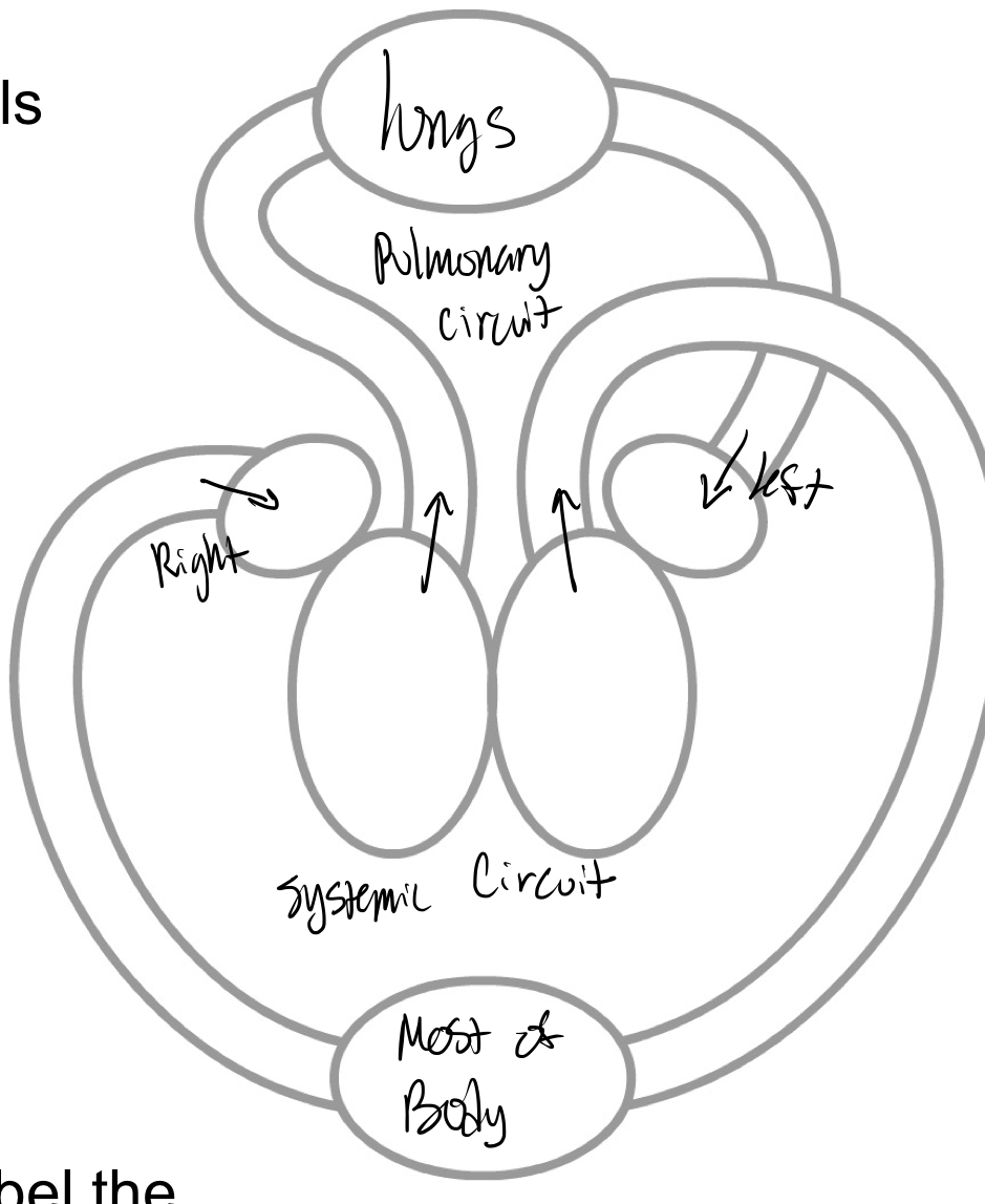
Circulatory routes
Pulmonary circuit= delivers blood to and from the lungs= for external respiration
Systemic circuit= delivers blood to and from the rest of the body= For internal respiration
Systemic circuit= delivers blood to and from the rest of the body= For internal respiration
51
New cards
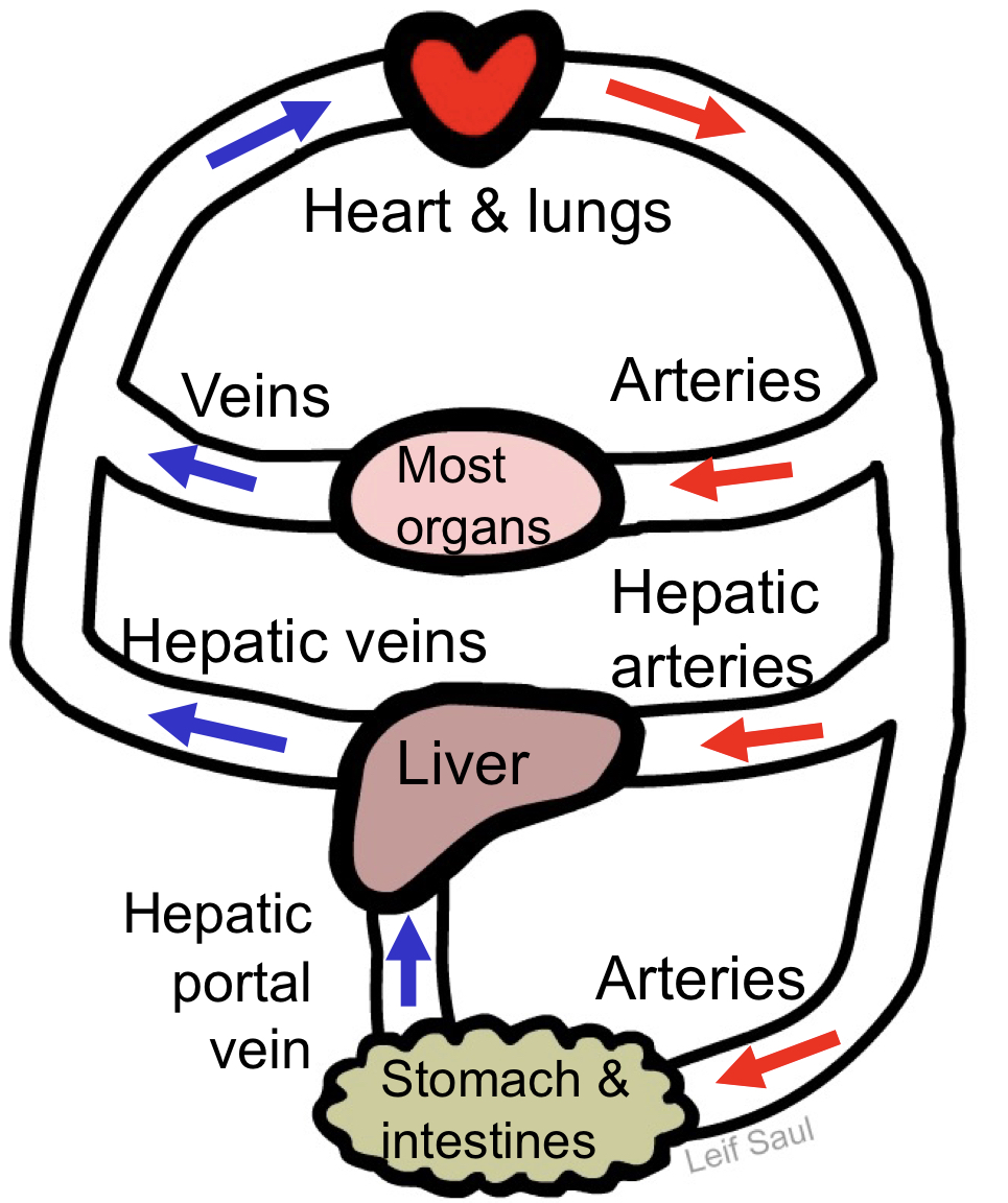
General circulatory principles
Capillary beds- where exchange takes place
Artery- delivers blood from the heart to capillary beds (oxygenated blood- most arteries not all)
Vein- delivers blood from capillary beds
Back to the heart (most veins)
Or to another capillary bed (these veins are portal veins)
Deoxygenated blood- most veins (not all)
Artery- delivers blood from the heart to capillary beds (oxygenated blood- most arteries not all)
Vein- delivers blood from capillary beds
Back to the heart (most veins)
Or to another capillary bed (these veins are portal veins)
Deoxygenated blood- most veins (not all)
52
New cards
Hepatic portal system
53
New cards
Chambers and vessels overview
54
New cards
Chambers and vessels
Atria (receive blood from veins)
Right atrium: receives deoxygenated blood from: inferior and superior vena cava; coronary sinus- returns blood from heart tissue
Left atrium: receives oxygenated blood from: pulmonary veins
Ventricles (eject blood from heart)
Right ventricle: pumps deoxygenated blood to: pulmonary trunk -> pulmonary arteries
Left ventricle: pumps oxygenated blood to:
aorta- coronary arteries (supplying blood to heart tissues) are branches of aorta
55
New cards
Heart valves
Prevent backflow of blood
Atrioventricular valves
Semilunar valves
Mnemonic: tri before you bi
Atrioventricular valves
Semilunar valves
Mnemonic: tri before you bi
56
New cards
Atrioventricular valves (AV valves)
Between atrium and ventricle
Tricuspid (R AV) valve- between the right atrium and ventricle
Bicuspid (mitral, L AV) valve- between left atrium and ventricle
These valves held in place by chordae tendinae, which are anchored to papillary muscles- prevents eversion (prolapse)
Tricuspid (R AV) valve- between the right atrium and ventricle
Bicuspid (mitral, L AV) valve- between left atrium and ventricle
These valves held in place by chordae tendinae, which are anchored to papillary muscles- prevents eversion (prolapse)
57
New cards
Semilunar valves (SL valves)
Between great arteries and ventricles
Aortic SL valve- between left ventricle and aorta
Pulmonary SL valve- between right ventricle and pulmonary trunk
Aortic SL valve- between left ventricle and aorta
Pulmonary SL valve- between right ventricle and pulmonary trunk
58
New cards
Heart sounds in each heartbeat
First heart sound (“lub”)= closing of both AV valves when L and R ventricles begin contracting
Second heart sound (“dup”)= closing of both SL valves when L and R ventricles begin relaxing
Second heart sound (“dup”)= closing of both SL valves when L and R ventricles begin relaxing
59
New cards
Lymphatic system function
A system of vessels and nodes that returns excess tissue fluid to the blood
Needed because plasma tends to leak out of blood capillaries
Note: most cells get oxygen and nutrients directly from interstitial fluid
Filters pathogens to be targeted by immune system
Needed because plasma tends to leak out of blood capillaries
Note: most cells get oxygen and nutrients directly from interstitial fluid
Filters pathogens to be targeted by immune system
60
New cards
Lymphatic system: Pathway of flow
Throughout most of the body, tissue fluid (interstitial fluid) enters lymphatic capillaries- The fluid is now called lymph
Moves through lymph vessels and lymph nodes
Returned to the blood at veins at base of the neck
Moves through lymph vessels and lymph nodes
Returned to the blood at veins at base of the neck
61
New cards
lympahtic system: Structures
Very low pressure system- uses valves to maintain flow direction
Lymphatic capillaries: Wall= Endothelium (simple squamous epithelium); Minivalves formed by loose edges of cells
Larger lymph vessels- has valves (similar to those of veins)
Lymph nodes- contains reticular CT (with reticular fibers); lymph flows through; pathogens are filtered out by immune cells
Lymphatic capillaries: Wall= Endothelium (simple squamous epithelium); Minivalves formed by loose edges of cells
Larger lymph vessels- has valves (similar to those of veins)
Lymph nodes- contains reticular CT (with reticular fibers); lymph flows through; pathogens are filtered out by immune cells
62
New cards
lymphatic system Pathology
Lymphedema= accumulation of interstitial fluid due to poor lymphatic drainage
Example (extreme): elephantitis- caused by parasitic worm
Example (extreme): elephantitis- caused by parasitic worm
63
New cards
Functional classification
Sensory (afferent) neurons
Carries signal to CNS
Make up the sensory division of PNS
Note- axon terminal is (typically) in CNS
Motor (efferent) neurons
Carries signal from CNS to effector
Make up the motor division of PNS
Note- cell body is (typically) in CNS
Interneurons- between sensory and motor neurons- found entirely within CNS
64
New cards
Structural classification
Multipolar: many processes (many dendrites, 1 axon), most neurons (“typical”)
Bipolar: One axon, One fused dendrite, found in some sensory organs
Unipolar (pseudounipolar): typical sensory neurons, receptive endings; no dendrites
Axon with peripheral and central processes
Bipolar: One axon, One fused dendrite, found in some sensory organs
Unipolar (pseudounipolar): typical sensory neurons, receptive endings; no dendrites
Axon with peripheral and central processes
65
New cards
Nervous system: Gross anatomy
Nerve= group of axons traveling together in PNS
Endoneurium= surrounds axon
Perineurium= surrounds each subgroup of axons
Epineurium= wraps around outside of nerve
Fascicle= subgroup of axons
Endoneurium= surrounds axon
Perineurium= surrounds each subgroup of axons
Epineurium= wraps around outside of nerve
Fascicle= subgroup of axons
66
New cards
Nerves versus neurons
Each nerve contains the axons of many neurons
Axons are also called nerve fibers
Axons carry signals toward the axon terminals
Most nerves carry signals in both directions- because contain neurons oriented in both directions
The only exception are some cranial nerves
Axons are also called nerve fibers
Axons carry signals toward the axon terminals
Most nerves carry signals in both directions- because contain neurons oriented in both directions
The only exception are some cranial nerves
67
New cards
Ganglion
Collection of neuron cell bodies in the PNS
68
New cards
Gray and white matter
Two general types of nervous tissue in the CNS
White matter= myelinated axons
Gray matter= everything else (Unmyelinated axons, dendrites, cell bodies)
White matter= myelinated axons
Gray matter= everything else (Unmyelinated axons, dendrites, cell bodies)
69
New cards
Tract
Collection of axons traveling together inside the CNS
White matter
White matter
70
New cards
Nucleus
Collection of neuron cell bodies inside the CNS
Gray matter
Gray matter
71
New cards
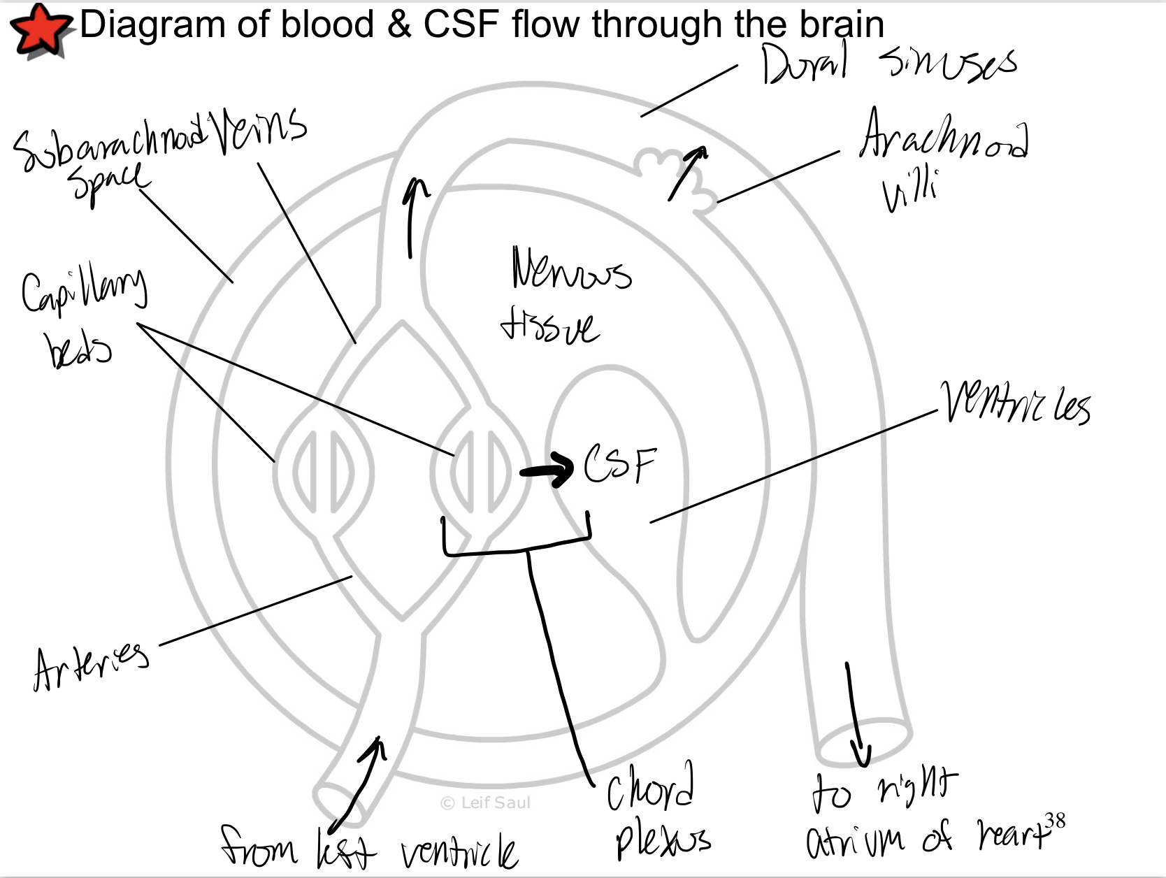
Diagram of Blood and CSF flow through the brain