sound and hearing
1/67
There's no tags or description
Looks like no tags are added yet.
Name | Mastery | Learn | Test | Matching | Spaced |
|---|
No study sessions yet.
68 Terms
sound
collisions created when objects vibrate; in gas (air), it is changed in pressure (compression and rarefaction)
frequency
the number of times per second that a pattern of pressure repeats; related to wavelength of sound: wavelength=speed/frequency; related to pitch - perceptual quality of how “high” or “low” a sound is
amplitude
the magnitude of displacement of sound pressure waves; - related to loudness - the perceived intensity of a sound
loudness
the perceived magnitude of sound intensity
decibel
a unit for measuring sound intensity - represent the log of the ratio of sound pressure level (spl) of the measured sound to that of that of the absolute threshold (barely detectable) sound
sine waves
pure tones; the waveform for which variation as a function of time is a sine function
fourier analysis
breaking down complex signals or functions into simpler components
harmonic spectrum
the spectrum of a complex sound in which energy is at integer multiples of the fundamental frequency
fundamental frequency
the lowest-frequency component of a complex period sound
timbre
the psychological sensation by which a listener can judge that two sound with the same loudness and pitch are different (quality conveyed by harmonics and other high frequencies
outer ear, middle ear, inner ear
peripheral auditory system
pinna and ear canal
outer ear
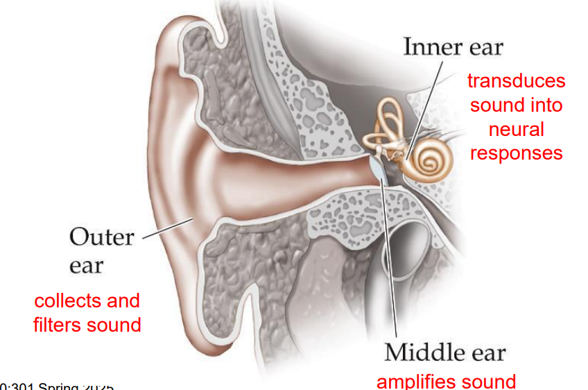
ossicles (bones)
middle ear

cochlea and auditory nerve
inner ear

outer ear
collects and filters sound
middle ear
amplifies sound
inner ear
transduces sound into neural responses
purpose of pinnae
– Sounds are first collected from the environment by the pinnae
– The directional sound filtering properties of the pinnae are very important for sound localization
– The pinnae funnel sound waves into the ear canal
purpose of ear canals
– The length and shape of the ear canal enhances certain sound frequencies
– To collect sound waves and funnel them to the tympanic membrane
– To insulate and protect the tympanic membrane
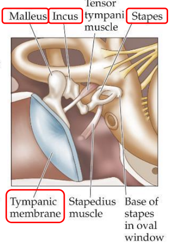
tympanic membrane (ear drum)
the border between outer ear and middle ear; thin sheet of skin at the end of the outer ear canal, vibrates in response to sound
ossicles
smallest bones in your body, located in the middle ear; malleus, incus, stapes

malleus
hammer; receives vibrations from the tympanic membrane and is attached to the incus

incus
anvil; the middle ossicle; transmits vibrations from the malleus to the stapes

stapes
stirrup; connected to the incus on one end and the oval window

oval window
the border between the middle ear and inner ear
organ of corti
spiral structure of inner ear
vestibular canal, tympanic canal, middle canal
three canals in the cochlea
vestibular canal
extends from oval window at base of cochlea to helicotrema at the apex - canal closest to ossicles and through which pressure waves move first

tympanic canal
extends from the helicotrema at the apex to the round window at the base of the cochlea


middle canal
sandwiched between the vestibular and tympanic canals and contains the cochlear partition
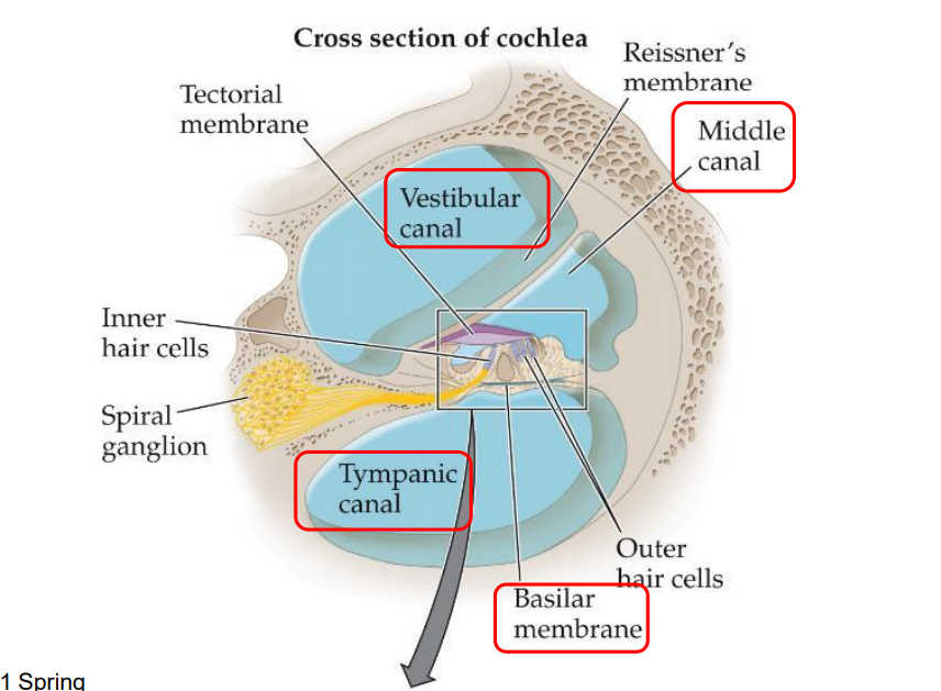
basilar membrane
plate of fibers that forms the base of the cochlear partition and separates the middle and tympanic canals in the cochlea s

hair cells
cells that support the stereocilia, which transduce mechanical movement in the cochlea into neural activity sent to the brain stem - some hair cells also receive input from the brain - arranged in four rows that run down length of basilar membrane
stereocilia
hairlike extensions on the tips of the hair cells in the cochlea that initiate the release of neurotransmitters when they are flexed; the tip of each stereocilium is connected to the side of its neighbor by a tiny filament called a tip link
tectorial membrane
extends into the middle canal of the ear, floating above inner hair cells and touching outer hair cells
inner hair cells
convey almost all info about sound waves to the brain (using afferent fibers)
outer hair cells
convey info from the brain (using efferent fibers)
outer ear
pinna → ear canal → tympanic membrane
middle ear
malleus → incus → stapes
inner ear
oval window → vestibular canal → helicotrema → tympanic membrane → round window
place code
tuning of different parts of the cochlea to different frequencies (info about the particular frequency of an incoming sound wave is coded by the place along the cochlear partition with the greatest mechanical displacement, info about amplitude (intensity) at a particular frequency is coded by the firing rate of neurons tuned to that frequency)
coding of frequency
info of incoming sound wave coded along the place along the cochlear partition with the greatest mechanical displacement
coding of amplitude
info about sound intensity at a particular frequency coded by the firing rate of neurons tuned to that frequency
threshold tuning curve
a function plotting the response threshold of a neuron as a function of frequency
characteristic frequency
frequency to which the neuron is most sensitive
high spontaneous fibers
respond more easily to low SPL, but reach saturation more quickly
rate saturation
nerve fiber is firing as quickly as possible and thus incapable of increasing the firing rate
low spontaneous fibers
require more SPL to fire and continue increasing firing rate to higher SPLs
phase locking
tendency of a neuron to fire at particular phases of an ongoing periodic sound waveform - hair cells are only depolarized when moved in one direction, not in the other
temporal coding
auditory systems ability to process and encode info based on the timing of soundwaves rather than just their frequency and intensity
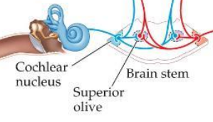
cochlear nucleus
first synapse of the AN fibers in the brain (in brain stem)

superior olive
brain stem region in the auditory pathway where inputs from both ears first converge
inferior colliculus
midbrain nucleus in auditory pathway
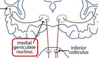
medial geniculate nucleus (MGN)
part of the thalamus that relays auditory signals to the cortex (analogous to the LGN in vision) s

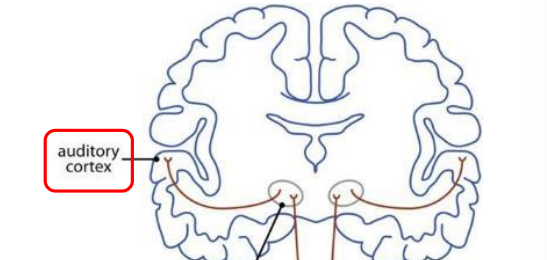
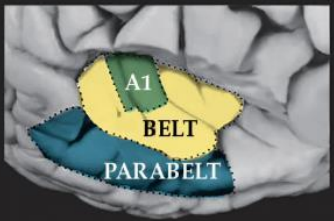
primary auditory cortex (a1)
first cortical area for processing audition (located in temporal lobe)
belt area
region of cortex directly adjacent to a1 (and receives its inputs) where neurons respond to more complex sound characteristics

parabelt area
region of cortex adjacent to belt area where neurons respond to more complex characteristics of sounds as well as input from other senses
volley principle
explains how the auditory system encodes high frequency sounds by having groups of neurons fire action potentials slightly out of sync with each other - synchronized firing creates a “volley” of impulses that, when combined, effectively represent frequencies higher than what a single neuron could individually encode
locus of processing
large portion of processing occurs beyond v1
topographic organization
tonotopic (frequency - based) organization); starts in cochlea, maintained all the way through primary auditory cortex (a1)
psychoacoustics
study of the psychological correlates of the physical dimensions
physical properties
frequency, amplitude/intensity (SPL)
psychological percept
pitch, loudness
masking
using a second ( e.g., noise ) to make the detection of another sound more difficult
critical bandwidth
range of frequencies conveyed within a channel
conductive hearing loss
hearing loss caused by impairment of the mechanical transmission of sound waves to the cochlea
sensorineural hearing loss
hearing loss caused by damage to the cochlea or to the auditory nerve
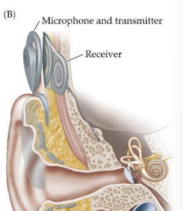
cochlear implant
tiny flexible coils with mini electrode contacts, surgeons thread implant through round window toward cochlear apex , tiny mic transmits radio signals to receiver int the scalp, computer chip performs Fourier transform and stimulates appropriate location in cochlea for each frequency
hearing aids
earliest devices were horns, today, electronic