Musculoskeletal System 1
1/100
There's no tags or description
Looks like no tags are added yet.
Name | Mastery | Learn | Test | Matching | Spaced |
|---|
No study sessions yet.
101 Terms
What is the function of the musculoskeletal system?
It provides form, stability, and movement to the human body
What are the main components of the musculoskeletal system?
The main components are muscles, bones, joints, cartilage, tendons, and ligaments.
What type of muscle is found in the musculoskeletal system?
Skeletal (or striated) muscles.
What do bones make up in the musculoskeletal system?
Bones make up the skeleton.
What are joints?
Joints are the union between two or more bones.
What is the role of cartilage in the musculoskeletal system?
Cartilage provides cushioning and support at the joints.
What is the function of tendons?
Tendons are fibrous bands that connect muscles to bones
What do ligaments do in the musculoskeletal system?
Ligaments are fibrous bands that connect bones to other bones
How are the skeletal and muscular systems connected?
They are connected by connective tissues, such as tendons and ligaments.
What is the skeleton composed of?
The skeleton is composed of bone and cartilage
What type of tissue is bone?
Bone is a living tissue and a highly specialised hard connective tissue.
What are the functions of bones?
Bones provide protection for vital structures, support for the body, a mechanical basis for movement (leverage), storage for salts (e.g. calcium), and a continuous supply of new blood cells.
What is the function of cartilage in the skeletal system?
Cartilage is a semi-rigid, resilient connective tissue that supports joints and provides cushioning
What does the muscular system consist of?
The muscular system consists of all the muscles of the body
What is the function of muscle cells (muscle fibres)?
Muscle cells (muscle fibres) produce contractions that move body parts, including internal organs.
What are the three types of muscle in the human body?
The three types of muscle are skeletal striated muscle, cardiac striated muscle, and smooth muscle (unstriated)
What is the function of skeletal muscle?
Skeletal muscle (voluntary) moves bones and other structures, such as the eyes.
Where is cardiac muscle found, and what type of muscle is it?
Cardiac muscle is involuntary and forms the walls of the heart and adjacent parts of the great vessels, such as the aorta.
What is the role of smooth muscle, and where is it found?
Smooth muscle (involuntary) forms part of the walls of vessels and hollow organs (viscera), such as the intestines.
How can skeletal muscles be classified?
Skeletal muscles can be classified according to their shape.
What is an example of a flat muscle, and where is it located?
An example of a flat muscle is the external oblique muscle, which covers the abdomen.
What is an example of a quadrate muscle?
An example of a quadrate muscle is the rectus abdominis (commonly known as the "six-pack")
What is an example of a fusiform muscle, and where is it located?
An example of a fusiform muscle is the biceps brachii, located in the arm.
What is an example of a circular (or sphincteral) muscle, and where is it located?
An example of a circular muscle is the orbicularis oris, which is located around the lips
What is an example of a unipennate muscle, and where is it located?
An example of a unipennate muscle is the extensor digitorum longus, located in the leg.
What is an example of a bipennate muscle, and where is it located?
An example of a bipennate muscle is the rectus femoris, located in the anterior thigh
What is the primary function of skeletal muscles?
Skeletal muscles produce movements of the skeleton
What is a tendon, and what is its role?
A tendon is the white, non-contractile portion of a muscle that attaches the muscle to the bone.
How do skeletal muscles function?
Skeletal muscles function by contracting (shortening); they pull and never push.
What is the muscle belly, and what is its function?
The muscle belly is the fleshy, red, contractile part of the muscle responsible for producing movement.
What does the term "muscle" refer to?
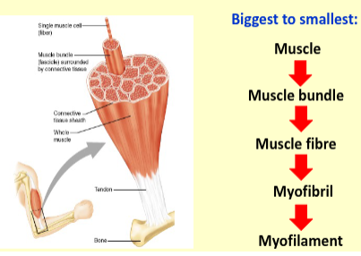
The term "muscle" refers to a number of muscle fibres bound together by connective tissue.
How are muscles usually linked to bones?
Muscles are usually linked to bones by tendons
What is a single skeletal muscle cell called?
A single skeletal muscle cell is known as a muscle fibre.
What is a motor unit?
a single motor neurone and the muscle fibres it innervates
What is a motor neuron pool?
A motor neuron pool is all the motor neurons that supply one complete muscle.
Where are the cell bodies of the motor neuron pool for a given muscle located?
The cell bodies of the motor neuron pool are located close to each other either in the spinal cord (for lower motor neurons) or in the brainstem (for upper motor neurons).
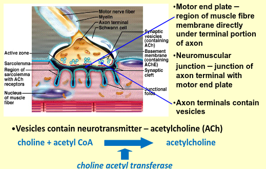
Neuromuscular junction
What is the resting potential of a neuron?
The resting potential is when the neuron is at rest, with a stable negative electrical charge
What is an action potential (AP)?
An action potential (AP) is a burst of electrical activity created by depolarising current, fired when depolarisation reaches a threshold.
When is an action potential fired?
An action potential is fired when the depolarisation of the neuron reaches a threshold
What happens during depolarisation?
During depolarisation, sodium (Na+) channels open, allowing Na+ ions to enter the neuron, making the inside of the neuron positively charged
What happens after depolarisation in the neuron?
Potassium (K+) channels open, allowing K+ ions to rush out, reversing the depolarisation
What occurs once potassium (K+) channels close?
Once K+ channels close, the action potential (AP) returns to a resting potential of approximately -80 mV
Recall the events that occur at the neuromuscular junction
1. Arrival of action potential (AP) at motor nerve terminals
2. AP at nerve terminal causes influx of Ca2+ into axon terminals
though Ca 2+ channels
3. Ca 2+ causes release of ACh from synaptic vesicles
4. Ach diffuses from axon terminals to motor end plate (MEP) in
muscle fibre
5. Ach binds to nicotinic receptors on MEP – activation of ion channels
and influx of Na + ions
6. Depolarisation of membrane and generation of action potential
What can block the ACh receptor and what effect does it have?
The ACh receptor can be blocked by curare (arrowhead poison), leading to muscle paralysis.
Is ACh binding to its receptor reversible?
Yes
What does MEP contain and what is its role?
MEP (Motor End Plate) contains acetylcholinesterase, which breaks down acetylcholine (ACh).
What happens to choline after ACh is broken down?
Choline is transported back to the axon terminals to be reused in the new synthesis of ACh.
What happens to ion channels when ACh is no longer bound to receptors?
Ion channels close when receptors no longer contain bound ACh.
Muscle hierarchy of organisation
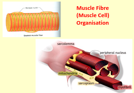
Muscle fibre (muscle cell) organisation
What characteristic pattern is seen in skeletal muscle fibres under a light microscope?
Skeletal muscle fibres display a characteristic striped pattern, known as striated muscle, consisting of a series of light and dark bands.
What causes the striated pattern in skeletal muscle fibres?
The striated pattern is caused by the arrangement of thick and thin filaments in the cytoplasm into cylindrical bundles called myofibrils.
How are thick and thin filaments arranged in myofibrils?
Thick and thin filaments are arranged in a repeating pattern within myofibrils.
What is one unit of the repeating pattern in myofibrils called?
One unit of the repeating pattern is called a sarcomere
What are thick filaments made of?
Thick filaments are composed of the contractile protein myosin
What are thin filaments made of?
Thin filaments are composed of the contractile protein actin.
What other proteins are found in thin filaments, and what is their function?
Thin filaments also contain the proteins troponin and tropomyosin, which regulate contraction
Arrangement of thick and thin filaments
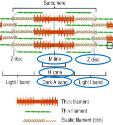
What is the dark band of thick filaments called?
A band
Where are the thin filaments anchored in a sarcomere?
The ends of thin filaments are anchored to interconnecting proteins at the Z line.
What is the I band in a sarcomere?
The I band is the portion of thin filaments that do not overlap with thick filaments, and it appears as a light band.
What is the H zone?
The H zone is a narrow, light band in the centre of the A band where thick filaments do not overlap with thin filaments
What is the M line?
The M line consists of proteins that link together the central region of thick filaments in a sarcomere
What bridges the space between overlapping thick and thin filaments?
The space between overlapping thick and thin filaments is bridged by projections called cross bridges.
How do myosin molecules extend from thick filaments?
Myosin molecules extend from the surface of the thick filament.
What happens during muscle contraction involving cross bridges?
During muscle contraction, cross-bridges make contact with thin filaments, exerting force on them to cause contraction.
What proteins regulate muscle contraction in the thin filament?
Troponin and tropomyosin regulate muscle contraction in the thin filament.
What is the structure of tropomyosin?
Tropomyosin is a rod-shaped molecule composed of two intertwined polypeptide chains
What is the structure and function of troponin?
Troponin is a small globular protein bound to both tropomyosin and actin, playing a key role in regulating contraction.
What do the two chains of tropomyosin on the thin filament regulate?
The two chains of tropomyosin regulate the access of cross-bridges to the actin binding sites
What happens when cross-bridges bind to actin?
When cross-bridges bind to actin, the tropomyosin molecules are moved away from their blocking positions on actin
When does the movement of tropomyosin occur?
The movement of tropomyosin occurs when Ca²⁺ binds to specific sites on troponin
What happens when Ca²⁺ binds to troponin?
The binding of Ca²⁺ causes a change in the shape of troponin, which drags tropomyosin away from the myosin binding site on actin.
What happens when Ca²⁺ is removed?
The removal of Ca²⁺ reverses the process, causing tropomyosin to return to its blocking position on actin.
What is rigor mortis?
Rigor mortis is the stiffening of skeletal muscles that begins several hours after death and is complete after approximately 12 hours.
Why does rigor mortis occur after death?
Rigor mortis occurs because ATP concentration in cells falls after death. The nutrients and oxygen required for ATP production are no longer supplied by circulation.
What happens to the actin and myosin links in the absence of ATP?
Without ATP, the link between actin and myosin does not break, causing the thick and thin filaments to remain bound by immobilised cross-bridges, resulting in muscle rigidity.
When does the stiffness of rigor mortis disappear?
The stiffness of rigor mortis disappears 48-60 hours after death due to the disintegration of muscle tissue
What is muscle contraction?
Muscle contraction is the activation of force-generating sites within muscle fibres, known as cross-bridges
How does skeletal muscle fibre shorten during contraction?
During contraction, the overlapping thick and thin filaments move past each other, propelled by the cross-bridges.
Do the lengths of thick or thin filaments change during muscle contraction?
No, the lengths of thick or thin filaments do not change; the sliding filament mechanism causes them to overlap more, shortening the muscle.
What happens to the H zone during muscle contraction?
During contraction, the H zone (the area where thick and thin filaments do not overlap) is reduced.
What happens to sarcomeres during muscle contraction?
Sarcomeres shorten during muscle contraction.
How is the I band affected during muscle contraction?
The I band (the light band where only thin filaments are present) is reduced during contraction.
What happens during the shortening of muscle fibres in relation to myosin cross-bridges?
During shortening, each myosin cross-bridge attached to a thin filament (actin) moves in an arc
How do multiple cross-bridges contribute to muscle contraction?
The swivelling motion of many cross-bridges forces thin filaments attached to successive Z lines towards the centre of the sarcomere, thereby shortening the sarcomere
What initiates the binding of cross-bridges to actin during muscle contraction?
The binding of calcium ions (Ca²⁺) initiates the binding of cross-bridges to actin.
What is the cross-bridge cycle?
The cross-bridge cycle is the sequence of events between the time a cross-bridge binds to the thin filament, moves, and then is set to repeat.
What are the four steps in the cross-bridge cycle?
Attachment of the cross-bridge to the thin filament.
Movement of the cross-bridge, producing tension in the thin filament.
Detachment of the cross-bridge from the thin filament.
Energising the cross-bridge so that it can attach to a thin filament and repeat the cycle.
What happens when myosin (M) is at rest with ADP and Pi?
it is energised by the splitting of ATP, producing ADP and Pi.
What occurs during the activation step of the cross-bridge cycle?
During activation, actin (A) binds to the myosin (M) cross-bridge, forming the complex A.M.ADP.Pi
What happens during cross-bridge movement?
During cross-bridge movement, the complex A.M.ADP.Pi undergoes a conformational change, generating force and tension.
What causes cross-bridge dissociation from actin?
Cross-bridge dissociation from actin occurs when ATP binds to the myosin (M), forming A + M.ATP
What happens after cross-bridge dissociation in terms of ATP?
After dissociation, ATP is hydrolyzed into ADP and Pi, energising the cross-bridge to prepare for the next cycle.
Sarcoplasmic reticulum
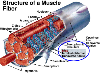
What happens during the activation of contractile machinery in muscle fibres?
the concentration of Ca²⁺ in the sarcoplasm rises, which activates the contractile machinery
How is Ca²⁺ released during muscle contraction?
Ca²⁺ is released from the sarcoplasmic reticulum (SR) in response to the depolarisation of T-tubules.
What occurs after contraction in terms of Ca²⁺?
After contraction, the activation of the contractile machinery declines, and Ca²⁺ is pumped back into the SR, preparing for future contractions.
What is required for Ca²⁺ to be pumped back into the SR?
The process of pumping Ca²⁺ back into the SR is active and requires the splitting of ATP