Neurophysiology
1/60
There's no tags or description
Looks like no tags are added yet.
Name | Mastery | Learn | Test | Matching | Spaced | Call with Kai |
|---|
No analytics yet
Send a link to your students to track their progress
61 Terms
PNS is broken into what 2 divisions
Sensory (afferent) and motor (efferent)
Motor is broken into what 2 divisions
Autonomic (involuntary/visceral) and Somatic (voluntary/skeletal muscles)
Autonomic is broken into what 2 divisions
Sympathetic and Parasympathetic
What is the criteria for Neural Regeneration to occur and why is it important
when axons are severed, they can repair themselves if:
- damage occurs outside the CNS
- does not affect neuron's body
*mature neurons are amitotic (do not divide)
*if damage is severe or is close to cell body, then the whole neuron may die and others stimulated by its axon may die too
Neural Regeneration Steps
1. Wallerian degeneration - distal end (further from body) of axon dies
2. proximal end of axon dies back toward cell body
3. As the two ends seal themselves, substances being transported along the axon accumulate and the ends swell
4. Schwann cells phagocytize the disintegrating axon and myelin.
5. proximal end of axon gives rise to a growth cone - grows out to effector organs
6. Schwann cells form cellular cords that guide the regenerating axon - "sprouts" across the gap and to their original contacts.
7. Schwann cells release growth factors (nerve growth factor = NGF)
8. Inhibitory factors released to tell axon to stop growing
Regeneration in the CNS never occurs in distances more than
1mm
Why is damage in the CNS views as irreversible?
- the microglial cells and astrocytes rid the area of deteriorating cell debris
- the oligodendrocytes die and fail to oversee and guide fiber regrowth
- the dead oligodendrocytes are replaced with a glial scar which blocks sprouting axons
- the myelin sheaths of nearby axons contain growth inhibiting
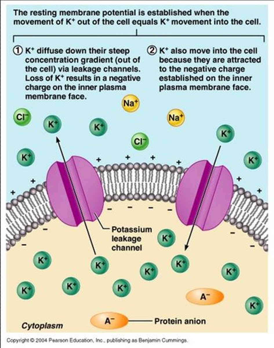
Resting Membrane Potential
- 70mV (more negative inside than outside)
- K+ more inside so it diffuses out
- Na+ more outside so it diffuses in
- non diffusible protein anions inside
- ATPase pump ejects 3 Na+ out for every 2 K+ it pumps in (establishes concentration gradient)
what are graded potentials
- voltage changes in membrane potential that by themselves are insufficient to trigger an action potential
- over short distances/in small area
- usually at cell body/dendrites
- fade as they go (short lived = decrimental)
how are graded potentials triggered and what are the types
- stimulus in the neurons environment that causes chemically gates ion channels to open
- Anything that changes the membrane permeability to any type of ion
- Anything that alters ion concentrations on the two sides of the membrane
- depolarization: more positive (ligand gated Na+ open and flows in)
- hyperpolarization: more negative (ligand gates K+ open and flow out)
what do graded potentials allow
short range communication and prime the neuron for generation of action potential
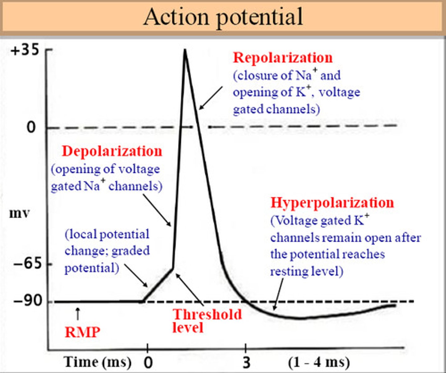
Action Potential Phases
1. resting state: all gated Na+ and K+ channels closed (Na+ activation gates closed and inactivation gates open)
2. depolarization - threshold reaches (-55mV) and Na+ voltage gated channels open at hillock due to graded potential and enters axon
* positive feedback: Na+ enters axon, further depolarizes, opens more channels, more Na+ enters (all or nothing response)
* K+ gates are closed
3. repolarization - Na+ gates close and voltage gates K+ channels open and K+ rush out of cell
4. hyperpolarization - K+ channels from the repolarization phase close much more slowly, so there is a small period of undershoot
What restores RMP
Activation of the Sodium/Potassium pump: sodium is pumped out of the axon and potassium is pumped into the axon. The axon is now ready to be depolarized again
When does Propagation or conduction of the AP occur; in what direction and why, and what's different from muscle cells
- as nearby sodium voltage gates open
- Depolarization of adjacent membrane areas in the forward direction only, triggers the AP on down the axon
- Conduction is always in one direction in nerve cells, away from the point of origin due to refractory periods (different in muscle cells, can move in either direction)
- Sodium gates open in "front" of the depolarization and close at the "back" of depolarization
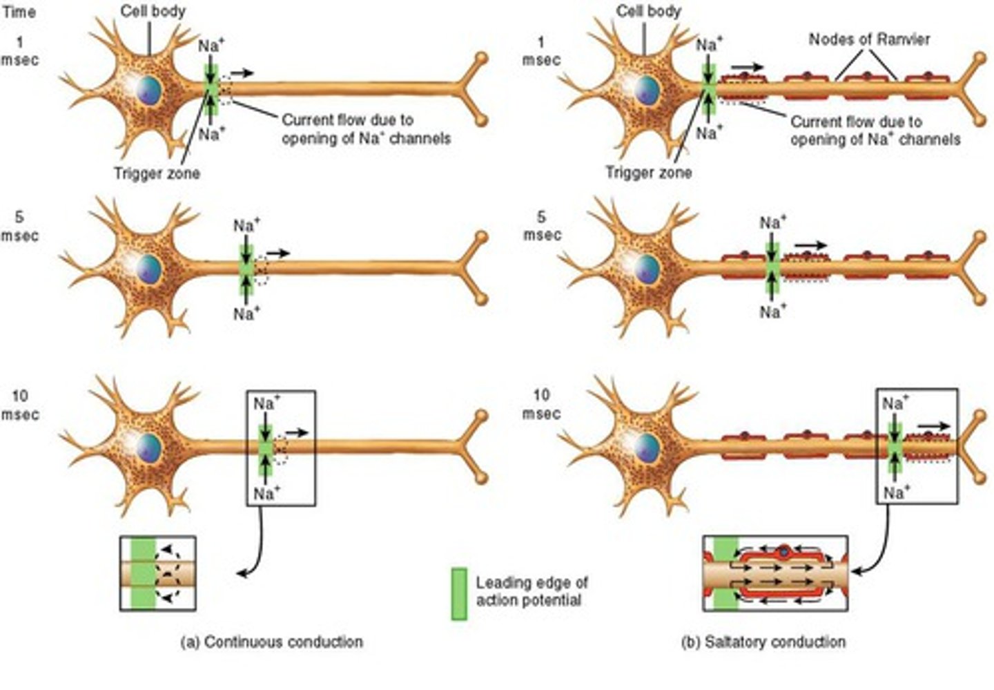
Local (passive) conduction
- spread of depolarization from site of stimulation to adjacent segments of the axon
- unmyelinated
- spreads gradually and decays with distance along length of axon
Saltatory conduction
- current passes through membrane at nodes of ranvier
- depolarization jumps from node to node
- myelinated axons (prevents leakage of charge from axon)
C-fibers
action potential is regenerated at each segment of the neuron membrane moving in a wave-like pattern. Occurs along unmyelinated axons and dendrites (slow)
A-fibers and what does it supply
- Saltatory Conduction
- Myelinated axons
- When an AP is generated, depolarization , moves to the next node
- covers less surface area
- Found in large diameter nerves that supply the skeletal muscles, joints, and large sensory nerves
B-fibers and what does it supply
smaller diameter myelinated neurons that have slower conduction velocities than A fibers. Found in nerves that conduct impulses from the viscera to the brain and spinal cord and the ANS
absolute refractory period
The minimum length of time after an action potential during which another action potential cannot begin.
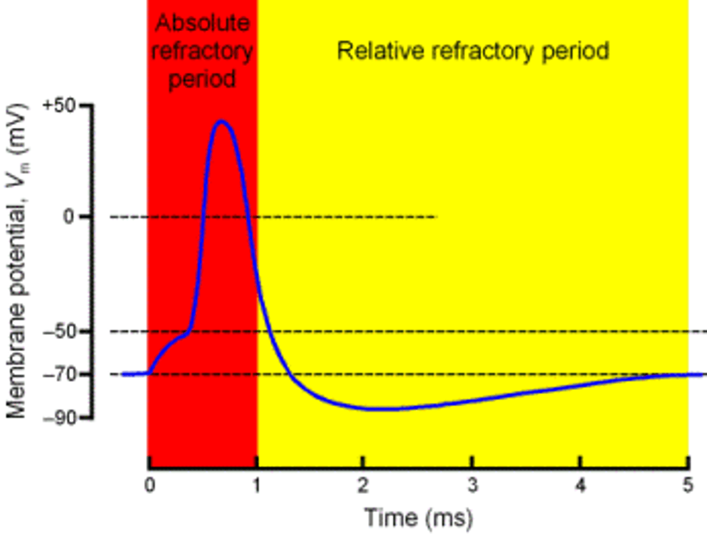
relative refractory period
A period after firing when a neuron is returning to its normal polarized state and will fire again only if the incoming message is much stronger than usual

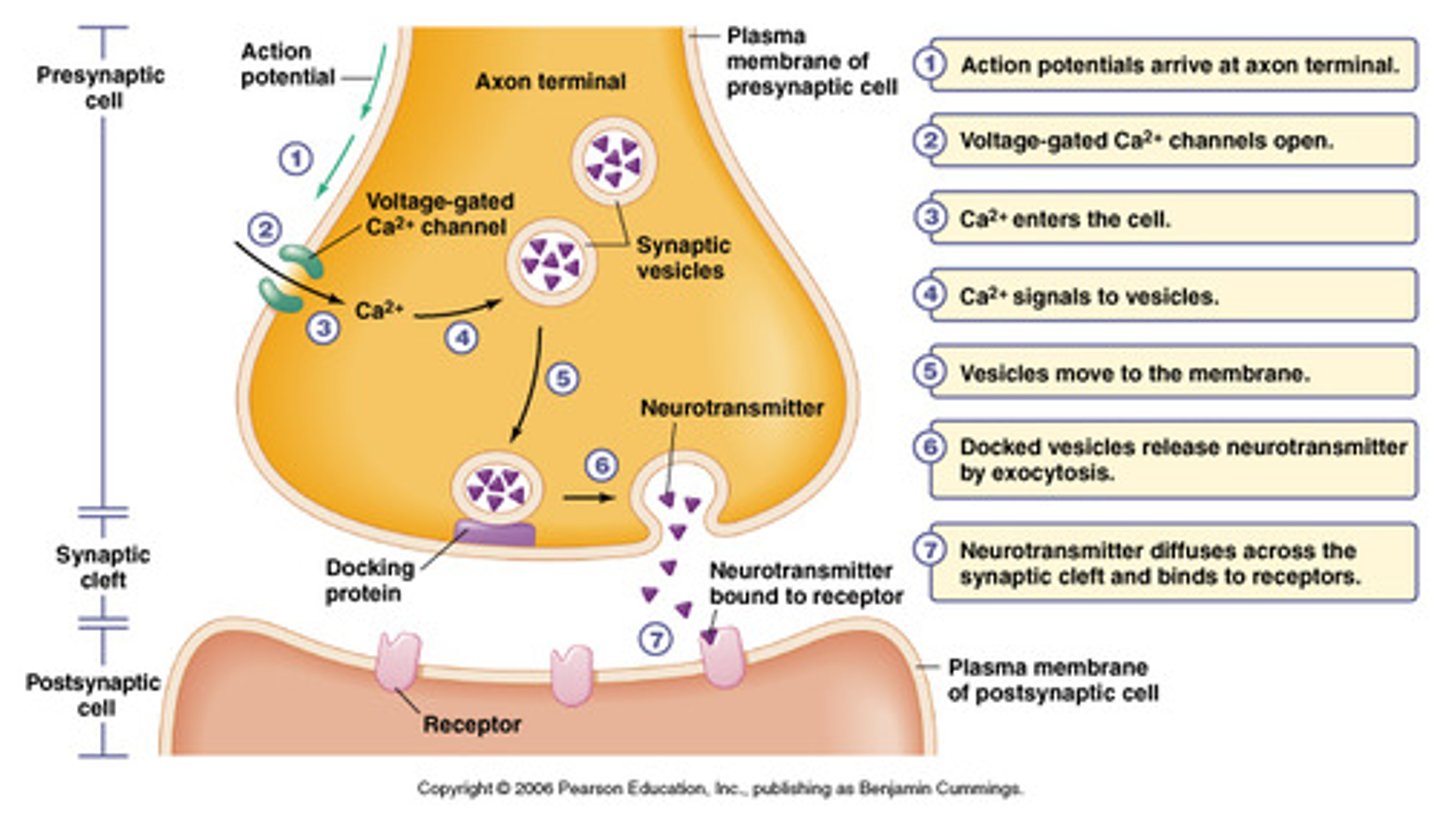
The Synapse Steps
1. action potential arrives at axon terminal (at end of presynaptic neuron)
2. depolarization opens voltage-gated Ca2+ channels and Ca2+ enters axon terminal
3. vesicles release neurotransmitters via exocytosis into synaptic cleft
4. neurotransmitters bind to receptors (usually ligand gated) on post-synaptic dendrites or cell body
5. ligand gated channels open or close causing graded potentials which can be excitatory or inhibitory
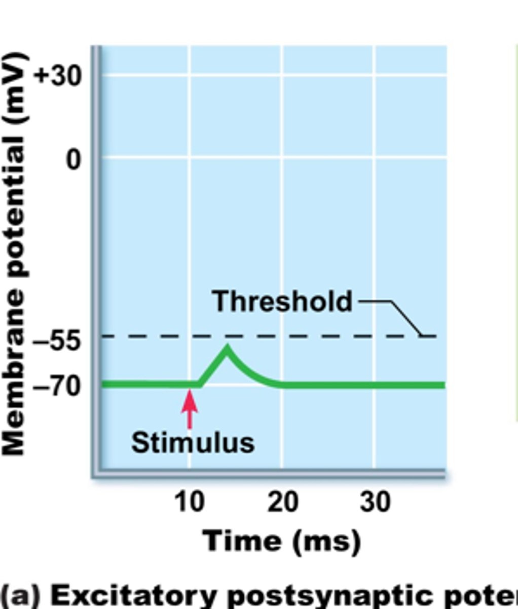
EPSP (excitatory postsynaptic potential)
- neurotransmitter opens ligand gated Na+ channels
- Na+ moves into post-synaptic cell
- causes depolarization (more positive) of post-synaptic cell (closer to threshold and more likely to have an action potential if there are enough)
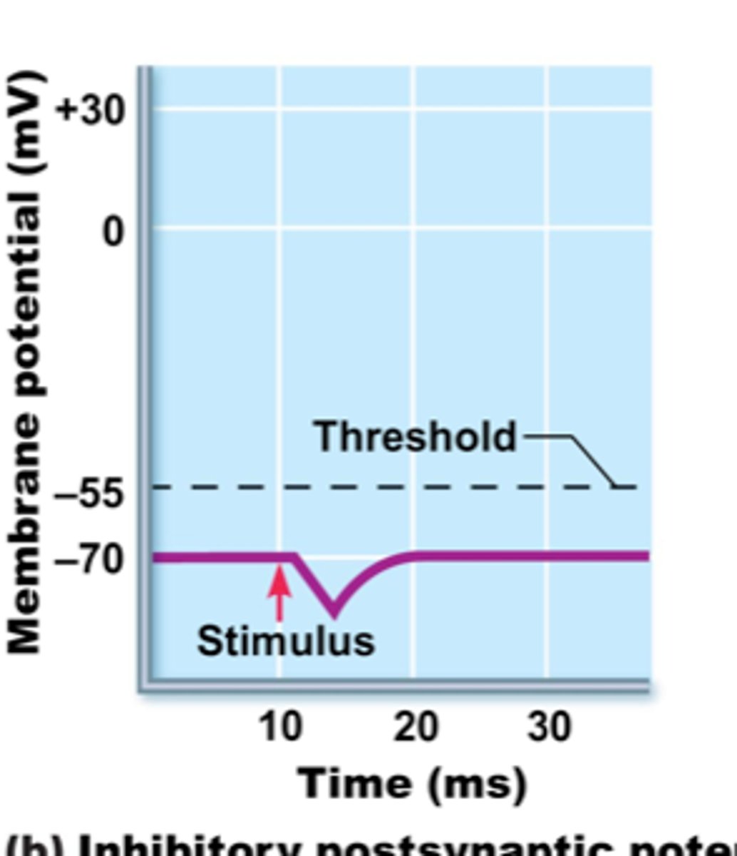
IPSP (inhibitory postsynaptic potential)
- neurotransmitter binds to K+ or Cl- ligand gated channels
- K+ moves out of post-synaptic cell or Cl- moves into cell
- causes hyperpolarization (more negative) of post-synaptic cell (further from threshold and less likely to have an action potential)
what is the success or failure of the action potential origination
The amount of neurotransmitter bound is due to the intensity of the stimulus and the concentration of the neurotransmitter in the cleft, which directly affects the intensity of the local or graded potential it creates
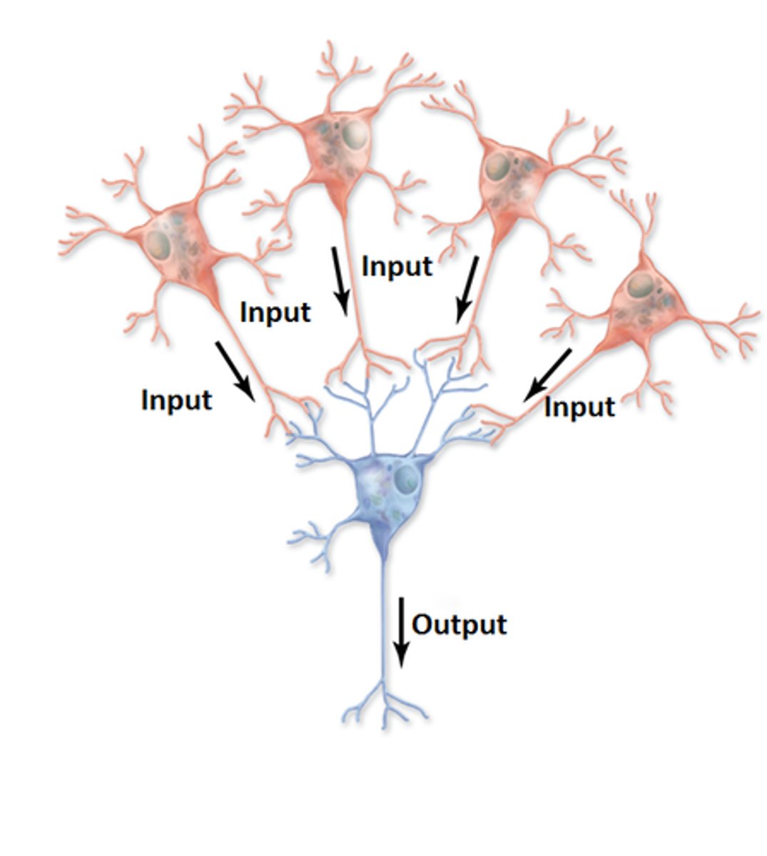
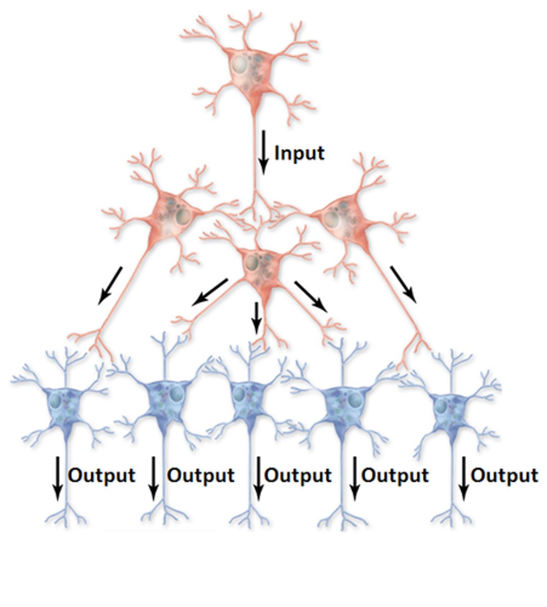
convergent circuit
thousands of synapses from many different presynaptic cells can affect a single postsynaptic cell
divergent circuit
a single presynaptic cell can send branches to affect many other postsynaptic cells
summation
graded potentials added up created by several neurotransmitters on the postsynaptic membrane
What happens to the Neurotransmitter once the message has been sent?
The neurotransmitter
(1) is actively transported back into the presynaptic axon terminal, called reuptake
(2) will diffuse away from the receptor site
(3) is broken down by enzyme to an inactive form and reabsorbed by the presynaptic neuron to end the transmission.
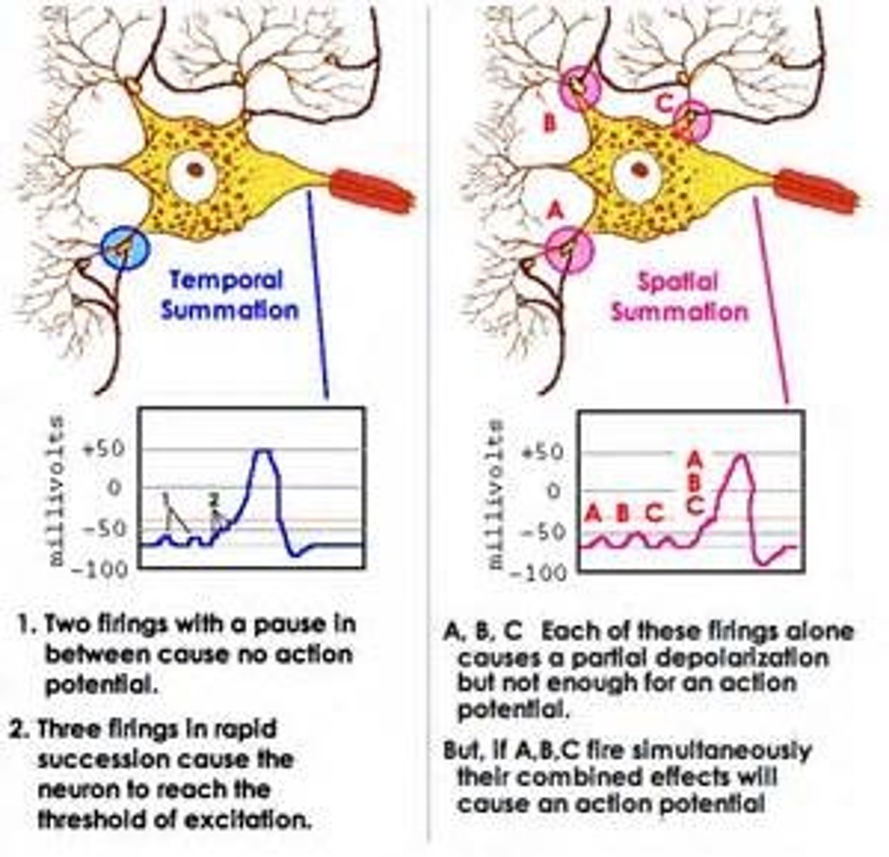
temporal summation
- stimulating the cell repeatedly at the same location
- If the releases come fast enough that the graded potential from one release has not faded before the next, then the graded potentials might sum to reach threshold
- effects of neurotransmitters released from a single presynaptic neuron that is having multiple action potentials add together in an action on the postsynaptic neuron
- timing is critical
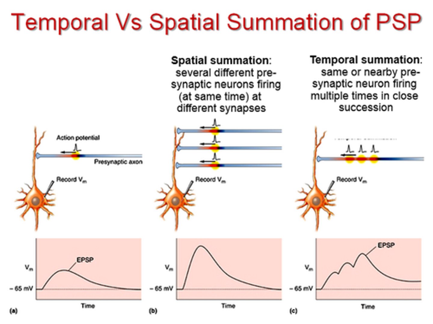
spatial summation
- graded potentials that started at different synapses come together and add up
- stimulating the cells at multiple locations at the same time
- have to spread and come close together enough to add together
- the effects of neurotransmitters from several presynaptic neurons add together to result in an action on the postsynaptic neuron
- spacing is critical
channel linked receptor
- direct transmitter action
- these chemicals can DIRECTLY affect their target cell and bring about a fast response
- They are able to bind to a receptor in the cell membrane and immediately, an effect of depolarization or hyperpolarization is seen
Excitatory receptor sites
(Nicotinic, Ach and glutamate), allows sodium, potassium, calcium to pass (depolarization)
Inhibitory receptor sites
(GABA, glycine) allows potassium, chloride to pass (hyperpolarization)
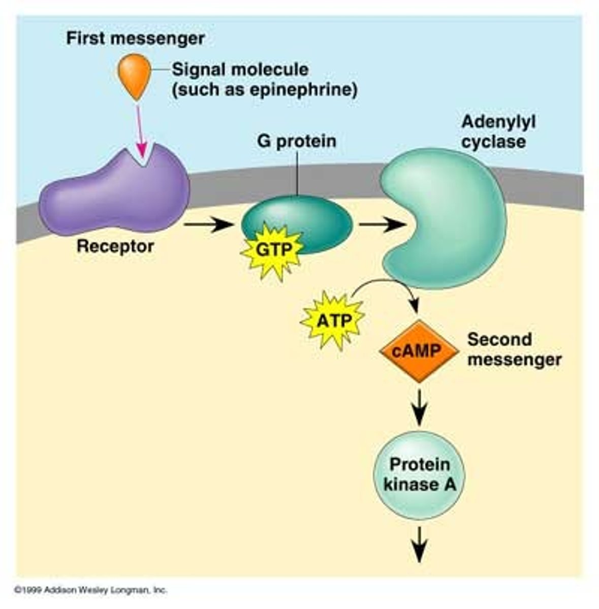
G-protein linked receptors steps
1. NT binds to G-protein linked receptor
2. G-Protein is activated
3-4. G-protein will activate cyclic AMP, cyclic GMP or calcium ions to use as second messengers to bring about the effect on the target cell.(open or close ion channels to create change in membrane potential)
G-protein linked: examples and what kind of activity (direct or indirect)
(Muscarinic, Substance P, NE, E)
- indirect, slow, complex, prolonged and diffuse (ideal for some learning processes)
- cannot directly affect their target cell, but must rely on another protein (second messenger) to bring about a response
- So takes longer
What kinds of drugs increase the action of the neurotransmitter?
- agonist
- synaptic enzyme inhibitors
- reupatake inhibitors
What kinds of drugs decrease the action of the neurotransmitter?
antagonists (blockers)
agonist
Drugs that bind to a receptor and produce similar responses to the normal activation of that neurotransmitter and/or receptor. (mimetic)
Antagonist
Drugs that bind to the receptor but are unable to activate it. By occupying the receptors, antagonists prevent binding of the normal neurotransmitter at the synapse.
Reuptake inhibitors
Drugs that interfere with the reuptake of neurotransmitters in the synapse so that a greater amount remains in the synapse for binding
Synaptic enzyme inhibitors
Inhibit the enzymes that break down the neurotransmitter so that a greater amount remains in the synapse for binding
Acetylcholine
1. what is it and what does it do
2. receptor subtype
3. agonist
4. antagonist
5. enzyme that breaks it down
1. neurotransmitter that stimulates digestion, slows heart rate, and contracts skeletal muscle
2. nicotine and muscarinic
3. nicotine and muscarinic
4. curare and atropine
5. acetylcholinesterase
Cholinergic neurons
- release Ach
- first to die in prefrontal or motor areas in Alzheimer's disease
Cholinergic Receptors (Ach): two types
muscarinic and nicotinic
muscarinic receptor (where and what do they do)
- G protein receptor located within the heart, digestive tract, and iris
- Parasympathetic effects (digestion, decrease heart rate, vasodilation and decreased BP), and constriction of pupils
- effects can be blocked by atropine
Nicotinic Receptors (where and what do they do)
- located within the motor end plate of skeletal muscles. Stimulation causes skeletal muscle contraction
- Also found in many parts of the CNS, where they are important in cognitive function and behavior
biogenic amines
small molecules that are synthesized from amino acids. (Most common biogenic amines are epinephrine, norepinephrine, dopamine, serotonin and histamine) The first 3 are classified as catecholamines
what roles do EPI and NOREPI play a role in and what drugs slow the breakdown of them
- emotional behavior, alertness, blood pressure regulation and the biological clock (hormone release). Imbalances of these neurotransmitters are associated with mental illness (Bipolar disorder)
- MAO inhibitors
Norepinephrine
1. what is it
2. receptor subtype
3. agonist
4. antagonist
1. neurotransmitter
2. alpha and beta receptor
3. phenylephrine and isoproterenol
4.phenoxybenxamine and propranolol
Alpha Receptors (where and what does it do there)
*a1= in iris and blood vessels of smooth muscle -> dilaiton, vasoconstriction, and increases BP
*a2= in presynaptic neurons -> decrease release of NOREP
Beta receptors (where and what does it do there)
*B1= in heart -> increase HR and contraction of heart muscle
*B2= in bronchial tree and skeletal muscles and their blood vessels -> bronchodilator, increased strength of skeletal muscle, vasodilation of blood vessels in skeletal muscle
dopamine: what does it do and where are the neuros that contain this neurotransmitter located
- influences movement, emotional response, and ability to experience pleasure and pain
- clustered in basal nuclei
Parkison's disease (what is it and treatment)
- the dopaminergic neurons in the basal nuclei die so the brains of people with Parkinson's disease contain almost no dopamine
- L-DOPA, a dopamine agonist relieves symptoms by stimulating dopamine receptors
What are dopamine antagonists used to treat
- Schizophrenia (can cause overactive dopamine system)
- prevent or reverse the actions of dopamine by keeping dopamine from attaching to receptors
Serotonin and where does it have an excitatory effect vs inhibitory
- A neurotransmitter that affects hunger, sleep, arousal, and mood.
- excitatory effect on pathways that control muscles muscles and an inhibitory effect on pathways that mediate sensation
- serotonin reuptake inhibitors thought to aid in the treatment of depression (zoloft) but can make you eat more
Glutamate and what happens after a brain injury
- Excitatory neurotransmitter that helps with learning and memory
- after a brain injury, neurons disintegrate due to lack of oxygen -> increases glutamate levels -> toxic to surrounding neurons
Monosodium Glutamate (MSG)
- food additive ("flavor enhancer")
- dissociates into free Na+ and glutamate ions in water
Substance P
Neuropeptide (string of amino acids) transmits pain information to CNS
Endorphins / Enkephalins
- Inhibitory (endogenous opioids) act as natural opiates, analgesics or euphorics, reducing our perception of pain under certain stressful conditions
- Inhibit substance P
- Morphine/opiates attach to the same receptors that bind the natural opiates
- May also have an effect on eating, drinking, regulating the cardiovascular system and mood and emotion.
gamma amino butyric (GABA)
(Primary inhibitory neurotransmitter in the brain. Slows things down) decreased binge eating, increased sleeping, decreased aggression and ventilation