Sensory Systems
1/39
Earn XP
Name | Mastery | Learn | Test | Matching | Spaced | Call with Kai |
|---|
No analytics yet
Send a link to your students to track their progress
40 Terms
Sensory Systems
Detect external events
The 3 discussed in class are organized according to a common anatomical plan
Visual, auditory, somatosensory
Sensory Receptors/ Sensory Epithelium
Specialized cell types/ parts of a cell that are in the periphery of the body- exposed to external events and stimuli
These receptors are specialized to transduce environmental energy (modality) into chemical or electrical signals into the nervous system
Transduction
The conversion of stimulus energy to a neural signal
Accomplished by receptor cells
Receptor Cells
A cell whose axon or dendrite is capable of transduction in a particular sensory modality
Specialized to transduce a particular form of environmental energy (modality) into a change in membrane potential - receptor potential
Grouped together in sheets referred to as ‘sensory epithelium’
Receptor Potential
Change in membrane potential at the site of transduction
Causes action potentials to be generated in the receptor cell or its downstream target
The rate and timing of action potentials carry information about the stimulus to the brain
Relay Nuclei
Groups of neurons located in the central nervous system that process signals from receptor neurons and transmit signals to the thalamus
Thalamus
Obligatory relay of visual, auditory, and somatosensory information to primary cortices
Groups of neurons organized into nuclei within the thalamus, that process signals from relay nuclei and transmit signals to the cerebral cortex
Primary Cerebral Cortex
Anatomical target of the thalamus
The first stop of sensory information on its way to cognitive processing
Conscious perception occurs when information reaches
Project to higher levels of cortex
Secondary Cerebral Cortex
Anatomical area that processes signals from primary sensory cortex
Transmits signals to association cortex, motor cortex, and subcortical structures
Where multimodal and other perceptions are formed
Modality Specificity
Category of stimuli to which receptor is sensitive
Receptive Field
Location on the sensory surface within which a stimulus (of the appropriate modality) can influence the activity of a sensory neuron
Range of locations on the sensory surface that, when stimulated, alter neurons activity
Lateral Inhibition
Inhibition of adjacent neurons in a map, which facilitates localization of stimuli
Sharpens the receptive field by inhibiting channels to its side
Winner take all
Acuity
Perceptual ability to discriminate between different parameter values
Ability to discriminate 2 similar but not identical sensory stimuli
Depends on the receptor density and receptive field
Spatial Organization of the Sensory Surface
Maintained at higher levels of the brain
Topographic maps
Think of the topographic axonal projections as labelled lines
Pupil (visual system)
Constriction and dilation allow less or more light to come through the eye
How/ where is light focused? (visual system)
Light is focused by the lens on the back of the eye which houses the retina
2D camera trained on your visual field
Visual Field (visual system)
The full range of what you can see
Retina (visual system)
Site of transduction
Fovea (visual system)
Center of the retina and sigh of highest acuity
Optic Nerve (visual system)
Axons from output cells of the retina gather together and form the optic nerve, which heads towards the thalamus
Blind spot
What is the path of light? (visual system)
Light passes through the retinal circuitry and is absorbed by photoreceptors, rods and cones, they absorb photons and generate membrane potential
Rods (visual system)
Sensitive to low light levels- scotopic
Don’t distinguish between different wavelengths
Low acuity
Peripheral field vision
Cones (visual system)
Sensitive to bright light levels- photopic
Distinguish between different wavelengths- 3 types, red, green, blue
High acuity
Central filed of vision
High spacial density in fovea
How is light absorbed in rods and cones? (visual system)
Light is absorbed by photopigments which activate a G-protein cascade that enzymatically cleaves cGMP
In the dark, cGMP holds open ligand gated Na+ channels which depolarizes the cell leading to the release of an inhibitory neurotransmitter that suppresses the downstream neuron
In the light, cGMP concentration decreases, closing Na+ channels and stops the neurotransmitter release and the circuit is disinhibited
The downstream cell is activated, causing the retinal ganglion cells to fire spikes
Retinal ganglion cells gather together and leave the retina at the optic disc
Where to retinal ganglion cells project? (visual system)
To the thalamus
The medial axons cross the midline once
Information content of the optic nerve, chiasm, and tract are different
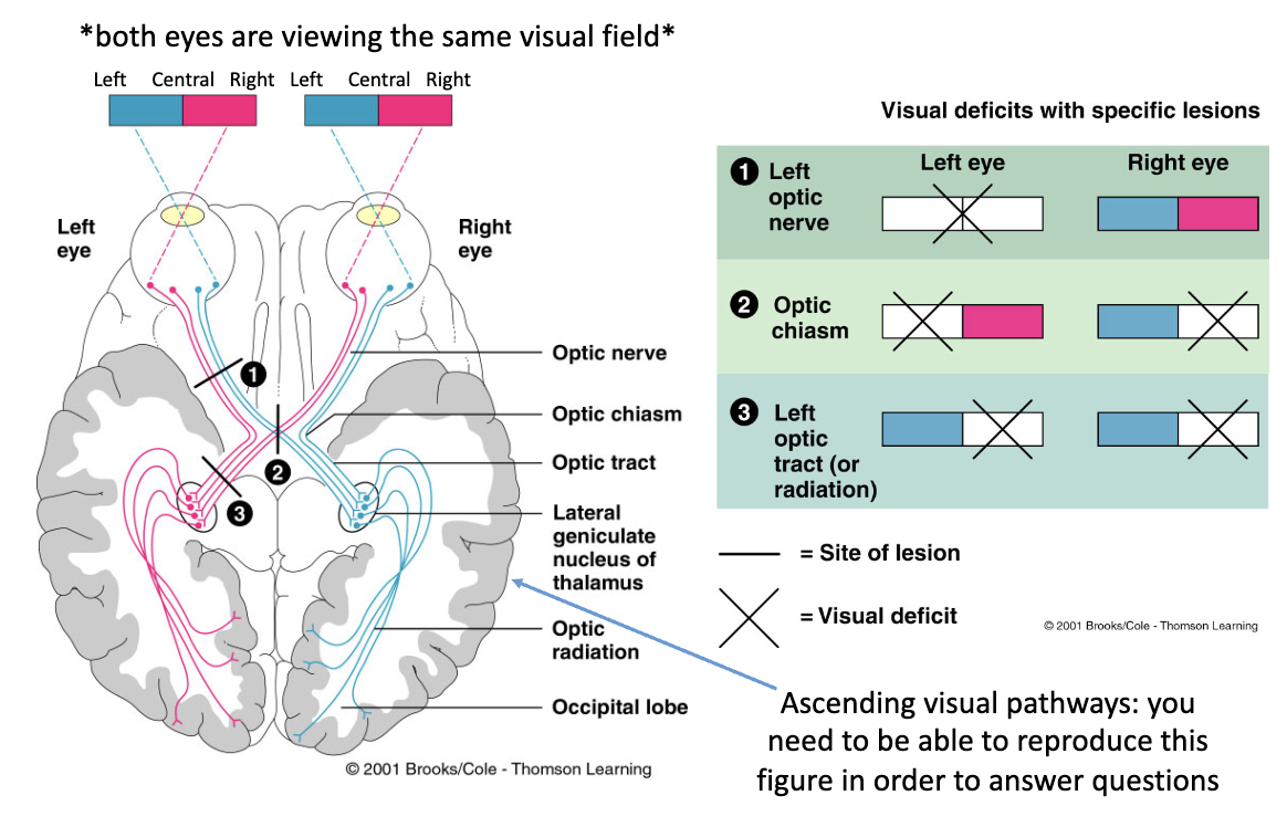
Optic Nerve (visual system)
Information across the visual field from one eye
If severed, you lose all vision from that eye
Optic Chiasm (visual system)
Information that crosses the midline
Left visual field from the left eye
Right visual field form the right eye
Optic Tract (visual system)
Information from the contralateral visual field
If severed you lose vision from the contralateral visual field on the side it was damaged
Left side damaged= right visual field lost
Retinotopic/ Visuotopic Map (visual system)
The thalamus projects to the primary visual cortex which contains a topographic map of the retinal surface
That is also a map of where the photons cam from, your visual scene
Visual activity percolates out form the primary cortex along ‘what’ and ‘where’ pathways, involved in building complex perception (not covered in lecture)
Sound (auditory system)
A wave with alternating cycles of compression and rarefaction of particles in a medium (air or water)
Pinna (auditory system)
Part of the external ear
Reflects sound into the ear canal
Tympanic Membrane (auditory system)
Eardrum, compression waves (sound) causes it to vibrate
Ossicles (auditory system)
Bones in the middle ear
Mechanically efficient conduit of vibration
Oval Window (auditory system)
Receives vibrations from the ossicles
Set up fluid vibrations in the inner ear causing sound transduction
Fluid movement within the cochlea
Basilar Membrane (auditory system)
There is a gradient in the physical properties that makes different locations resonate with different frequencies of sound
Narrow, stiff end near the oval window best resonates in response to high frequencies
Broad, compliant end near the helicotrema best resonates in response to low frequencies
The entire length is populated by hair cells
Their apical stereocilia are embedded in the tectorial membrane, who’s pivot point is offset compared to the basilar membrane
Explain how the auditory system works (auditory system)
Sound is reflected by the pinna into the ear canal, causing the tympanic membrane to vibrate
Vibrations are conducted via the ossicles (mechanically efficient) to the oval window which causes fluid movement within the cochlea
This causes the basilar membrane to move up and down
The tectorial membrane also vibrates
This creates a shearing force that bends the stereocilia forward and backward with each sound cycle
The stereocilia membranes have mechanically gated ion channels that open with each cycle of sound and depolarize the hair cell (concentration gradients are flipped, K+ is higher outside the cell and causes the depolarization)- this is receptor potential
It causes transmitter release form the hair cells to the primary afferent fibers which head towards the brain
A map of the cochlear surface is maintained up through the primary cortex via labelled line projection- map of tones, not sound source locations
Pacinian Copuscle (somatosensory system)
A type of touch receptor
Their membranes contain stretch-activated channels that open in response to membrane deformation
Phasic Signaling (somatosensory system)
In response to a sustained stimulus, encapsulated receptor types rapidly adapt, meaning they exhibit brief ‘on’ and ‘off’ response
Rapidly adapting receptor types are partially responsible for percepts
Tonic Signaling (somatosensory system)
Non-encapsulated receptor types exhibited sustained responses
Explain how pacinian corpuscle touch receptors work (somatosensory system)
When the stretch-activated channels are opened in response to membrane deformation, the cell depolarizes, triggering spikes that propagate towards the spinal cord
Rapid adaptation in pacinian corpuscles is due to slow mechanical separation of the overlying connective layers
Sensory information crosses the midline exactly once on its journey to the cortex
In the cortex there is a topographic map of the sensory surface/ hpmunculus