AP 1, Unit 3, Ch 9 - Muscle Histology and Physiology
1/67
There's no tags or description
Looks like no tags are added yet.
Name | Mastery | Learn | Test | Matching | Spaced |
|---|
No study sessions yet.
68 Terms
Organization of a muscle, from simple to complex
-myofilament
-myofibril
-muscle fiber
-muscle fascicle
-whole muscle
What are the functions of the muscular ssystem?
1) movement of the body
2)maintenance of posture
3) respiration
4) production of body heat
5) communication
6) constriction of organs and vessels
7) contraction of the heart
What are the three types of muscle?
1)skeletal
2)smooth
3)cardiac
where is skeletal muscle located?
attached to bones
where is smooth muscle located?
walls of hollow organs, blood vessels, eyes, glands, and skin
where is cardiac muscle located?
the heart
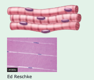
striated appearance
skeletal muscle
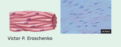
spindle-shaped appearance
smooth muscle
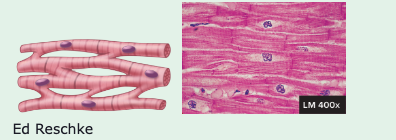
striated but less muclei
cardiac muscle
What are the four properties of muscle tissue? (explain)
Contractability - ability to shorten/contract
excitability - ability to receive an electrical stimulus
extensibility - the ability to stretch beyond normal length
elasticity - the ability to return to normal length after stretching
What is a muscle cell called?
a muscle fiber
what are the three layers of CT in skeletal muscle? (explain)
epimysium - forms a CT sheath that surrounds each skeletal muscle
perimysium - separates whole muscles into bundles of muscle fibers
endomysium - separates individual muscle fibers
What nerve cells are responsible for stimulating skeletal muscle contraction
motor neurons
What is hypertrophy in skeletal muscles?
The increase in size of muscle fibers, not in number of muscle fibers.
What are the two main components in a skeletal muscle?
electrical
mechanical
What are the electrical components of a skeletal muscle? (explain)
sarcolemma - plasma membrane of muscle fibers
transverse tubules - inwards folds of the sarcolemma, forms T tubules that carry electric signals into the muscle so everything contracts at once
sarcoplasmic reticulum - smooth ER that releases ca2+ as a “switch” for contraction
What is a “triad” in electrical components of skeletal muscle?
the terminal cisternae at the ends of the sarcoplasmic reticulum that run alongside T tubules, critical component of muscle contraction
sarcoplasm
the cytoplasm of a muscle fiber
What are the mechanical components of a muscle fiber? (explain)
myofibril - bundles of protein filaments which interact to shorten the muscle during contraction
myofilaments - two different types in each myofibril: actin myofilaments (thin filaments) and myosin myofilaments (thick filaments) arranged into ordered units called sarcomeres
sarcomere
join end to end to form the myofibril
What are Z disks in a sarcomere?
delicate structure found at the end of a sarcomere to which myofilaments attach
What are the three regions of a sarcomere? (explain)
I band, darker A band, I band. Each I band includes a Z disk, I bands have only actin myofilaments.
What is in the A band? (explain)
H zone, which has a darkened line called the M line. A band is made up of actin and myosin filaments, but H zone is only myosin filaments. M line holds myosin filaments in place.
What does the protein titin do in skeletal muscle?
give ability to stretch (extensibility) and recoil (elasticity)
Actin myofilament structure (explain)
Globular (G) actin - chain of 200 Gs creates fibrous (f) actin. Each G subunit has a active site for myosin myofilaments to bind during contraction
tropomyosin - long fibrous protein that lies in the groove along F actin. In relaxed muscle, it covers G binding sites.
troponin - consists of a subunit that anchors to actin, subunit that prevents tropomyosin from uncovering G binding sites, and another subunit that binds Ca2+
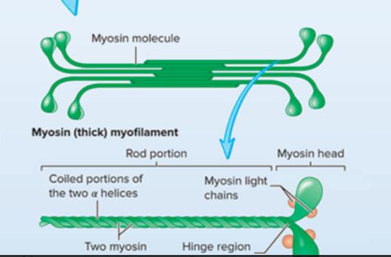
What are myosin myofilamets composed of?
Myosin head, rod portion
action potentials
electrical signals from motor neurons which cause action potentials in the muscle fibers. Occurs when an excitable cell is stimulated, changes the membrane potential so that the inside of the membrane becomes positively charged compared to the outside.
What is in a neuromuscular junction?
axon terminals and the area of the muscle fiber sarcolemma they innervate
neuromuscular junction and synapses
the point of contact between a motor neuron and muscle fiber
presynaptic terminal
anlarged axon terminal or terminal bouton
synaptic cleft
space between the presynaptic terminal and the muscle fiber
postsynaptic membrane/ motor-end plate
membrane around the muscle fiber at the neuromuscular junction
synaptic vesicles
released from the presynaptic membrane and diffuse across the synaptic cleft to the muscle fiber
acetylcholine (ACh)
neurotransmitter for motor neurons
sliding filament model
model for shortening or contracting muscle (H zone dissapears)
resting membrane potential (explain)
the charge in an unexcited cell. The concentration of K+ inside the membrane is higher than outside, the concentration of Na+ outside is higher than inside, and the membrane is more permeable to K+ than Na+. (sodium-potassium pump, ch 3)
What are the two phases of an action potential?
depolarization
repolarization
explain “depolarization” in an action potential
depolarization is when the inside of the membrane becomes more positive, and when the membrane reaches a certain threshold, an action potential is triggered
What is the “treshold” in action potential?
the membrane potential at which voltage gated Na+ channels open
explain “repolarization” in an action potential
the return of the membrane potential to its resting value after opening of voltage gated Na+ channels, depolarization, then opening of K+ channels.
all-or-none principle of action potentials
when a stimulus is applied to a cell, either an action potential is produced or it is not, a cell will either contract all the way or it won’t
action potential frequency
the number of action potentials produced in a certain amount of time, the greater the stimulus to a neuron/muscle fiber the more action potentials will fire
acetylcholinesterase
breaks down ACh so it doesn’t accumulate in the synaptic cleft
excitation-contraction coupling (explain)
begins with neuromuscular junction, action potential propagated along sarcolemma and T tubules. At T tubules, APs cause voltage gated Ca2+ channels to open in terminal cisternae, when they open Ca2+ rapidly diffuses out into sarcoplasmic reticulum. In the sasrcoplasm, , Ca2+ binds to troponin and frees up binding sites for myosin heads to attach to actin myofilaments, muscles then contract when those cross bridges move.
cross-bridge cycling (explain)
Ca2+ binds to troponin and active sites on G heads are exposed. This forms a cross-bridge and triggers movement of myosin heads at their hinge region. this movement is called the power stroke. As long as Ca2+ is present, the cycle repeats.
muscle twitch
the response of a muscle fiber to a single action potential
What are the three phases of a muscle twitch
lag (latent phase)
contraction
relaxation
What is the lag phase of a muscle twitch?
the gap in time between the time of stimulus application to the motor neuron and the beginning of the contraction. The time in which the AP is traveling along the axon, NM junction, and travels the sarcolemma.
What is the contraction phase of a muscle twitch?
starts when Ca2+ is released and the cross bridge cycling occurs
What is the relaxation phase of a muscle twitch?
Ca2+ is pumped from sarcoplasm, Ca2+ levels decline and cross bridge cycling stops
What are the two types of muscle contractions?
isometric
isotonic
isometric contractions
the muscle does not shorten. Tension in muscle is increased but the length stays the same. (ie lifting something that is too heavy for your or standing straight with erect posture)
isotonic contractions
the muscle shortens. When you lift an object or move your limbs.
motor unit
a single motor neuron and all the muscle fibers it innervates
passive tension
tension applied to the load when a muscle stretches but is not stimulated
total tension
the sum of active and passive tension
muscle tone
when muscles stay contracted for long periods of time
slow twitch muscle fibers
contract slowly, have better developed blood supply, more mitochondria, and more fatigue resistant. Respond slowly to nervous stimulation.
fast twitch
respond rapidly to nervous stimulation, they are able to break down ATP more rapidly. Whitish in appearance bc of worse blood supply.
What are the ATP dependent enzymes in muscle contraction?
the myosin head
the Na+-K+ pump to maintain membrane potential
Ca2+ reuptake pump
Anaerobic respiration
does NOT require O2
Aerobic respiration
Does require O2.
fatigue
temporary state of reduced work capacity
caveolae
shallow invagination in smooth muscle that performs a function similar to T tubules aand sarcoplasmic reticulum
How does smooth muscle contraction work?
Ca2+ enters sarcoplasm and binds to protein calmodulin
calcium-calmodulin activates an enzyme myosin kinase
myosin-kinase transfers a phosphate group from ATP to heads of myosin molecules
cross bridge cycling occurs but slower
relaxation occurs when myosin phosphate removes phosphate group from myosin molecules
latch state
sustained tension in smooth muscle
What are the two types of smooth muscle?
visceral - occurs in sheets. Includes digestive, reproductive, and urinary tracts
multiunit - walls of blood vessels, arrector pili, small bundles, iris of the eye
intercalated disks
cell-to-cell attachments in cardiac muscle include gap junctions for communications