Oral Pathology LO 10 part 2.
1/39
There's no tags or description
Looks like no tags are added yet.
Name | Mastery | Learn | Test | Matching | Spaced | Call with Kai |
|---|
No analytics yet
Send a link to your students to track their progress
40 Terms
Secondary polycythemia
The increase in RBCs is caused by a physiologic response to decreased oxygen. A decrease in blood oxygen causes an increase in erythropoietin by the kidney. (Erythropoietin is a hormone to produce RBCs). May be due tp pulonary disease, heart disease, living at a high altitude, or elecation in carbon monoxide: associated with tobacco smoking.
Relative polycythemia
This is caused by a decrease in plasma volume. Causes of acute ————— ————————-: diuretics, vomiting, diarrhea, excessive sweating. Causes of chronic or stress ————————: most patients are middle aged Caucasian men under physiological stress, slightly overweight, hypertensive, heavy smokers.
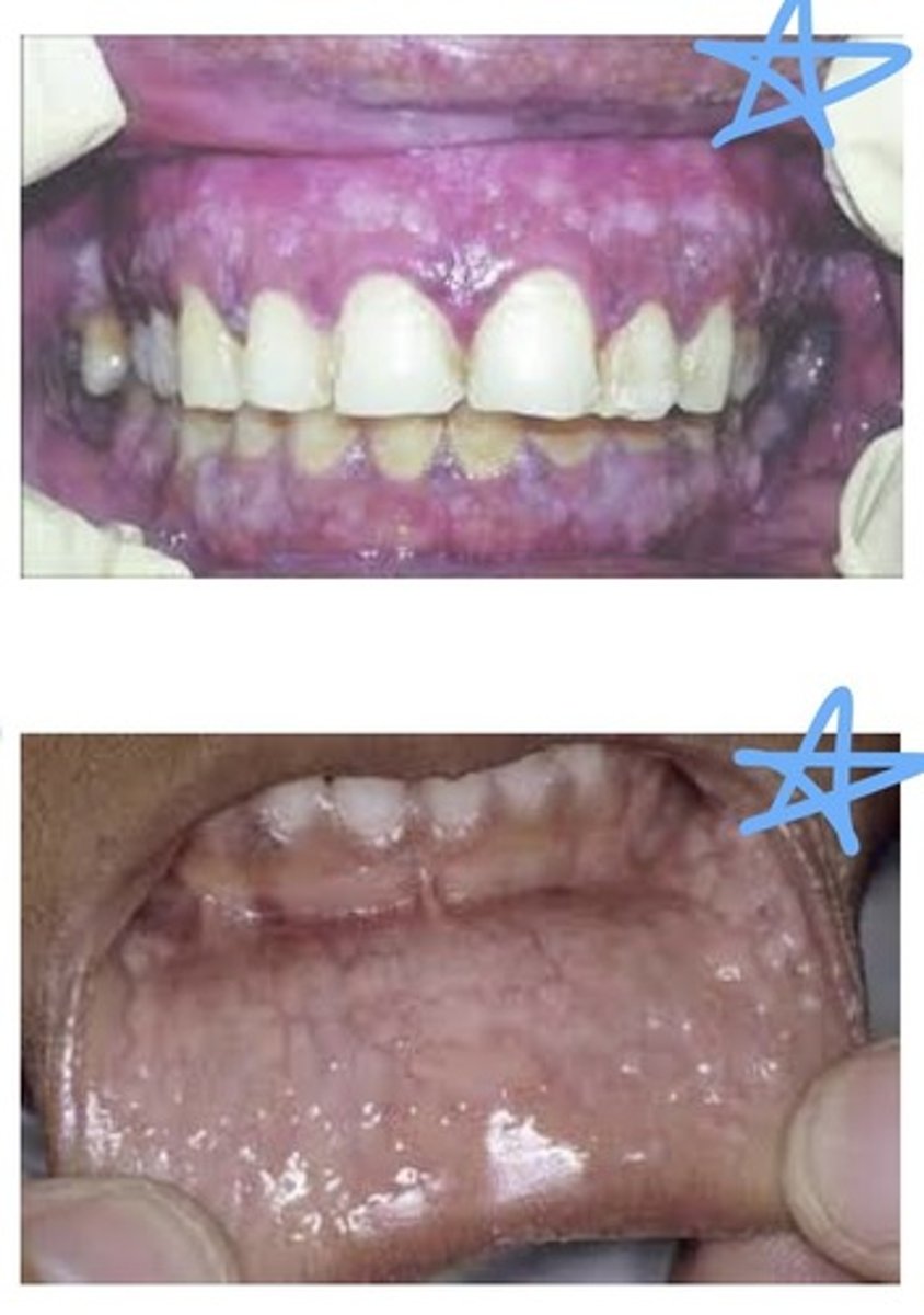
Polycythemia
Oral manifestations of ————————:
Oral mucosa may appear deep red to purple, gingiva may be edematous and bleed easily, Submucosal petechiae, Ecchymosis, hematoma formation. Caution: increased bleeding after oral surgery.
Granulocytes, lymphocytes, monocytes
The 3 groups of WBCs found in circulation:
Granulocytes (granules in cytoplasm)
Polymorphonuclear (PMNs), leukocytes, or neutrophils, eosinophils, and basophils are all types of this WBC:
Agranulocytosis
A significant reduction in circulating neutrophils (WBCs). All of these terms mean immunosupression. This can result from a problem in development of neutrophils or accelerated destruction of neutrophils. Primary: the cause is unknown; may be an immunologic disorder.
Secondary: a result of chemicals or drugs.
Clinical features: sudden onset of fever, chills, jaundice, weakness, sore throat, oral infection, oral necrotizing ulcerations, excessive oral bleeding, rapid destruction of tooth-supporting structures, lymphadenopathy.
Leukopenia
Abnormally low white blood cell count.
Neutropenia
A reduction in the number of circulating neutrophils.
Cyclic neutropenia
A cyclic decrease in the number of circulating neutrophilic leukocytes. Cause: mutation of gene. Oral manifestations: gingival inflammation, ulceration of tongue, ulceration of mucosal tissue.
Leukemia
Malignant neoplasms of hematopoietic (blood-forming) stem cells. Characterized by an excessive number of abnormal WBCs in circulating blood. WBCs are non-functional so client is immunosupressed. Unknown causes, genetic link?
Cancer in the blood producing stem cells.
Acute leukemias
Type of leukemia characterized by very immature lymphocyte cells. ————— lymphoblastic leukaemia (ALL): involves immature lymphocytes, primarily affects children and young adults, good prognosis. ————— myeloblastic leukemia (AML): involves immature granulocytes, primarily effects adolescents and young adults, prognosis is not as good. Clinical features: sudden and dramatic onset, weakness and fatigue caused by anemia, fever caused by infections, immunosuppression, enlargement of lymph nodes, bleeding caused by decrease in platelets.
Celiac disease
Chronic disorder associated with sensitivity/intolerance to dietary gluten. Oral manifestations include painful burning tongue, atrophy of papillae of the tongue, ulceration of oral mucosa, diarrhea, and paresthesia (numbness) of extremities. Resulting in ‘cobblestoning’ due to repeated trauma/healing. Patients should adhere to a gluten-free diet.
Hemostasis
The cessation of bleeding.
Platelets (thrombocytes)
These aggregate to form a temporary clot.
Fibrin
An insoluble protein essential to blood clotting.
Clotting factors (coagulation factors)
These convert fibrinogen to fibrin.
Thrombocytopenia
This is the term for a platelet count less than 100,000/mm3. Normal is 200,000-400,000/mm3.
Spontaneous gingival bleeding
This may occur if the platelet count is less than 20,000/mm3.
1-6min
Bleeding time is used to provide an assessment of the adequacy of platelet function, not number. Normal bleeding time is between — and — minutes.
5-10
Prolonged bleeding time in patients with platelet abnormalities is greater than — or —— minutes.
Prothrombin time (PT)
The ability to form a clot, normal is between 11 and 16 seconds.
International normalized ratio (INR)
This is the ratio of PT to thromboplastin activity. Clients taking blood thinners such as warfarin may be required to provide —.—.—. Numbers to ensure they are safe for dental treatment.
Partial thromboplastin time (PTT).
Measures the other way by which clot formation occurs. Used to monitor heparin therapy. A normal —.—.—. Is usually 25-40 seconds.
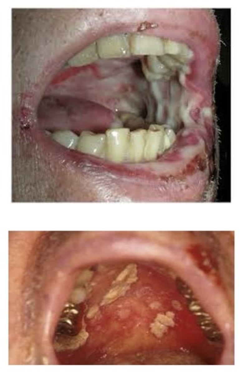
Purpura
A reddish-blue or purplish discolouration of skin or mucosa from spontaneous extravasation of blood. May be due to a defect or deficiency in blood platelets. May be due to capillary fragility. Blood may ooze from gingival margins, without the presence of gingivitis or inflammation.
Thrombocytopenic purpura
This is a bleeding disorder that results from a severe reduction in circulating platelets. 3 types: idiopathic —————————— (unknown cause), immune —————————— (an autoimmune type of process), and secondary —————————— ————— (often associated with drugs).
Clinical features: spontaneous purpuric or hemorrhagic lesions on the skin, pts bruise easily, may have blood in urine, frequent nose bleeds, spontaneous gingival bleeding,
Petechiae, clusters of petechiae or purpuric spots, ecchymosis.
Nonthrombocytopenic purpura
Bleeding disorders that can result from either a defect in capillary walls or disorders of platelet function. Vitamin C deficiency and infections or chemicals and allergy may be the cause of alterations in vascular walls. Drugs, allergy, and autoimmune disease may cause disorders of platelet function. Oral manifestations: spontaneous gingival bleeding, petechiae, ecchymosis, and hemorrhagic blisters.
Hemophilia
A disorder of blood coagulation. Results in severely prolonged clotting time, genetic link, can be fatal. Caused by a deficiency in plasma proteins involved in coagulation. There are types an and b. Inherited as X-linked diseases through an unaffected carrier daughter to a son.
Hemophilia type A
This type of hemophilia is caused by a deficiency in plasma thromboplastinogen or factor VIII (8).
Hemophilia type B
This type of hemophilia is known as Christmas disease. Less common; the clotting defect is plasma thromboplastin or factor IX (9).
Mucositis
This condition begins about the second week of radiation therapy and subsides a few weeks after its completion. Painful. Appears as erythematous and ulcerated mucosa. The patients may have difficulty eating, pain on swallowing, and loss of taste.
Rampant caries and candidiasis
Destruction of major salivary glands during radiation therapy may result in Xerostomia, patients are prone to these 2 conditions related to severe xerostomia. How the DH can help: fluoride application, patient ed, frequent follow up appts.
Basal cells
Drugs used for cancer chemotherapy affect ————— cells of the epithelium. Mucositis and oral ulceration are common complications. A decrease in all blood cells may occur.
Xerostomia
Blood pressure drugs, antianxiety meds, antipsychotics, and antihistamines can cause:
Prednisone
This corticosteroid suppresses the immune system and can lead to candidiasis and oral infections.
Phenytoin (Dilantin), nifedipine (Procardia) and cyclosporine
These 3 medications can cause gingival enlargement.
Medication-related osteonecrosis of the jaw (MRONJ)
The clinical diagnosis of this is masked on history and physical examination; radiographic findings are nonspecific. May be considered to have this if: current or previous tx with antiresorpitive or antiangiogenic agents (BISPHOSPHONATES) often prescribed for osteoporosis or used in cancer therapies, exposed bone or bone that can be probed through an intraoral or extraoral fistula in the maxillofacial region that has persisted at least 8 weeks, no history of radiation therapy to the jaws.
Stage 1
Exposure is limited and is typically painless. Patients in this stage of MRONJ are given antibacterial mouth rinse.
Stage 2
This stage of MRONJ is similar to stage 1 except lesions are typically painful and inflamed. Chlorhexidine mouth rinse, ABs, and the area may be debrided.
Stage 3
In this stage of MRONJ, lesions have exposed and necrotic bone present, pain, infections, and other conditions. Mouthrinses, antibiotics, and surgical debridement of lesion.
Mucosal discolouration (darkening)
Phenolphthalein, Minocycline, Antimalarial medications and Chemotherapeutic agents can all cause: