♋ Lecture 17: Cancer Continued

0.0(0)
Studied by 1 person
call kaiCall Kai
learnLearn
examPractice Test
spaced repetitionSpaced Repetition
heart puzzleMatch
flashcardsFlashcards
GameKnowt Play
Card Sorting

1/13

encourage image

There's no tags or description

Looks like no tags are added yet.

Last updated 6:51 AM on 4/1/26
Name
Mastery
Learn
Test
Matching
Spaced
Call with Kai

No analytics yet

Send a link to your students to track their progress

14 Terms

1
New cards

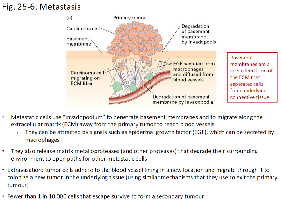
Metastatic Cell Migration

Invadopodia Formation
 Metastatic cells form invadopodia to penetrate basement membranes and migrate along the extracellular matrix (ECM) away from the primary tumor

Chemotactic Attraction
 Cells can be attracted by signals such as epidermal growth factor (EGF), sometimes secreted by macrophages

Matrix Degradation
 Metastatic cells release matrix metalloproteases (MMPs) and other proteases to degrade the ECM, creating paths for themselves and other tumor cells

Extravasation
 Tumor cells adhere to blood vessel linings in a new location and migrate through the vessel wall to colonize underlying tissue
  Uses similar mechanisms as invasion of the primary tumor

Survival Rate
 Fewer than 1 in 10,000 cells that escape survive to form a secondary tumor

Basement Membranes
 Specialized ECM structures that separate cells from underlying connective tissue

<p><strong>Invadopodia Formation</strong><br> Metastatic cells form <strong>invadopodia</strong> to <strong>penetrate basement membranes</strong> and migrate along the <strong>extracellular matrix (ECM)</strong> away from the primary tumor</p><p><strong>Chemotactic Attraction</strong><br> Cells can be attracted by signals such as <strong>epidermal growth factor (EGF)</strong>, sometimes secreted by <strong>macrophages</strong></p><p><strong>Matrix Degradation</strong><br> Metastatic cells release <strong>matrix metalloproteases (MMPs)</strong> and other proteases to <strong>degrade the ECM</strong>, creating paths for themselves and other tumor cells</p><p><strong>Extravasation</strong><br> Tumor cells <strong>adhere to blood vessel linings</strong> in a new location and <strong>migrate through the vessel wall</strong> to colonize underlying tissue<br>  Uses similar mechanisms as <strong>invasion of the primary tumor</strong></p><p><strong>Survival Rate</strong><br> Fewer than <strong>1 in 10,000 cells</strong> that escape survive to form a <strong>secondary tumor</strong></p><p><strong>Basement Membranes</strong><br> Specialized <strong>ECM structures</strong> that <strong>separate cells from underlying connective tissue</strong></p>
2
New cards

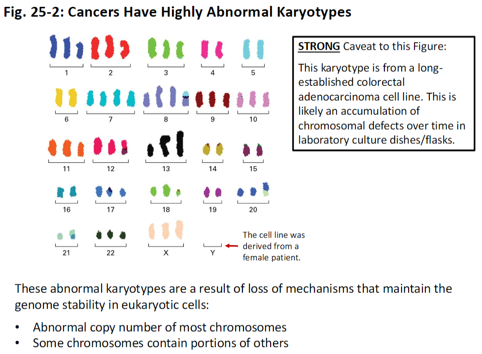
Cancer Cell Karyotypes

Abnormal Karyotypes
 Cancer cells often exhibit highly abnormal karyotypes due to loss of genome stability mechanisms

Characteristics
Abnormal copy number of most chromosomes
 Some chromosomes contain portions of other chromosomes

Caveat
 The figure shown is from a long-established colorectal adenocarcinoma cell line
 Many chromosomal defects may have accumulated over time in culture
 The original cell line was derived from a female patient

<p><strong>Abnormal Karyotypes</strong><br> Cancer cells often exhibit <strong>highly abnormal karyotypes</strong> due to <strong>loss of genome stability mechanisms</strong></p><p><strong>Characteristics</strong><br> <strong>Abnormal copy number</strong> of most chromosomes<br> Some chromosomes contain <strong>portions of other chromosomes</strong></p><p><strong>Caveat</strong><br> The figure shown is from a <strong>long-established colorectal adenocarcinoma cell line</strong><br> Many chromosomal defects may have <strong>accumulated over time in culture</strong><br> The original cell line was <strong>derived from a female patient</strong></p>
3
New cards

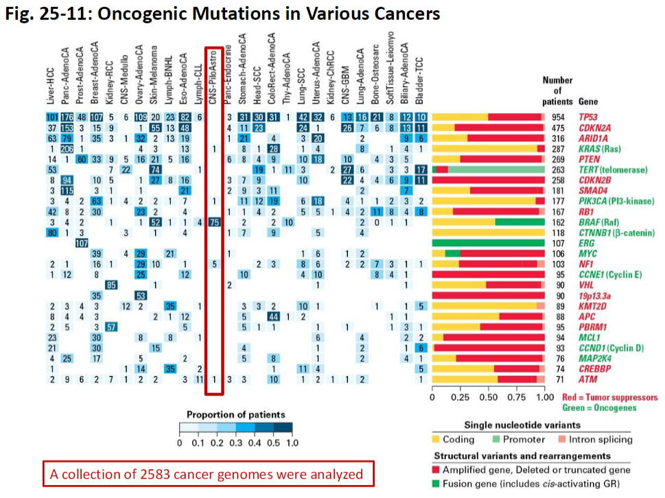
Oncogenic Driver Mutations in Cancer

Data Overview
 Analysis based on 2,583 cancer genomes

Key Concept
 Identifies oncogenic driver mutations that contribute to cancer development and progression
 Helps distinguish driver mutations from passenger mutations that do not confer growth advantage

<p><strong>Data Overview</strong><br> Analysis based on <strong>2,583 cancer genomes</strong></p><p><strong>Key Concept</strong><br> Identifies <strong>oncogenic driver mutations</strong> that contribute to cancer development and progression<br> Helps distinguish <strong>driver mutations</strong> from <strong>passenger mutations</strong> that do not confer growth advantage</p>
4
New cards

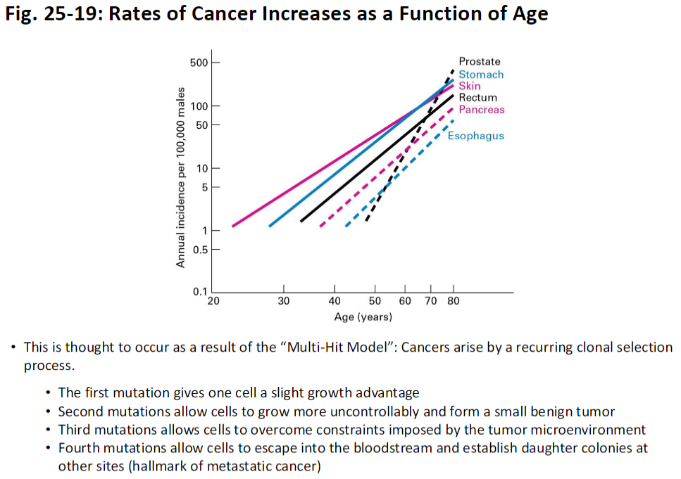
Cancer Incidence and Age – Multi-Hit Model

Concept
 Cancer rates increase with age due to the Multi-Hit Model, where tumors arise through a recurring clonal selection process

Mutation Progression
First Mutation
  Gives a cell a slight growth advantage

Second Mutation
  Allows cells to grow more uncontrollably and form a small benign tumor

Third Mutation
  Enables cells to overcome constraints imposed by the tumor microenvironment

Fourth Mutation
  Allows cells to enter the bloodstream and establish daughter colonies at other sites, a hallmark of metastatic cancer

<p><strong>Concept</strong><br> Cancer rates increase with <strong>age</strong> due to the <strong>Multi-Hit Model</strong>, where tumors arise through a <strong>recurring clonal selection process</strong></p><p><strong>Mutation Progression</strong><br> <strong>First Mutation</strong><br>  Gives a cell a <strong>slight growth advantage</strong></p><p> <strong>Second Mutation</strong><br>  Allows cells to <strong>grow more uncontrollably</strong> and form a <strong>small benign tumor</strong></p><p> <strong>Third Mutation</strong><br>  Enables cells to <strong>overcome constraints</strong> imposed by the <strong>tumor microenvironment</strong></p><p> <strong>Fourth Mutation</strong><br>  Allows cells to <strong>enter the bloodstream</strong> and <strong>establish daughter colonies</strong> at other sites, a hallmark of <strong>metastatic cancer</strong></p>
5
New cards

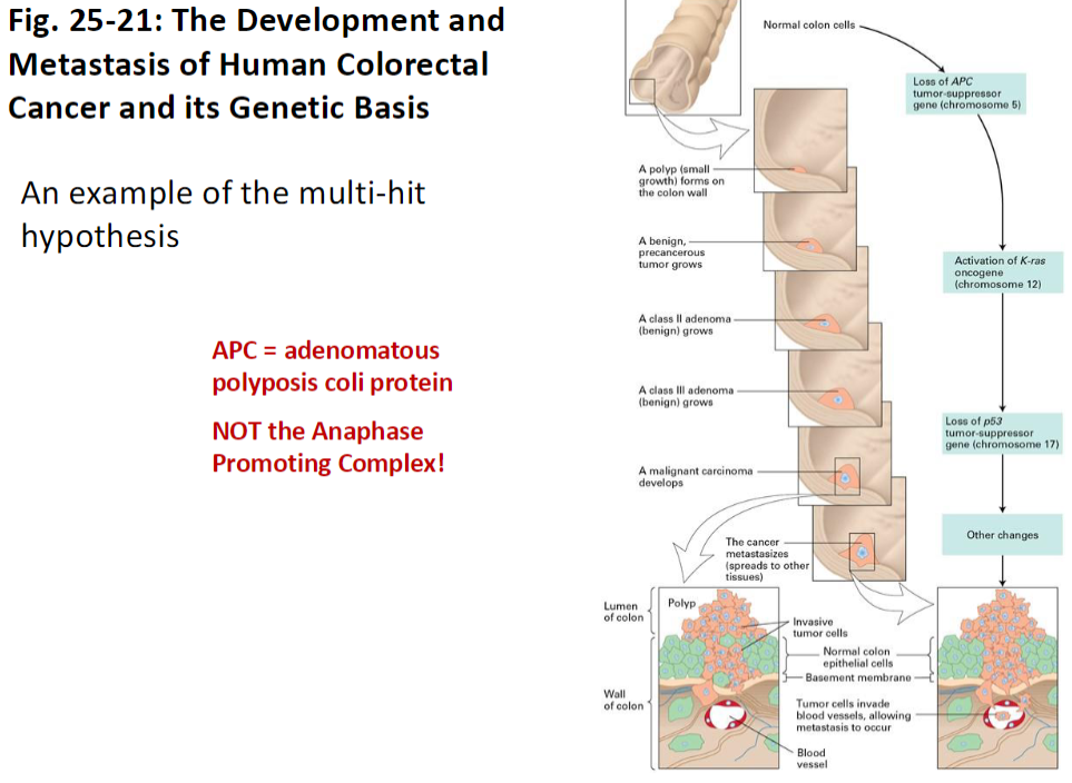
Development and Metastasis of Human Colorectal Cancer

Concept
 Example of the Multi-Hit Hypothesis explaining tumor development and progression

Genetic Basis
APC = adenomatous polyposis coli protein
  (Not to be confused with Anaphase Promoting Complex)

Key Idea
 Mutations accumulate in specific genes like APC, driving the formation of colorectal tumors and potentially metastasis

<p><strong>Concept</strong><br> Example of the <strong>Multi-Hit Hypothesis</strong> explaining tumor development and progression</p><p><strong>Genetic Basis</strong><br> <strong>APC</strong> = <strong>adenomatous polyposis coli protein</strong><br>  (Not to be confused with <strong>Anaphase Promoting Complex</strong>)</p><p><strong>Key Idea</strong><br> Mutations accumulate in <strong>specific genes</strong> like APC, driving the formation of <strong>colorectal tumors</strong> and potentially <strong>metastasis</strong></p>
6
New cards

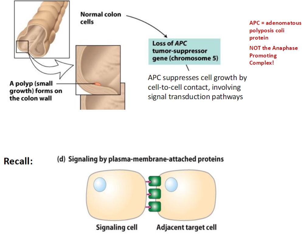
Colorectal Cancer Development – APC Loss

Normal Colon Cells
 Cells grow and function normally, maintaining tissue architecture

Polyp Formation
 A polyp (small growth) forms on the colon wall

APC Gene Loss
 Loss of APC (adenomatous polyposis coli protein, chromosome 5)
  (Not the Anaphase Promoting Complex)
APC function: suppresses cell growth via cell-to-cell contact and signal transduction pathways

Signaling Reminder
Signaling by plasma-membrane-attached proteins involves a signaling cell and an adjacent target cell

<p><strong>Normal Colon Cells</strong><br> Cells grow and function normally, maintaining <strong>tissue architecture</strong></p><p><strong>Polyp Formation</strong><br> A <strong>polyp</strong> (small growth) forms on the <strong>colon wall</strong></p><p><strong>APC Gene Loss</strong><br> Loss of <strong>APC</strong> (<strong>adenomatous polyposis coli protein</strong>, chromosome 5)<br>  (Not the <strong>Anaphase Promoting Complex</strong>)<br> <strong>APC function:</strong> suppresses <strong>cell growth</strong> via <strong>cell-to-cell contact</strong> and <strong>signal transduction pathways</strong></p><p><strong>Signaling Reminder</strong><br> <strong>Signaling by plasma-membrane-attached proteins</strong> involves a <strong>signaling cell</strong> and an <strong>adjacent target cell</strong></p>
7
New cards

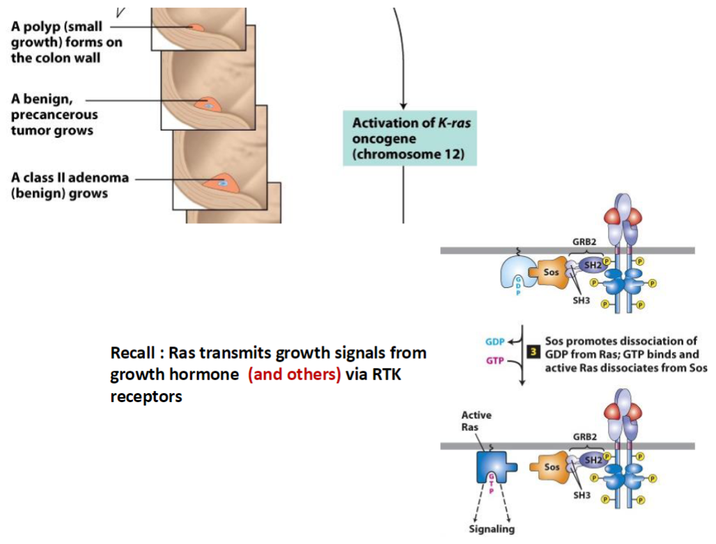
Colorectal Tumor Progression – K-ras Activation

Polyp Formation
 A polyp (small growth) forms on the colon wall
 Develops into a benign, precancerous tumor

K-ras Oncogene Activation
K-ras (chromosome 12) becomes activated, promoting growth of a class II adenoma (benign)

Ras Signaling Pathway
Ras transmits growth signals from growth factors via RTK (receptor tyrosine kinase) receptors
GRB2 binds activated receptor
Sos promotes dissociation of GDP from Ras
GTP binds Ras, activating it and allowing it to dissociate from Sos

Key Concept
 Activated Ras drives cell proliferation, contributing to tumor growth

<p><strong>Polyp Formation</strong><br> A <strong>polyp</strong> (small growth) forms on the <strong>colon wall</strong><br> Develops into a <strong>benign, precancerous tumor</strong></p><p><strong>K-ras Oncogene Activation</strong><br> <strong>K-ras</strong> (<strong>chromosome 12</strong>) becomes <strong>activated</strong>, promoting growth of a <strong>class II adenoma (benign)</strong></p><p><strong>Ras Signaling Pathway</strong><br> <strong>Ras</strong> transmits <strong>growth signals</strong> from <strong>growth factors</strong> via <strong>RTK (receptor tyrosine kinase) receptors</strong><br> <strong>GRB2</strong> binds activated receptor<br> <strong>Sos</strong> promotes dissociation of <strong>GDP</strong> from <strong>Ras</strong><br> <strong>GTP</strong> binds <strong>Ras</strong>, activating it and allowing it to <strong>dissociate from Sos</strong></p><p><strong>Key Concept</strong><br> Activated <strong>Ras</strong> drives <strong>cell proliferation</strong>, contributing to tumor growth</p>
8
New cards

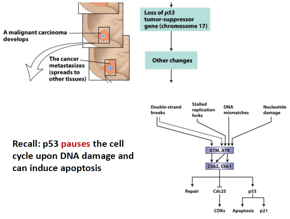
Colorectal Cancer Progression – Malignancy and Metastasis

Malignant Transformation
 A malignant carcinoma develops from the benign adenoma

Metastasis
 The cancer spreads to other tissues, establishing secondary tumors

Genetic Changes
Loss of p53 (tumor-suppressor gene, chromosome 17)
 Other genetic and epigenetic changes accumulate

p53 Function
p53 pauses the cell cycle in response to DNA damage
 Can induce apoptosis to prevent propagation of damaged cells

Key Concept
 Loss of p53 removes a critical growth checkpoint, allowing malignant progression and metastasis

<p><strong>Malignant Transformation</strong><br> A <strong>malignant carcinoma</strong> develops from the benign adenoma</p><p><strong>Metastasis</strong><br> The cancer <strong>spreads to other tissues</strong>, establishing secondary tumors</p><p><strong>Genetic Changes</strong><br> <strong>Loss of p53</strong> (<strong>tumor-suppressor gene, chromosome 17</strong>)<br> Other genetic and epigenetic changes accumulate</p><p><strong>p53 Function</strong><br> <strong>p53</strong> pauses the <strong>cell cycle</strong> in response to <strong>DNA damage</strong><br> Can induce <strong>apoptosis</strong> to prevent propagation of damaged cells</p><p><strong>Key Concept</strong><br> Loss of <strong>p53</strong> removes a critical <strong>growth checkpoint</strong>, allowing <strong>malignant progression</strong> and <strong>metastasis</strong></p>
9
New cards

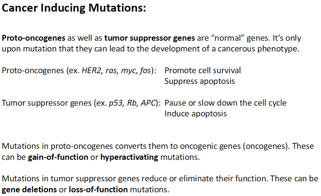
Cancer-Inducing Mutations

Normal Genes
Proto-oncogenes and tumor suppressor genes are normal genes that regulate cell growth and survival

Proto-Oncogenes
 Examples: HER2, Ras, Myc, Fos
  Promote cell survival
  Suppress apoptosis
 Mutation converts them into oncogenes
  Can be gain-of-function or hyperactivating mutations

Tumor Suppressor Genes
 Examples: p53, Rb, APC
  Pause or slow down the cell cycle
  Induce apoptosis
 Mutation reduces or eliminates their function
  Can be gene deletions or loss-of-function mutations

Key Concept
 Cancer arises when mutations disrupt the balance of cell proliferation and death, enabling uncontrolled growth

<p><strong>Normal Genes</strong><br> <strong>Proto-oncogenes</strong> and <strong>tumor suppressor genes</strong> are <strong>normal genes</strong> that regulate <strong>cell growth and survival</strong></p><p><strong>Proto-Oncogenes</strong><br> Examples: <strong>HER2, Ras, Myc, Fos</strong><br>  Promote <strong>cell survival</strong><br>  <strong>Suppress apoptosis</strong><br> Mutation converts them into <strong>oncogenes</strong><br>  Can be <strong>gain-of-function</strong> or <strong>hyperactivating mutations</strong></p><p><strong>Tumor Suppressor Genes</strong><br> Examples: <strong>p53, Rb, APC</strong><br>  Pause or <strong>slow down the cell cycle</strong><br>  Induce <strong>apoptosis</strong><br> Mutation reduces or eliminates their function<br>  Can be <strong>gene deletions</strong> or <strong>loss-of-function mutations</strong></p><p><strong>Key Concept</strong><br> Cancer arises when <strong>mutations</strong> disrupt the balance of <strong>cell proliferation and death</strong>, enabling <strong>uncontrolled growth</strong></p>
10
New cards

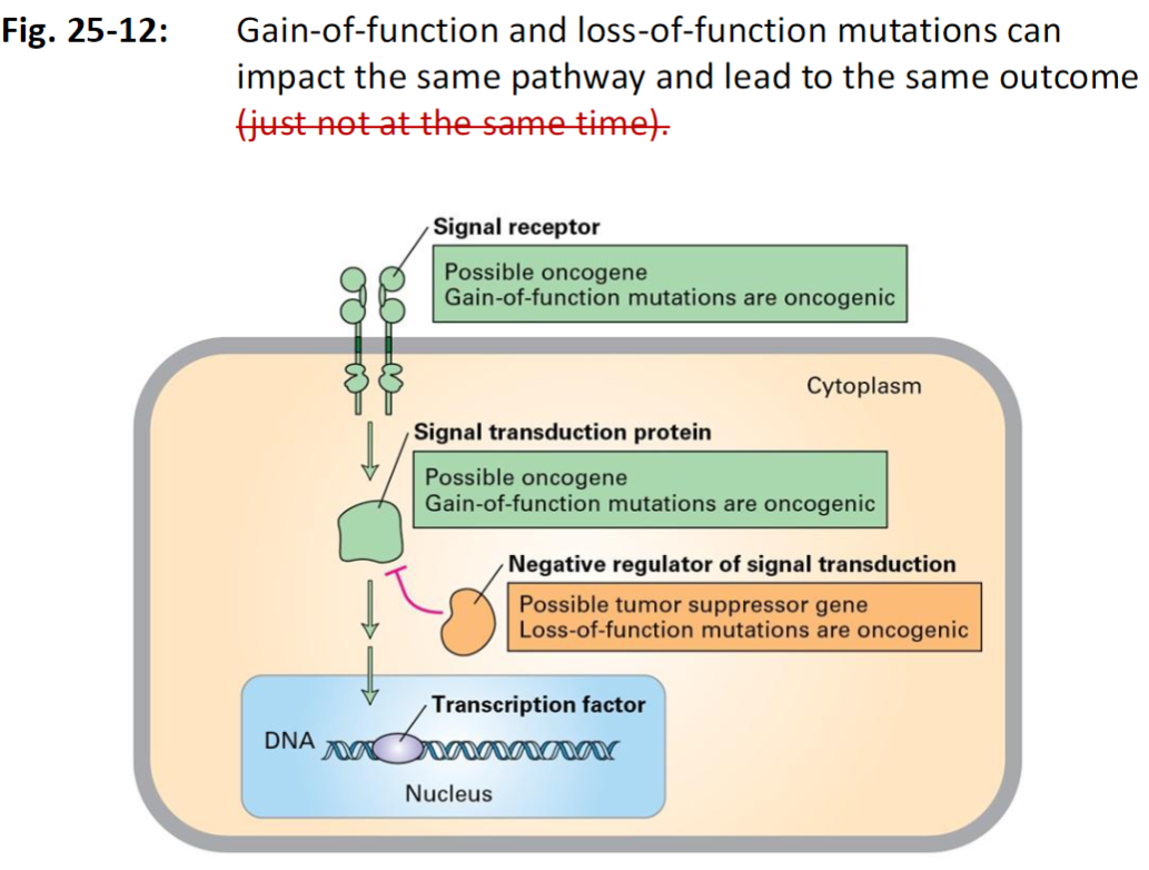
Mutations Affecting the Same Pathway

Signal Pathway Overview
 Mutations in different components of a signaling pathway can lead to similar oncogenic outcomes

Gain-of-Function Mutations
 Occur in proto-oncogenes such as:
  Signal receptors
  Signal transduction proteins
 These mutations are oncogenic, causing hyperactive signaling

Loss-of-Function Mutations
 Occur in negative regulators of signal transduction (tumor suppressor genes)
 These loss-of-function mutations remove inhibitory control, also resulting in oncogenic signaling

Outcome
 Both types of mutations disrupt normal cell growth control
 Leads to abnormal proliferation and potential tumor formation

<p><strong>Signal Pathway Overview</strong><br> Mutations in <strong>different components</strong> of a signaling pathway can lead to <strong>similar oncogenic outcomes</strong></p><p><strong>Gain-of-Function Mutations</strong><br> Occur in <strong>proto-oncogenes</strong> such as:<br>  <strong>Signal receptors</strong><br>  <strong>Signal transduction proteins</strong><br> These mutations are <strong>oncogenic</strong>, causing <strong>hyperactive signaling</strong></p><p><strong>Loss-of-Function Mutations</strong><br> Occur in <strong>negative regulators of signal transduction</strong> (<strong>tumor suppressor genes</strong>)<br> These <strong>loss-of-function mutations</strong> remove inhibitory control, also resulting in <strong>oncogenic signaling</strong></p><p><strong>Outcome</strong><br> Both types of mutations disrupt <strong>normal cell growth control</strong><br> Leads to <strong>abnormal proliferation</strong> and potential <strong>tumor formation</strong></p>
11
New cards

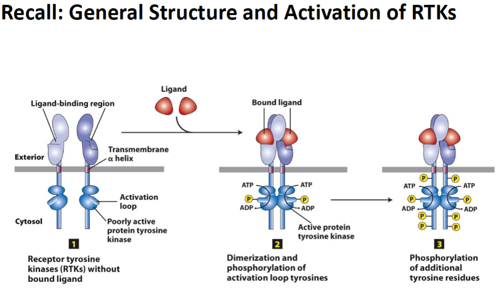
Receptor Tyrosine Kinases (RTKs) – Structure and Activation

Inactive State
RTKs exist without a bound ligand
ATP is present but the kinase is inactive

Activation Step 1: Dimerization
Ligand binding induces dimerization of RTKs
 Phosphorylation of activation loop tyrosines occurs
ATP → ADP, transferring phosphate groups (P)

Activation Step 2: Tyrosine Phosphorylation
Additional tyrosine residues in the receptor are phosphorylated
 Creates docking sites for downstream signal transduction proteins

Key Concept
 Activated RTKs initiate intracellular signaling pathways that regulate cell growth, survival, and differentiation

<p><strong>Inactive State</strong><br> <strong>RTKs</strong> exist without a <strong>bound ligand</strong><br> <strong>ATP</strong> is present but the kinase is <strong>inactive</strong></p><p><strong>Activation Step 1: Dimerization</strong><br> <strong>Ligand binding</strong> induces <strong>dimerization</strong> of RTKs<br> Phosphorylation of <strong>activation loop tyrosines</strong> occurs<br> <strong>ATP → ADP</strong>, transferring phosphate groups (<strong>P</strong>)</p><p><strong>Activation Step 2: Tyrosine Phosphorylation</strong><br> <strong>Additional tyrosine residues</strong> in the receptor are phosphorylated<br> Creates <strong>docking sites</strong> for downstream <strong>signal transduction proteins</strong></p><p><strong>Key Concept</strong><br> Activated <strong>RTKs</strong> initiate <strong>intracellular signaling pathways</strong> that regulate <strong>cell growth, survival, and differentiation</strong></p>
12
New cards

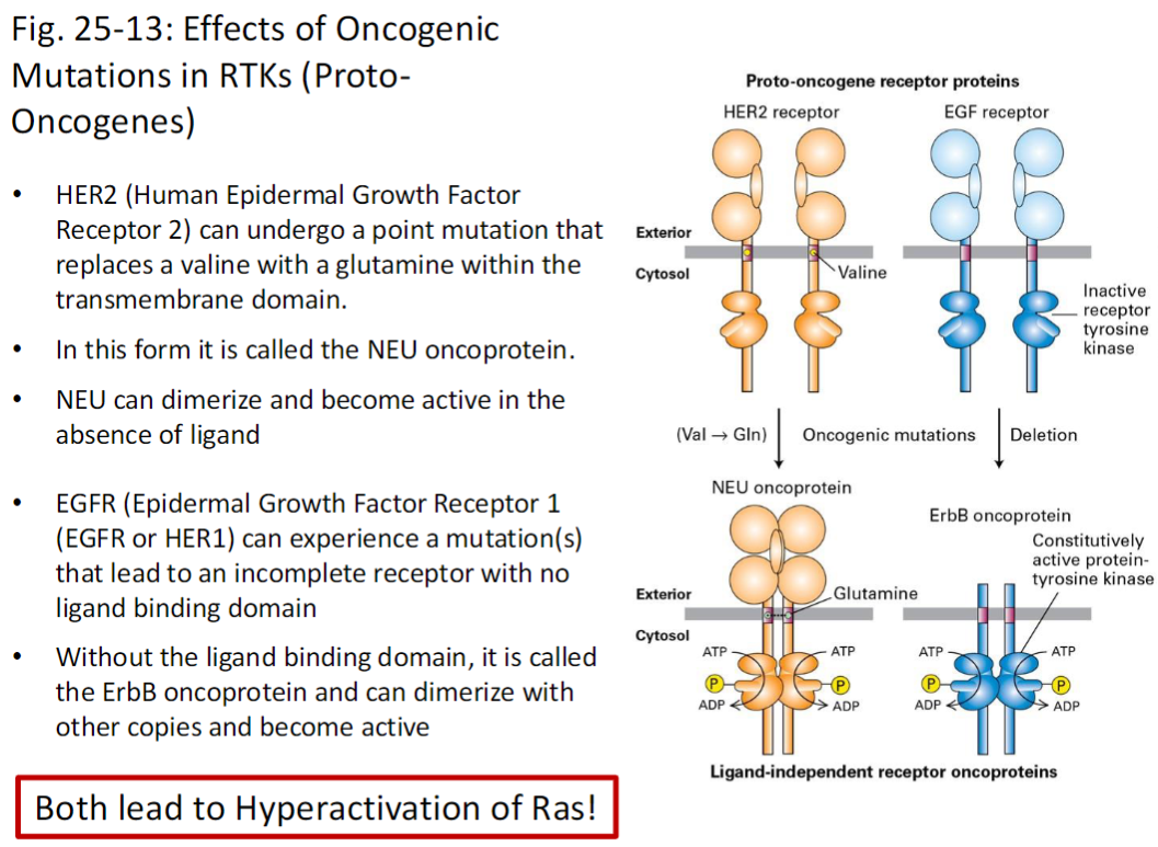
Oncogenic Mutations in RTKs (Proto-Oncogenes)

HER2 Mutation
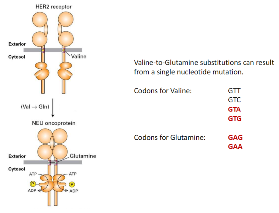
HER2 (Human Epidermal Growth Factor Receptor 2) can undergo a point mutation replacing valine with glutamine in the transmembrane domain
 Mutated form = NEU oncoprotein
NEU can dimerize and become active without ligand

EGFR Mutation
EGFR (Epidermal Growth Factor Receptor 1 / HER1) can mutate to produce an incomplete receptor lacking the ligand-binding domain
 Mutated form = ErbB oncoprotein
 Can dimerize with other copies and become active

Key Outcome
 Both mutations lead to hyperactivation of Ras, driving cell proliferation and potential tumorigenesis

<p><strong>HER2 Mutation</strong><br> <strong>HER2</strong> (<strong>Human Epidermal Growth Factor Receptor 2</strong>) can undergo a <strong>point mutation</strong> replacing <strong>valine</strong> with <strong>glutamine</strong> in the <strong>transmembrane domain</strong><br> Mutated form = <strong>NEU oncoprotein</strong><br> <strong>NEU</strong> can <strong>dimerize</strong> and become <strong>active without ligand</strong></p><p><strong>EGFR Mutation</strong><br> <strong>EGFR</strong> (<strong>Epidermal Growth Factor Receptor 1 / HER1</strong>) can mutate to produce an <strong>incomplete receptor</strong> lacking the <strong>ligand-binding domain</strong><br> Mutated form = <strong>ErbB oncoprotein</strong><br> Can <strong>dimerize with other copies</strong> and become <strong>active</strong></p><p><strong>Key Outcome</strong><br> Both mutations lead to <strong>hyperactivation of Ras</strong>, driving <strong>cell proliferation</strong> and potential <strong>tumorigenesis</strong></p>
13
New cards

Valine-to-Glutamine Substitution

Genetic Basis
 A single nucleotide mutation can convert valine to glutamine

Codons for Valine (Val)
GTT, GTC, GTA, GTG

Codons for Glutamine (Gln)
GAG, GAA

Key Concept
 This point mutation in the HER2 gene can create the NEU oncoprotein, leading to ligand-independent activation and hyperactive Ras signaling

<p><strong>Genetic Basis</strong><br> A <strong>single nucleotide mutation</strong> can convert <strong>valine</strong> to <strong>glutamine</strong></p><p><strong>Codons for Valine (Val)</strong><br> <strong>GTT, GTC, GTA, GTG</strong></p><p><strong>Codons for Glutamine (Gln)</strong><br> <strong>GAG, GAA</strong></p><p><strong>Key Concept</strong><br> This <strong>point mutation</strong> in the <strong>HER2 gene</strong> can create the <strong>NEU oncoprotein</strong>, leading to <strong>ligand-independent activation</strong> and <strong>hyperactive Ras signaling</strong></p>
14
New cards

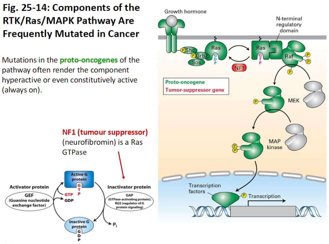
RTK/Ras/MAPK Pathway in Cancer

Pathway Components
 Includes RTKs, Ras, MAPK, and regulators like NF1 (tumor suppressor, neurofibromin)
NF1 functions as a Ras GTPase, inactivating Ras

Mutations
 Mutations in proto-oncogenes of this pathway often make the component hyperactive or constitutively active (always on)

Key Concept
 Constitutive activation of the RTK/Ras/MAPK pathway drives uncontrolled cell proliferation, a hallmark of cancer

<p><strong>Pathway Components</strong><br> Includes <strong>RTKs, Ras, MAPK</strong>, and regulators like <strong>NF1</strong> (<strong>tumor suppressor, neurofibromin</strong>)<br> <strong>NF1</strong> functions as a <strong>Ras GTPase</strong>, inactivating Ras</p><p><strong>Mutations</strong><br> Mutations in <strong>proto-oncogenes</strong> of this pathway often make the component <strong>hyperactive</strong> or <strong>constitutively active</strong> (<strong>always on</strong>)</p><p><strong>Key Concept</strong><br> Constitutive activation of the <strong>RTK/Ras/MAPK pathway</strong> drives <strong>uncontrolled cell proliferation</strong>, a hallmark of <strong>cancer</strong></p>

Explore top notes

note
Types of Attachment
Updated 536d ago
0.0(0)
note
Lam 2013, Reply to Stan Becker
Updated 1173d ago
0.0(0)
note
Microbiology Quiz 7 (BIO 210)
Updated 156d ago
0.0(0)
note
Ch 2- Hardware and software
Updated 1095d ago
0.0(0)
note
Chapter 18 The Great Depression
Updated 849d ago
0.0(0)
note
Chapter 8 - Emotions & moods
Updated 1354d ago
0.0(0)
note
Types of Attachment
Updated 536d ago
0.0(0)
note
Lam 2013, Reply to Stan Becker
Updated 1173d ago
0.0(0)
note
Microbiology Quiz 7 (BIO 210)
Updated 156d ago
0.0(0)
note
Ch 2- Hardware and software
Updated 1095d ago
0.0(0)
note
Chapter 18 The Great Depression
Updated 849d ago
0.0(0)
note
Chapter 8 - Emotions & moods
Updated 1354d ago
0.0(0)