Functional Anatomy Exam 3 Cards
1/49
There's no tags or description
Looks like no tags are added yet.
Name | Mastery | Learn | Test | Matching | Spaced | Call with Kai |
|---|
No analytics yet
Send a link to your students to track their progress
50 Terms
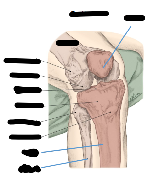
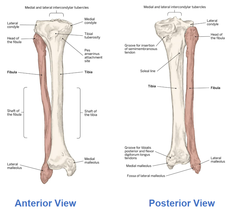
What 4 bones make up the knee
Femur, Tibia, Fibula, Patella
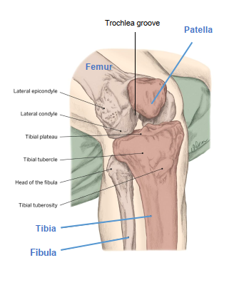
Purpose of Tibia and Fibula in knee joint
Tibia: Bear weight and articulate with femur
Fibula: No articulation, Serves as attachment for knee joint structures
Patella what kind of bone, what tendons attach it,where does it sit
Sesamoid floating bone, quadriceps and patellar tendon attach it, rests in trochlear groove.
Three joint of the knee
Tibiofemoral (knee joint proper), Patellofemoral, Tibiofibular (Not part of the knee)
Tibiofemoral joint type and size
Size: Biggest synovial in body, Type: ginglymus or hinge
Explain Q angle
Definition Intersection between the line of pull of the quads and line of pull of the patella (15 degrees in males and 20 degrees in females). Greater Q angle = larger lateral risk = greater injury risk
Four TIbiofemoral Ligaments and What motion they prevent
Anterior Cruciate Ligament (ACL): Prevents tibia from sliding anterior
Posterior Cruciate Ligament (PCL): Prevents Tibia from sliding posterior
Medial Cruciate Ligament (MCL): stabilizes valgus forces (inward)
Lateral Cruciate Ligament (LCL): stabilizes varus forces (outward)
Patellar Tendon and Ligament
Tendon: common attachment for quads superior to patella
Ligament: inferior to patella and connects to tibial tuberosity
Function of Bursa
Absorb shock and reduce friction
Is there Adduction and Abduction at the knee?
No
List all the biarticular muscles of the knee / ankle / hip (5)
Biceps femoris, Rectus Femoris, Semimebranosus, Semitendinosus, Gastronemius
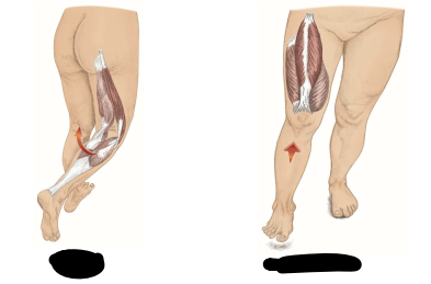
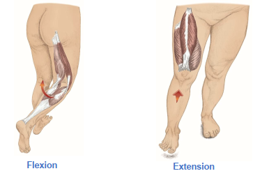
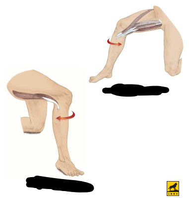
List primary knee flexors (3)
Hamstrings: Biceps Femoris, Semitendinosus, Semimembranosus
List primary knee extenders (4)
Quadriceps: Rectus femoris, Vastus Medialis, Vastus Lateralis, Vastus Intermedialis
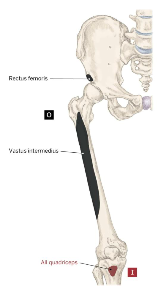
Rectus Femoris (A, O, I)
Action: Extend the Knee (tibiofemoral) , Flex the hip (coxal)
Origin: Anterior Inferior Illiac Spine (AIIS)
Insertion: Tibial Tuberosity
Vastus Medialis (A, O, I)
Action: Extend the knee (tibiofemoral)
Origin: Medial lip of linea aspera
Insertion: Tibial Tuberosity
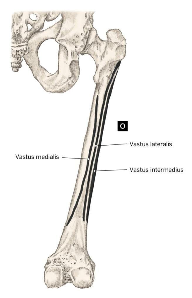
Vastus Lateralis (A, O, I)
Action: Extend the knee (tibiofemoral)
Origin: Lateral lip of linea aspera, gluteal tuberosity, greater trochanter
Insertion: Tibial tuberosity

Vastus Intermedialis (A, O, I)
Action: Extend the knee
Origin: Aterior and lateral shaft of the femur
Insertion: TIbial Tuberosity

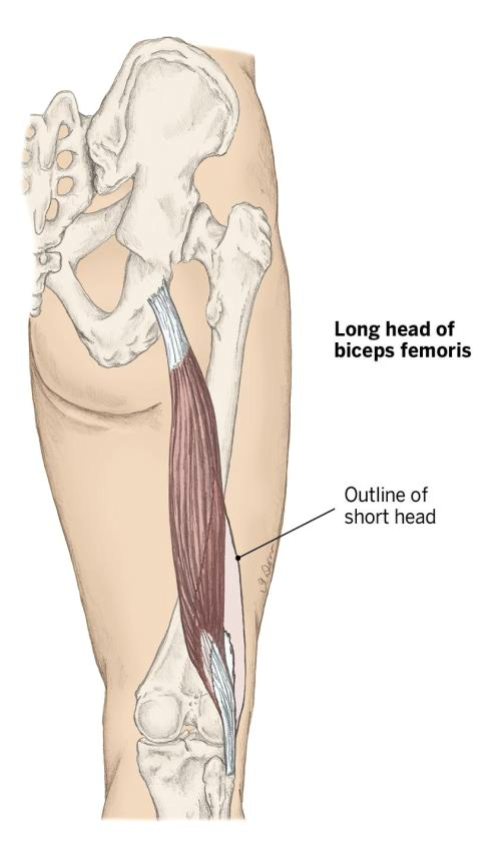
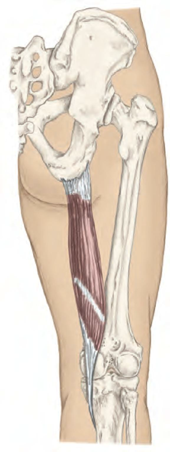
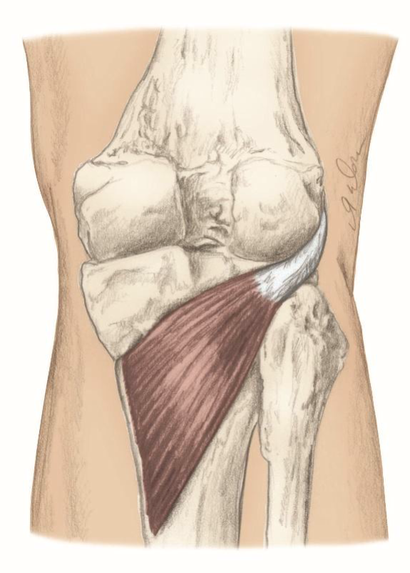
Biceps Femoris (A, O, I)
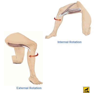
Action: Flex the knee, Laterally rotate flexed knee, tilt pelvis posteriorly,
Origin: Long head: ischial tuberosity, Short head: lateral lip of linea aspera
Insertion: Head of the fibula
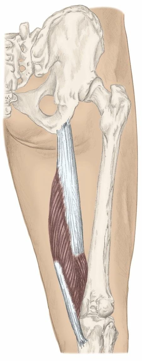
Semitendinosus (A, O, I)
Action: Flex the knee, medially rotate the flexed knee, extend the hip, tilt the pelvis posteriorly
Origin: Ischial tuberosity
Insertion: Proximal, medial shaft of tibia at pes anserinus tendon
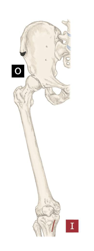
Semimembranosus (A, O, I)
Action: Flex the knee, medially rotate the flexed knee, extend the hip, tilt the pelvis posteriorly
Origin: ischial tuberosity
Insertion: posterior aspect of medial condyle of tibia
Sartorius (A, O, I)
Action: Felx the hip, laterally roatew hip, Abduct the hip, Flex the knee, medially rotate the flexed knee
Origin: Anterior Superior Iliac Spine (ASIS)
Insertion: Proximal, medial shaft of the tibia at pes anserinus tendon
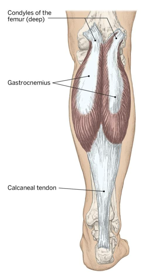
Gastronemius (A, O, I)
Action: Flex the knee (tibiofemoral), plantar flex the ankle (talocrural)
Origin: Condyles of the femur posterior surface
Insertion: Calcaneus via the calcaneal / achilles tendon
Popliteus (A, O, I)
Action: Medially roate flexed knee, Flex the knee
Origin: Lateral condyle of the femur
Insertion: Proximal posterior aspect of the tibia
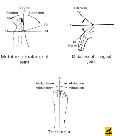
How many bones in the foot
14 phallanges, 7 tarsals, 5 metatarsals, 2 sesamoids under great toe
list joints of ankle and foot (3)
Talocrural, Subtalar, Transverse tarsal joints
Tibiofibular joint type of joint and amount of movement
Syndesmotic amphiarthrodial, minimal movement, attached proximal to distal
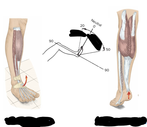
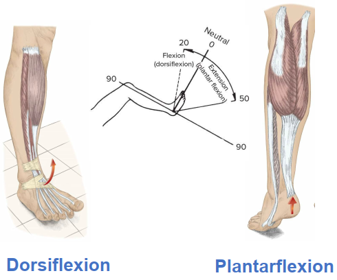
Talocrural joint
Made up of Fibula, tibia, talus
Hinge, ginglymus
Greater dorsiflexion ROM when knee flexed
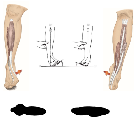
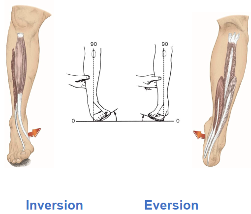
Ligaments of the Ankle (2 groups 5 total)
Lateral Collateral Ligaments: Anterior talofibular (ATFL), Calcaneofibular (CFL), and Posterior Talofibular (PTFL)
Deltoid ligament complex: Medial malleolus
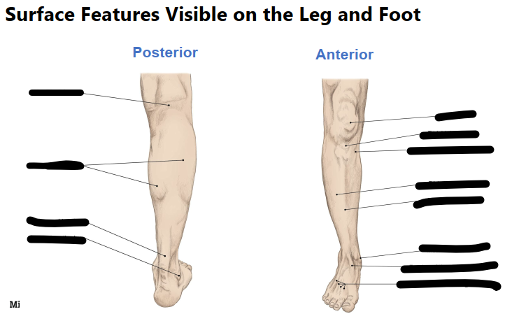
Three arches of the foot
Transverse arch, medial longitudinal arch, lateral logitudinal arch
Plantar Fascia (Plantar aponeurosis)
Broad structure ligament fromt he medial calcaneal tuberosity to proximal phallanges. Stabiolizes medial arch.
Anterior Compartment of Lower leg (4)
Tibialis Anterior, Fibularis (peroneus) Tertius, Extensor Digitorum Longus, Extensor Hallucis Longus
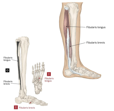
Lateral Compartment of the lower leg (2)
Fibularis (peroneus) Longus and Fibularis (peroneus) brevis
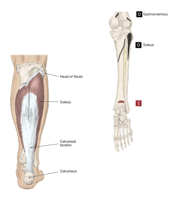
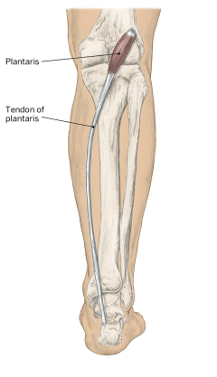
Superficial (3) and Deep (4) Posterior compartments of lower leg
Superficial: Gastronemius, soleus, Plantaris
Deep: Flexor digitorum longus, Flexor hallucis longus, popliteus, tibialis posterior
Soleus (A, O, I)
Action: Plantar flex the floot
Origin: Soleal line, proximal posterior surface of the tibia, and the posterior head of the fibula
Insertion: Calcaneus via the calcaneal or achilles tendon
Plantaris (A, O, I)
Action: Weak plantar flexion of the ankle and flexion of the knee
Origin: lateral supracondyle line of the femur
Insertion: calcaneus by calcaneal tendon
Fibularis longus (A, O, I)
Action: Evert foot, Plantar flex assist
Origin: Head of the fibula and proximal 2/3 of the lateral fibula
Insertion: Base of first metatarsal and medial cuneiform

Action:
Origin:
Insertion: