Lab exam 2
1/70
There's no tags or description
Looks like no tags are added yet.
Name | Mastery | Learn | Test | Matching | Spaced | Call with Kai |
|---|
No analytics yet
Send a link to your students to track their progress
71 Terms
Muscle Nomenclature
■ XX: Some muscles are named by their location, such as the tibialis anterior
■ XX: The adductor magnus is the largest of the adductor muscles. The gluteus minimus is the smallest of the gluteal muscles
■ XX: The deltoid muscle is shaped like a deltoid, or triangle
■ XX: The transverse abdominis has fibers that are oriented horizontally, or in a transverse direction
■ XX: The infraspinatus muscle attaches to the infraspinous fossa of the
scapula
■ XX of XX: The triceps brachii has 3 heads
■ XX: The extensor digitorum is a muscle that extends the fingers
■ XX Muscles with the term longus or brevis are named for the overall length of the muscle.
Muscle Nomenclature
■ Location: Some muscles are named by their location, such as the tibialis anterior
■ Size: The adductor magnus is the largest of the adductor muscles. The gluteus
minimus is the smallest of the gluteal muscles
■ Shape: The deltoid muscle is shaped like a deltoid, or triangle
■ Orientation: The transverse abdominis has fibers that are oriented horizontally, or in
a transverse direction
■ Attachments: The infraspinatus muscle attaches to the infraspinous fossa of the
scapula
■ Number of Heads: The triceps brachii has 3 heads
■ Action: The extensor digitorum is a muscle that extends the fingers
■ Length: Muscles with the term longus or brevis are named for the overall length of
the muscle
Muscles of the Face: Actions
■ XX: Raises eyebrows, wrinkles forehead, tenses, and retracts scalp
■ XX XX: Retracts and elevates upper lip
■ XX XX Retracts and elevates corner of mouth
■ XX: Draws corner of mouth to the side
■ XX: Tenses skin of neck, depresses mandible
■ XX XX: Closes eye
■ XX: Elevates mandible and closes jaw
■ XX: compresses cheeks
■ XX XX: Compresses, purses lips
■ XX: Elevates and protrudes lower lip
Muscles of the Face: Actions
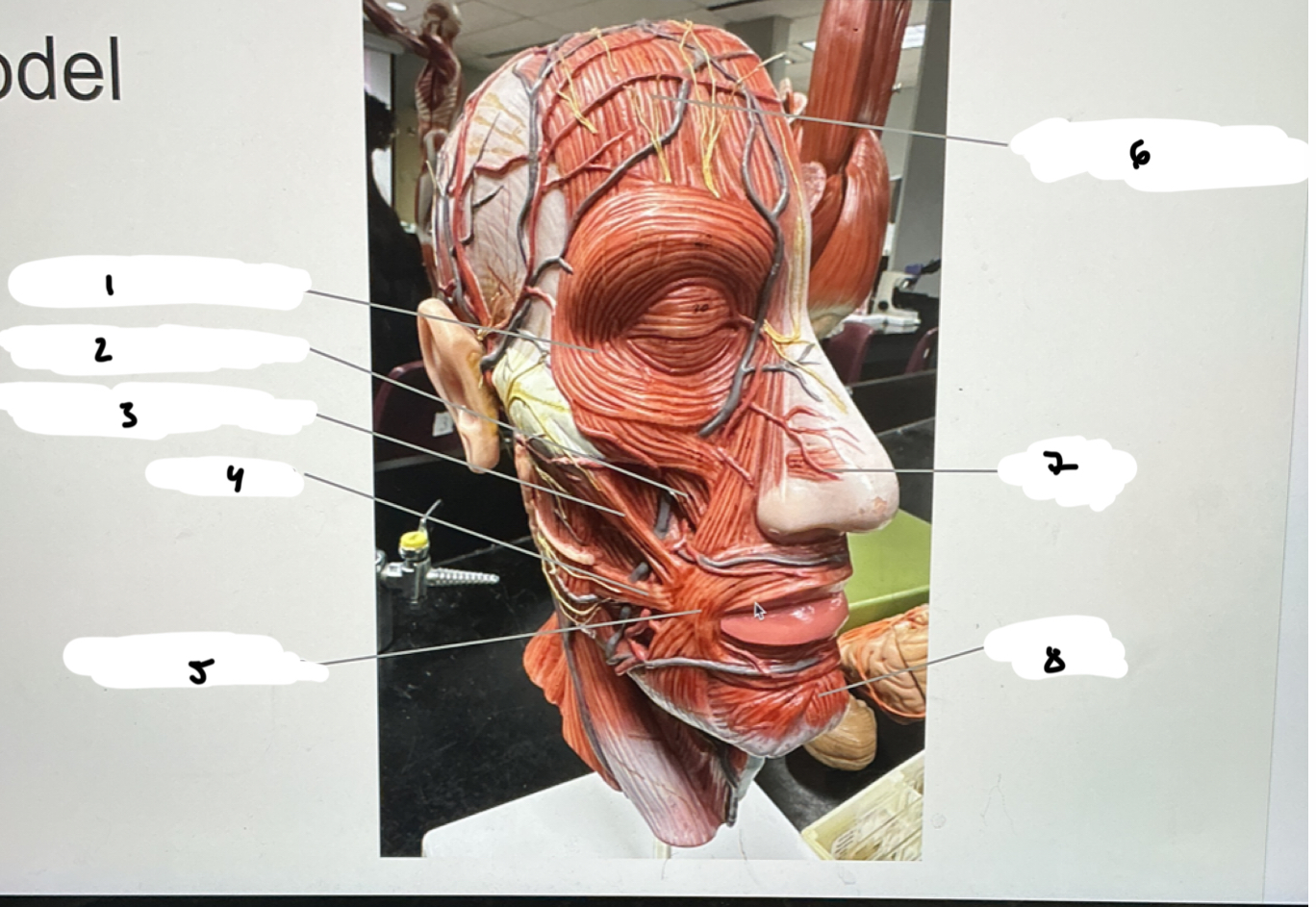
■ Occipitofrontalis: Raises eyebrows, wrinkles forehead, tenses, and retracts scalp
■ Zygomaticus Minor: Retracts and elevates upper lip
■ Zygomaticus Major: Retracts and elevates corner of mouth
■ Risorius: Draws corner of mouth to the side
■ Platysma: Tenses skin of neck, depresses mandible
■ Orbicularis Oculi: Closes eye
■ Masseter: Elevates mandible and closes jaw
■ Buccinator: compresses cheeks
■ Orbicularis Oris: Compresses, purses lips
■ Mentalis: Elevates and protrudes lower lip
1.XX
2.XX
3.XX
4.XX
5.XX
6.XX
7.XX
8.XX
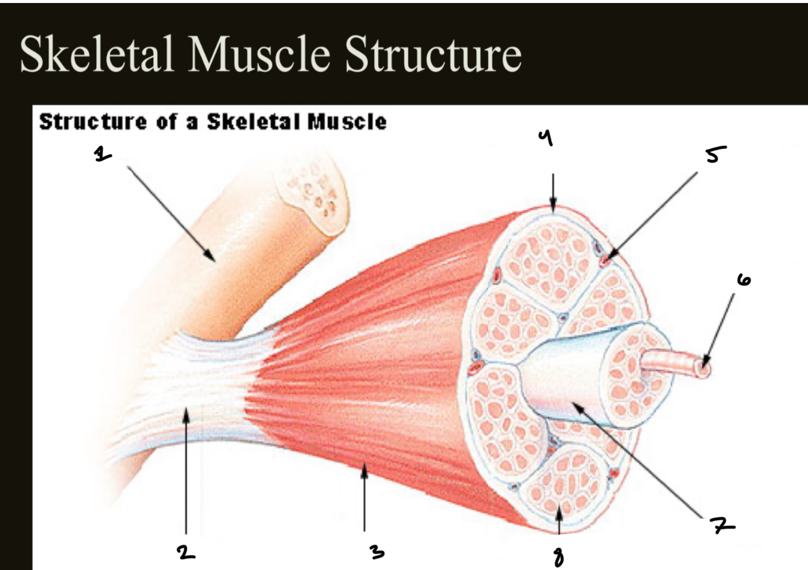
bone
Tendon
epimysium
perimysium
blood vessel
muscle fiber
fascicle
endomysium
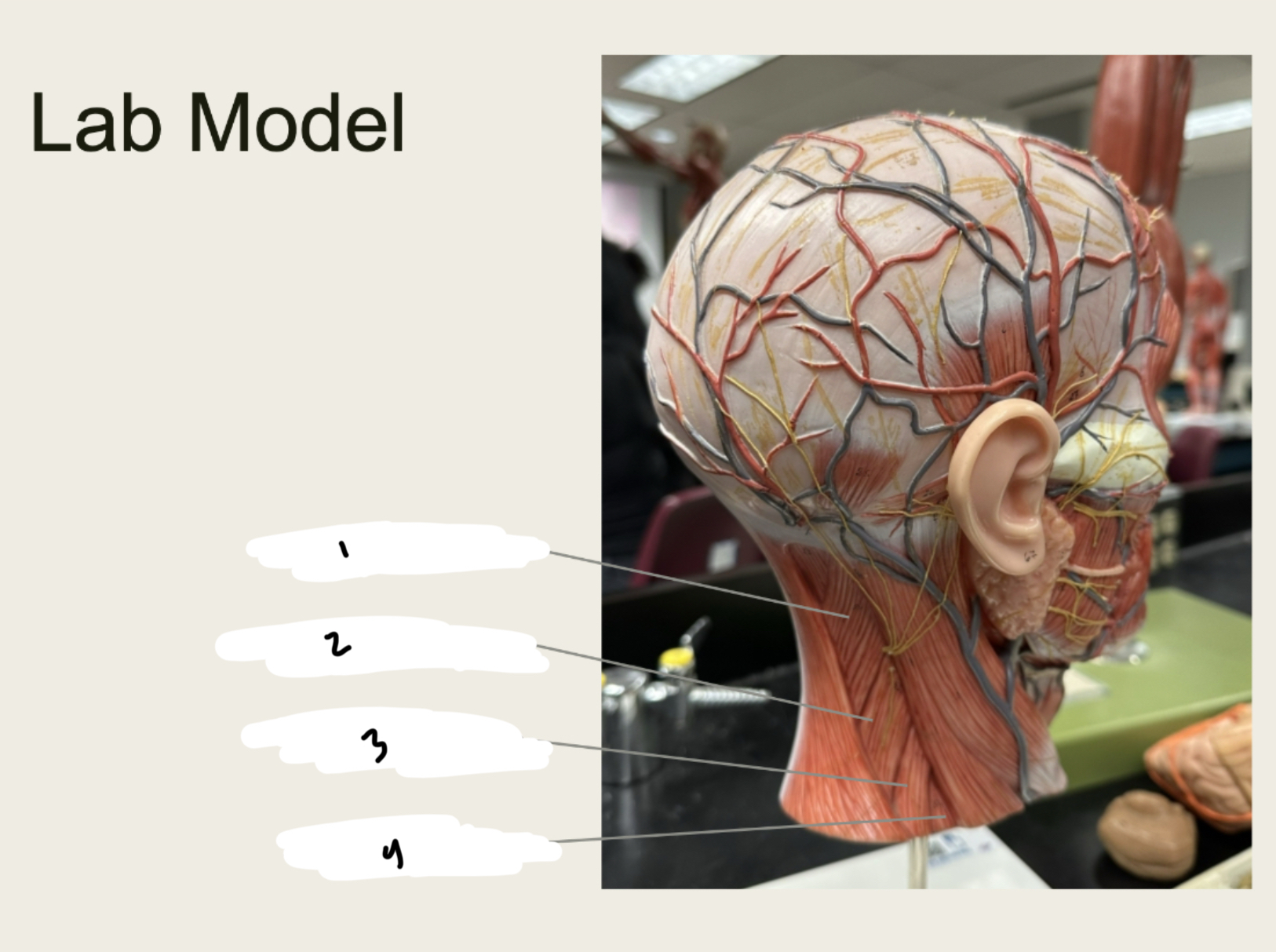
Muscles of the Neck: Actions
■ XX: Depressed mandible, and/or elevates larynx
■ XX: Flexes neck, bends neck towards shoulder, and turns face
to opposite side
■ XX: Depresses hyoid bone and larynx
■ XX: Depresses hyoid bone and larynx
■ XX: Depresses hyoid bone and larynx
■ XX XX: Elevates the scapula
■ XX: Flexes and rotates neck, elevates ribs 1 & 2
Digastric: Depressed mandible, and/or elevates larynx
■ Sternocleidomastoid: Flexes neck, bends neck towards shoulder, and turns face
to opposite side
■ Sternothyroid: Depresses hyoid bone and larynx
■ Sternohyoid: Depresses hyoid bone and larynx
■ Omohyoid: Depresses hyoid bone and larynx
■ Levator Scapulae: Elevates the scapula
■ Scalenes: Flexes and rotates neck, elevates ribs 1 & 2
1.XX
2.XX
3.XX
4.XX
5.XX
6.XX
7.XX
8.XX
1.orbicularis oculi
2.zygomaticus minor
3. zygomaticus major
4.risorius
5.orbicularis oris
6.occipitofrontalis
7.nasalis
8. mentalis
1.XX
2.XX
3.XX
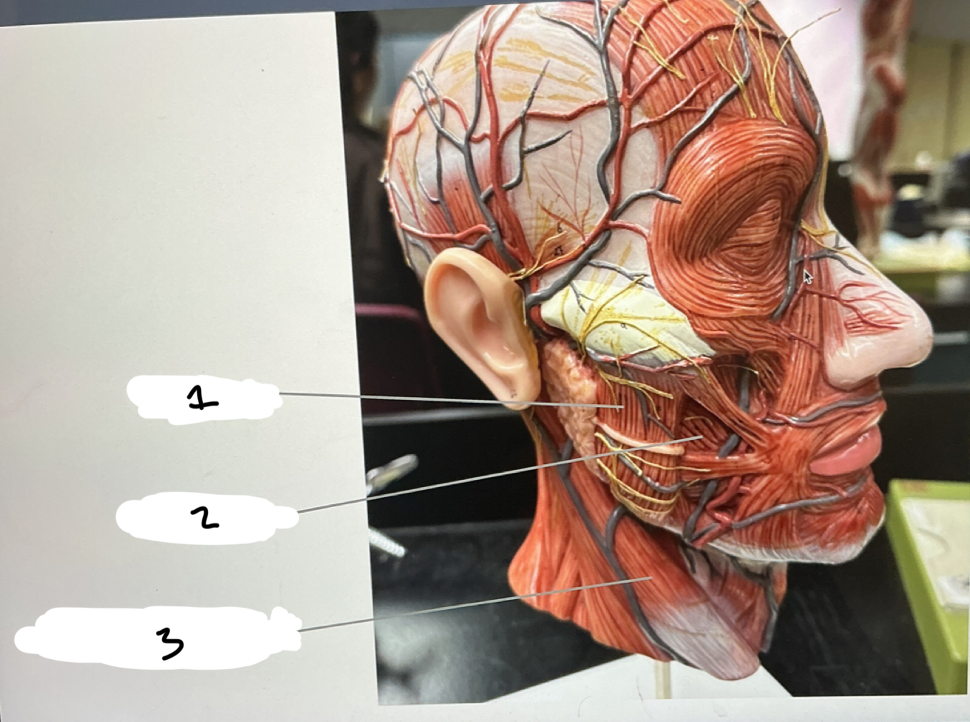
1.MASSETER
2.BUCCINATOR
3.STERNOCLEDIOMASTOID
1.XX
2.XX
3.XX
4.XX
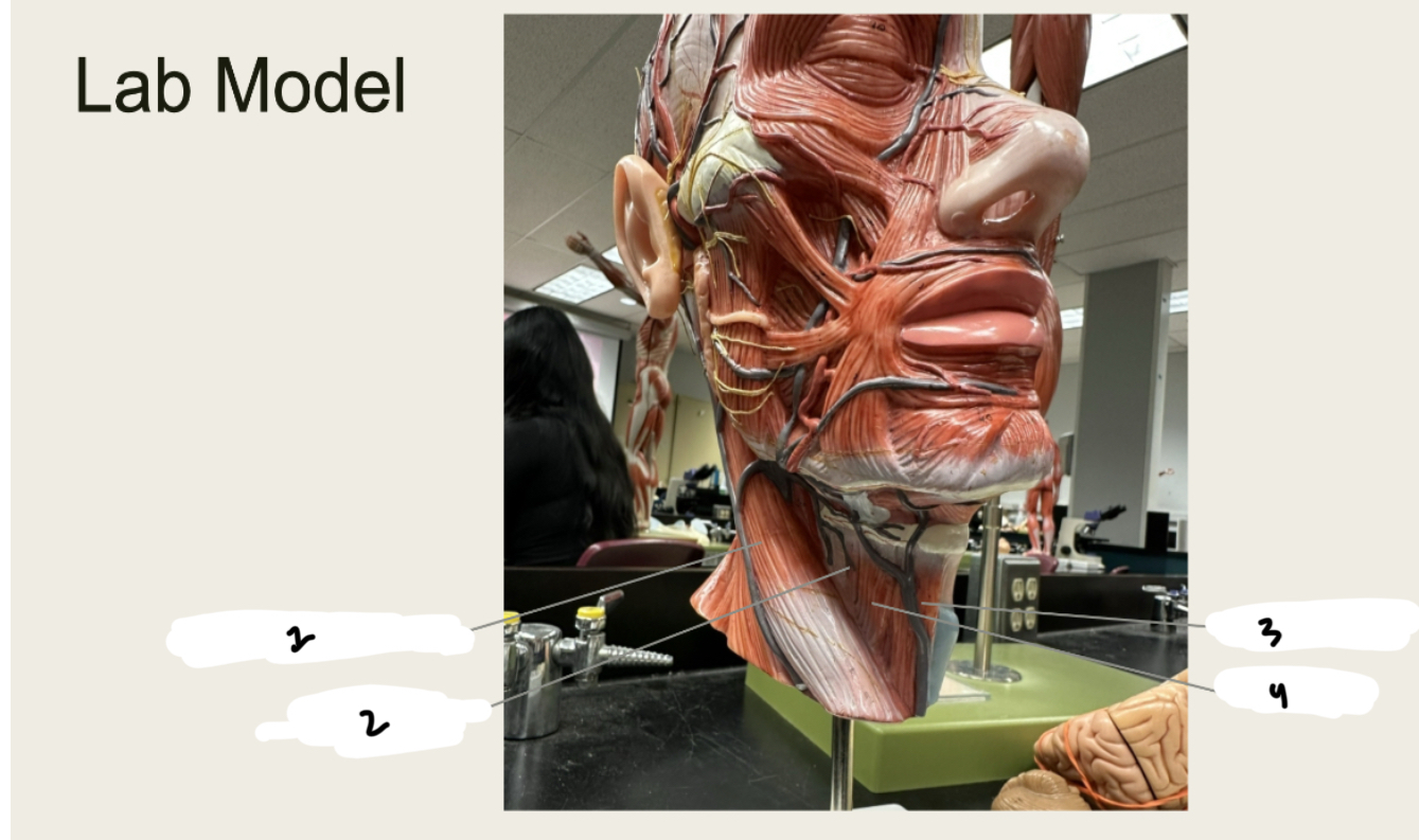
1.STERNOCLEIDOMASTOID
2.THYROHYOID
3.STERNOHYOID
4.OMOHYOID
1.XX
2.XX
3.XX
4.XX
1.LEVATOR SCAPULAE
2.POSTERIOR SCALENE
3.MIDDLE SCALENE
4.ANTERIOR SCALENE
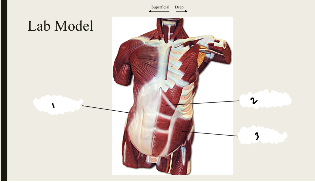
Muscles of the Abdomen: Actions
■ XX XX: Compresses abdomen
■XX XX: Compresses abdomen, depresses ribs, flexes vertebral
column, and rotates vertebral column to same side
■ XX XX: Compresses abdomen, depresses ribs, flexes vertebral
column, and rotates vertebral column to opposite side
■ XX XX: Depresses ribs, flexes vertebral column and compresses
abdomen
■XX XX: Depresses ribs
■ XX XX: Elevates Ribs
Muscles of the Abdomen: Actions
■ Transverse Abdominis: Compresses abdomen
■ Internal Oblique: Compresses abdomen, depresses ribs, flexes vertebral
column, and rotates vertebral column to same side
■ External Oblique: Compresses abdomen, depresses ribs, flexes vertebral
column, and rotates vertebral column to opposite side
■ Rectus Abdominis: Depresses ribs, flexes vertebral column and compresses
abdomen
■ Internal Intercostals: Depresses ribs
■ External Intercostals: Elevates Ribs
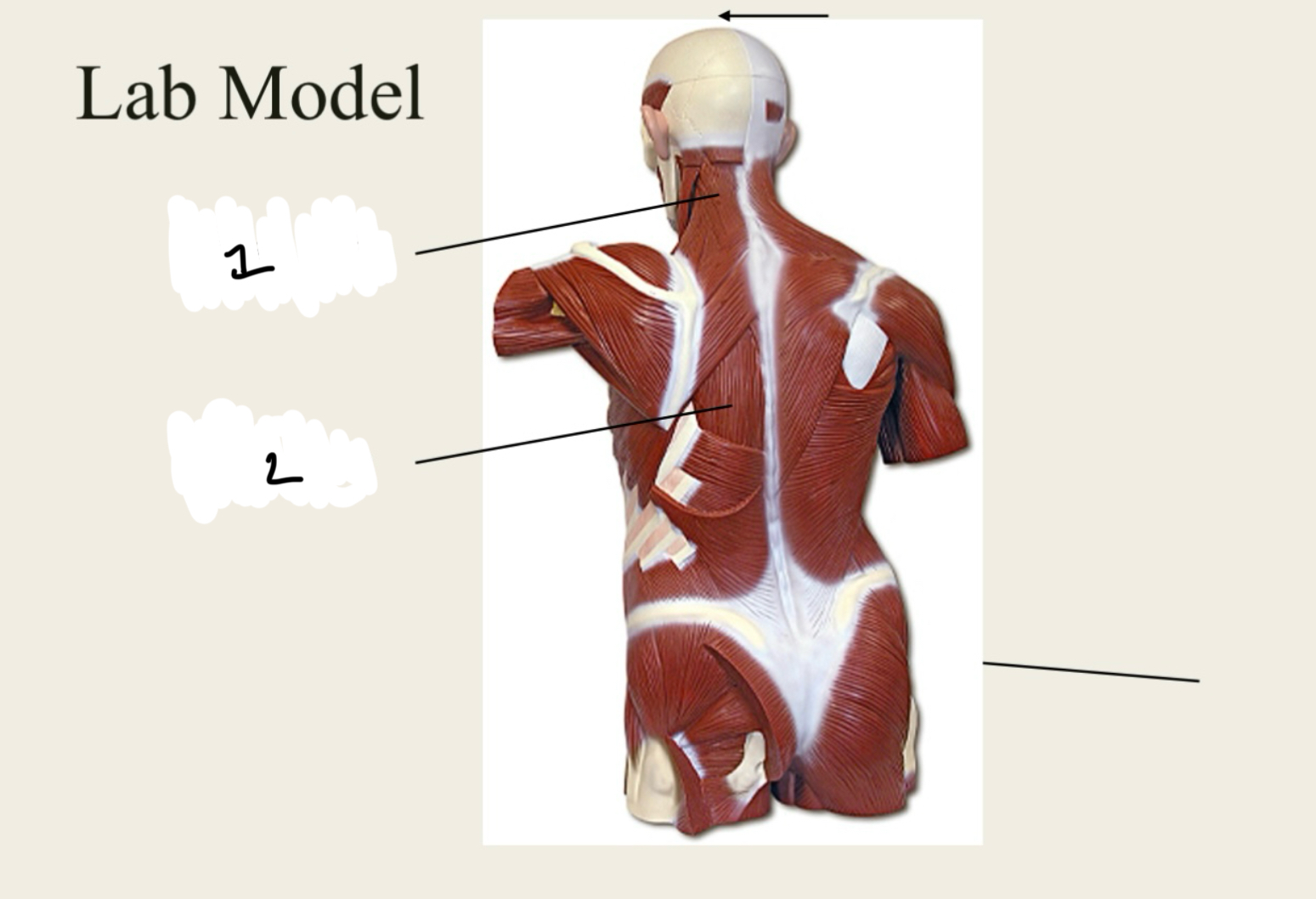
Muscles of the Back: Actions
■ XX XX: Extends and rotates vertebral column and head
– Iliocostalis
– Longissimus
– Spinalis
■ XX XX: Extends and rotates head
Muscles of the Back: Actions
■ Erector Spinae: Extends and rotates vertebral column and head
– Iliocostalis
– Longissimus
– Spinalis
■ Splenius Capitis: Extends and rotates head
1.XX
2.XX
3.XX
1.EXTERNAL OBLIQUE
2.RECTUS ABDOMINIS
3.INTERNAL OBLQIUE
1.XX
2.XX
1.Splenius Capitis
2.Erector Spinae
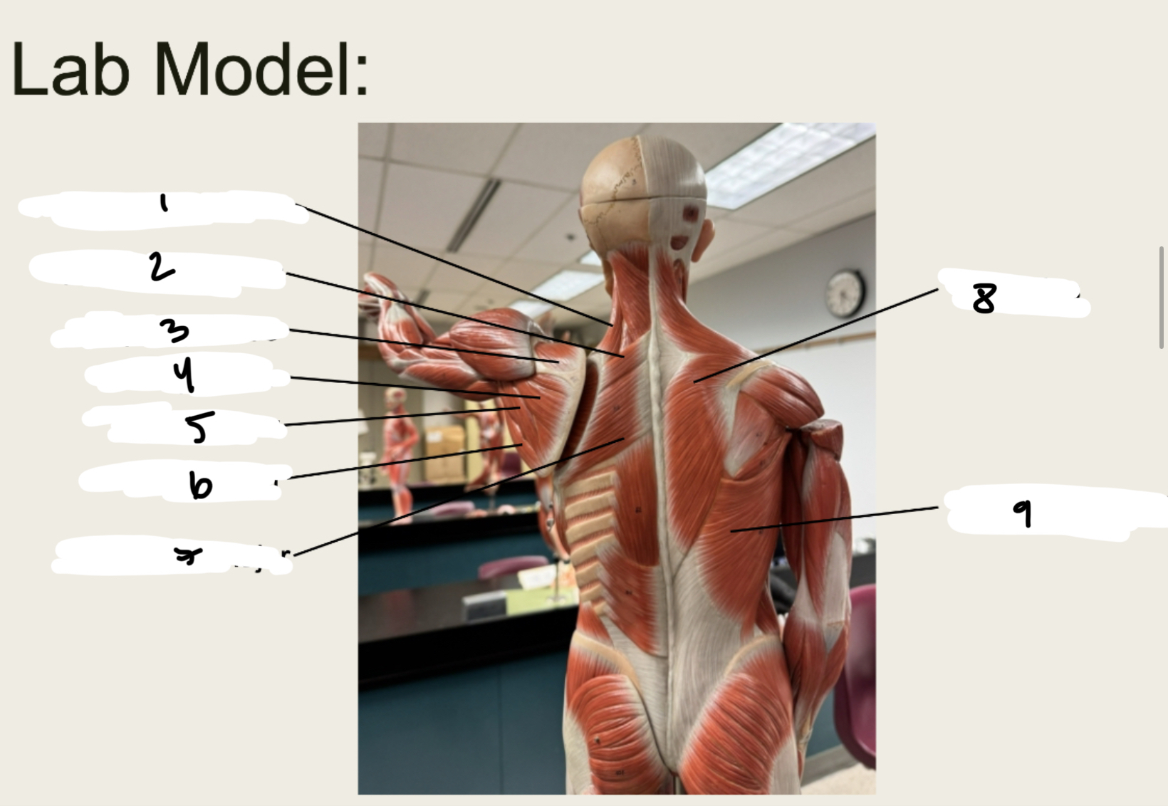
■ XX: Extends and abducts head, rotates and adducts scapula
■ XX XX: Elevates scapula
■ XX XX: Adducts scapula
■ XX XX: Adducts scapula
■ XX XX: Flexes, adducts, and medially rotates humerus
■ XX XX: Extends, adducts, and medially rotates arm
■ XX: Abducts arm, flexes, extends, medially and laterally rotates
arm
■ XX XX: Depresses glenoid cavity
■ XX XX: Extends and medially rotates arm
■ XX: Flexes and medially rotates arm
■ XX: Abducts arm, helps stabilize shoulder joint
■ XX: Laterally rotates arm, stabilizes shoulder joint
■ XX XX: Laterally rotates and adducts arm, stabilizes shoulder
joint
■XX: Medially rotates arm, stabilizes shoulder joint
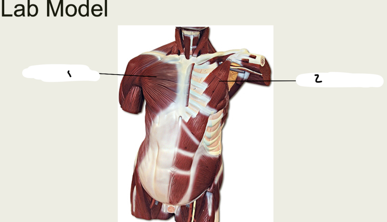
Trapezius: Extends and abducts head, rotates and adducts scapula
■ Levator Scapulae: Elevates scapula
■ Rhomboid Minor: Adducts scapula
■ Rhomboid major: Adducts scapula
■ Pectoralis Major: Flexes, adducts, and medially rotates humerus
■ Latissimus Dorsi: Extends, adducts, and medially rotates arm
■ Deltoid: Abducts arm, flexes, extends, medially and laterally rotates
arm
■ Pectoralis Minor: Depresses glenoid cavity
■ Teres Major: Extends and medially rotates arm
■ Coracobrachialis: Flexes and medially rotates arm
■ Supraspinatus: Abducts arm, helps stabilize shoulder joint
■ Infraspinatus: Laterally rotates arm, stabilizes shoulder joint
■ Teres Minor: Laterally rotates and adducts arm, stabilizes shoulder
joint
■ Subscapularis: Medially rotates arm, stabilizes shoulder joint
Muscles Acting on the Forearm:
Functions
■ XX: Flexes forearm
■ XX XX: Flexes arm, flexes forearm, supinates hand
■ XX XX: Extends and adducts arm, extends forearm
■ XX: Flexes forearm
■ XX XX: Pronates and flexes forearm
■ XX: Extends forearm
Muscles Acting on the Forearm:
Functions
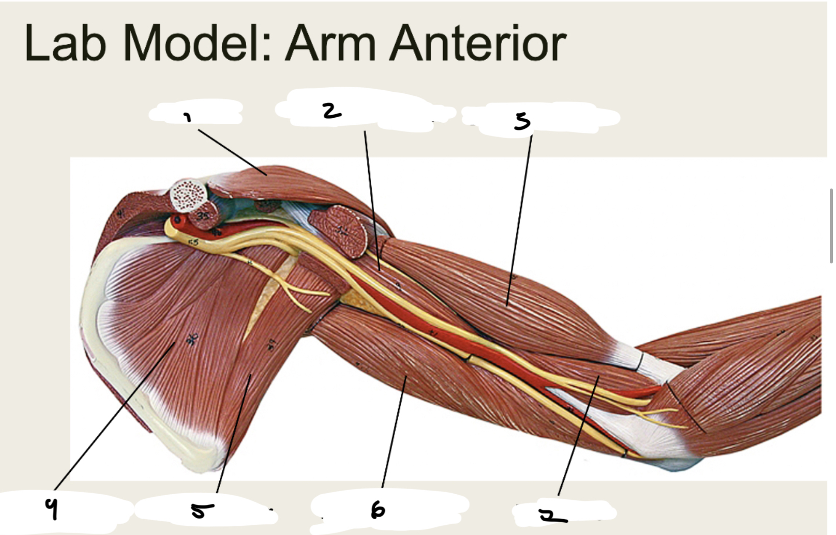
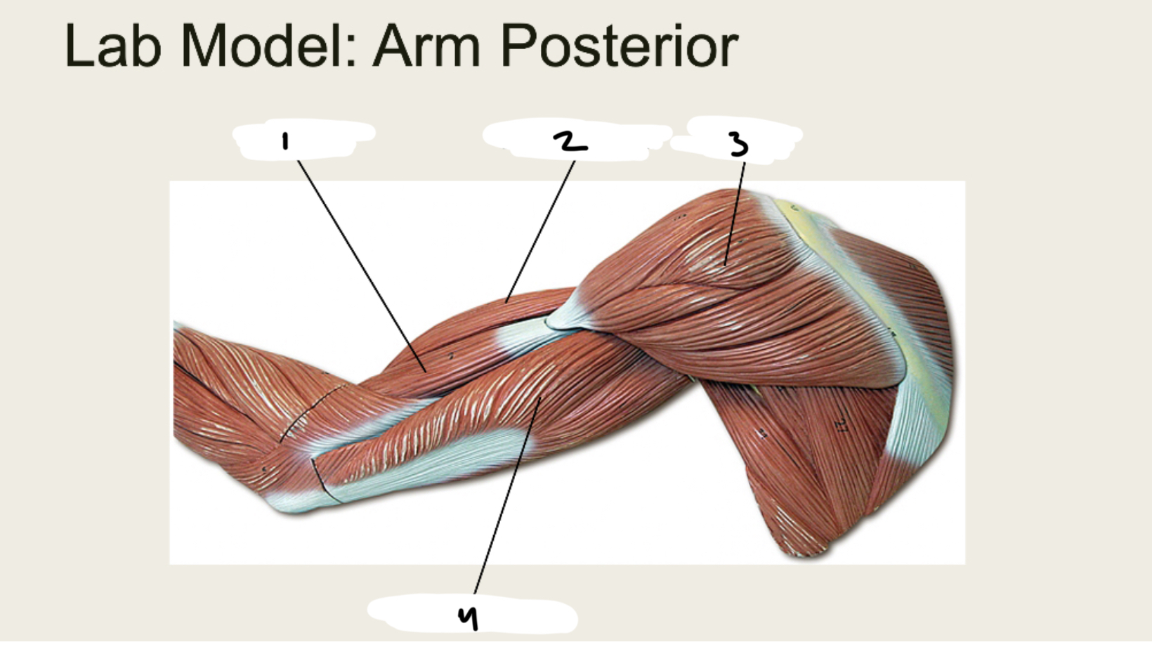
■ Brachialis: Flexes forearm
■ Biceps Brachii: Flexes arm, flexes forearm, supinates hand
■ Triceps Brachii: Extends and adducts arm, extends forearm
■ Brachioradialis: Flexes forearm
■ Pronator Teres: Pronates and flexes forearm
■ Anconeus: Extends forearm
Muscles Acting on the Wrist and
Hand: Function
■XX XX XX: Flexes and abducts wrists
■ XX XX XX: Flexes and adducts wrists
■ XX XX XX: Flexes proximal and middle phalanges, flexes
wrists
■ XX XX: Flexes wrist
■ XX XX XX XX: Extends and abducts wrists
■XX XX XX: Extends and adducts wrists:
■ XX XX XX: Abducts thumb
■ XX XX: Extends fingers and wrists
Muscles Acting on the Wrist and
Hand: Function
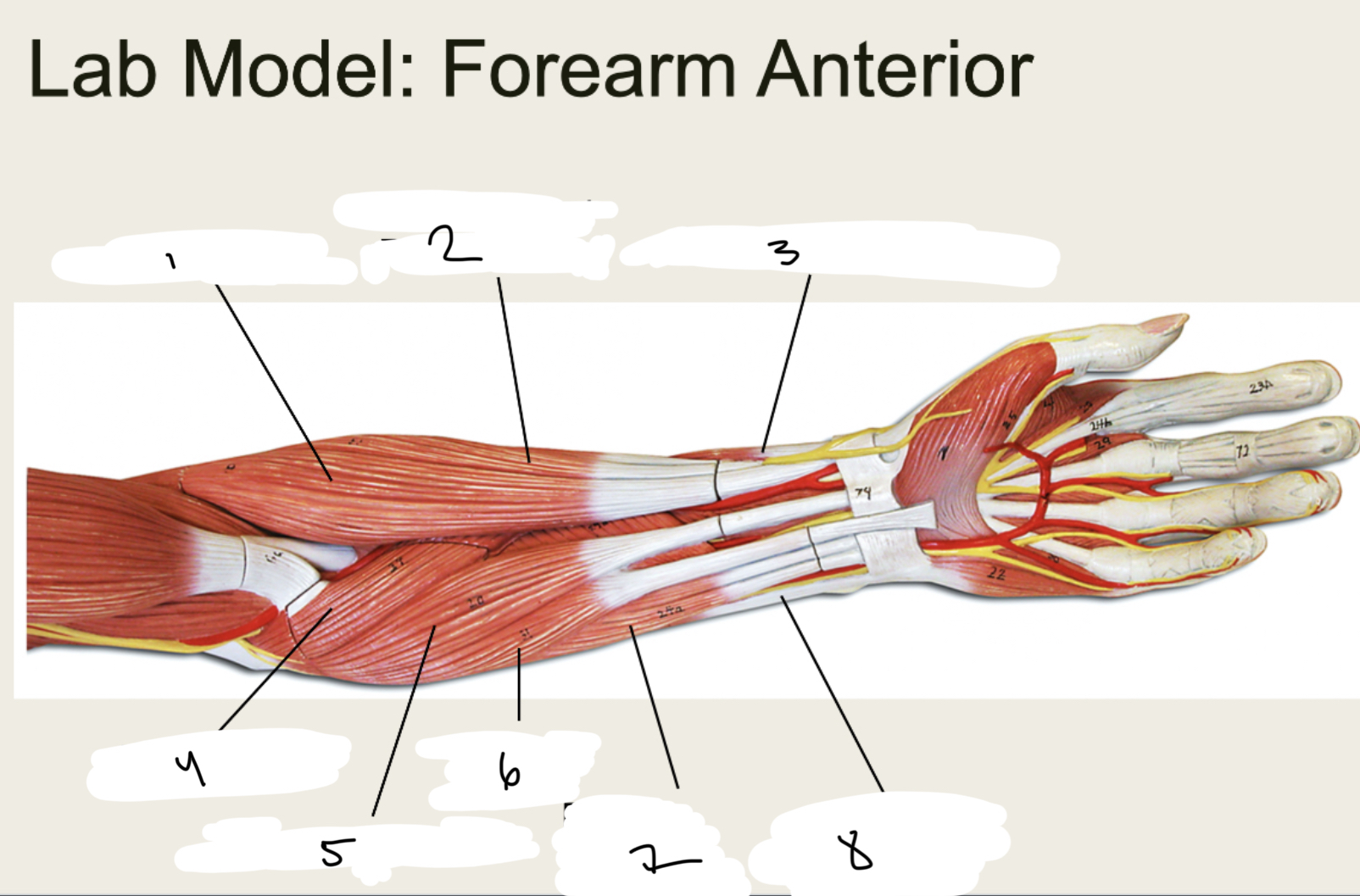
■ Flexor Carpi Radialis: Flexes and abducts wrists
■ Flexor Carpi Ulnaris: Flexes and adducts wrists
■ Flexor Digitorum Superficialis: Flexes proximal and middle phalanges, flexes
wrists
■ Palmaris Longus: Flexes wrist
■ Extensor Carpi Radialis Longus: Extends and abducts wrists
■ Extensor Carpi Ulnaris: Extends and adducts wrists:
■ Abductor Pollicis Longus: Abducts thumb
■ Extensor Digitorum: Extends fingers and wrists
1.XX
2.XX
3.XX
4.XX
5.XX
6.XX
7.XX
8.XX
9.XX
1.LEVATOR SCAPULAE
2.RHOMBOID MINOR
3.SUPRASPINATUS
4.INFRASPINATUS
5.TERES MINOR
6.TERES MAJOR
7.RHOMBOID MAJOR
8.TRAPZIUS
9.LATISSMUS DORSI
1.XX
2.XX
1.PECTORALIS MAJOR
2.PECTORALIS MINOR
1.XX
2.XX
3.XX
4.XX
5.XX
6.XX
7.XX
1.DELTOID
2.Coracobrachialis
3.BICEPS BRACHII
4.SUBSCAPULARIS
5.TERES MAJOR
6.TRICEPS BRACHII
7.BRACHIALIS
1.XX
2.XX
3.XX
4.XX
1.BRACHIALIS
2.BICEPS BRACHII
3.DELTOID
4.TRICEPS BRACHII
1.XX
2.XX
3.XX
4.XX
5.XX
6.XX
7.XX
8.XX
1.BRACHIORADIALIS
2.EXTENSOR CARPI RADIALIS LONGUS
3.ABDUCTOR POLLICIS LONGUS
4.PRONATER TERES
5.FLEXOR CARPI RADIALIS
6.PALMARIS LONGUS
7.FLEXOR DIGITORUM SUPERFICIALIS
8.FLEXOR CARPI ULNARIS
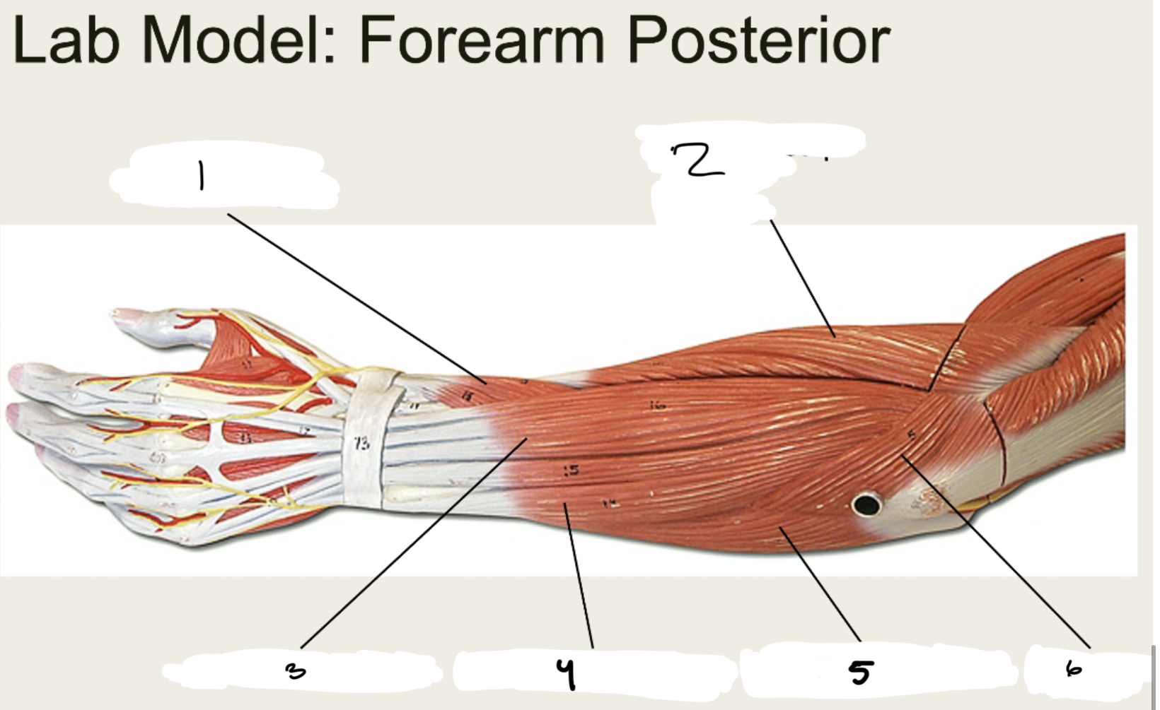
1.XX
2.XX
3.XX
4.XX
5.XX
6.XX
1.ABDUCTOR POLLICIS LONGUS
2.EXTENSOR CARPI RADIALIS LONGUS
3.EXTENSOR DIGITORUM
4.EXTENSOR CARPI ULNARIS
5.FLEXOR CARPI ULNARIS
6.ANCONEUS
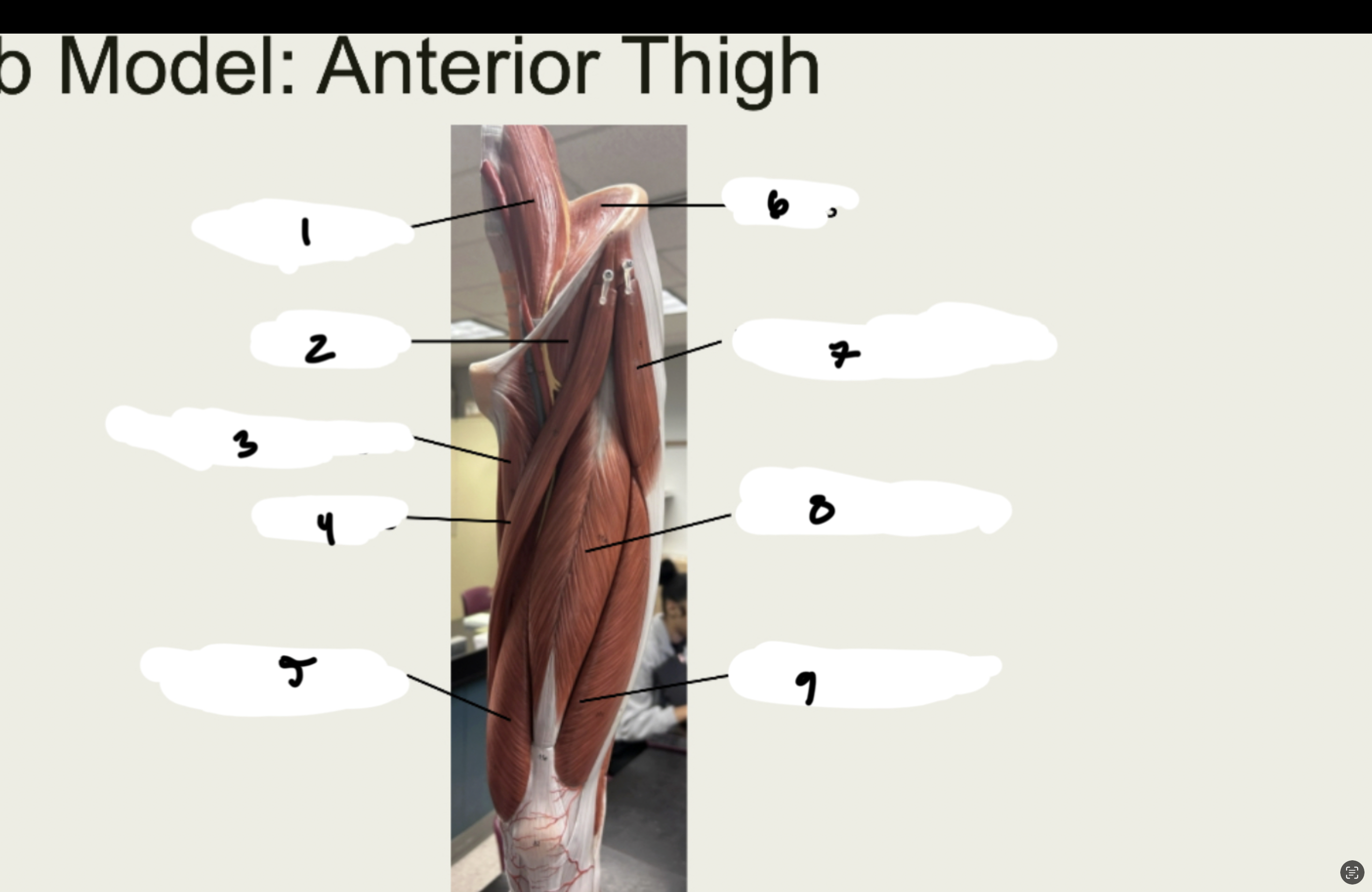
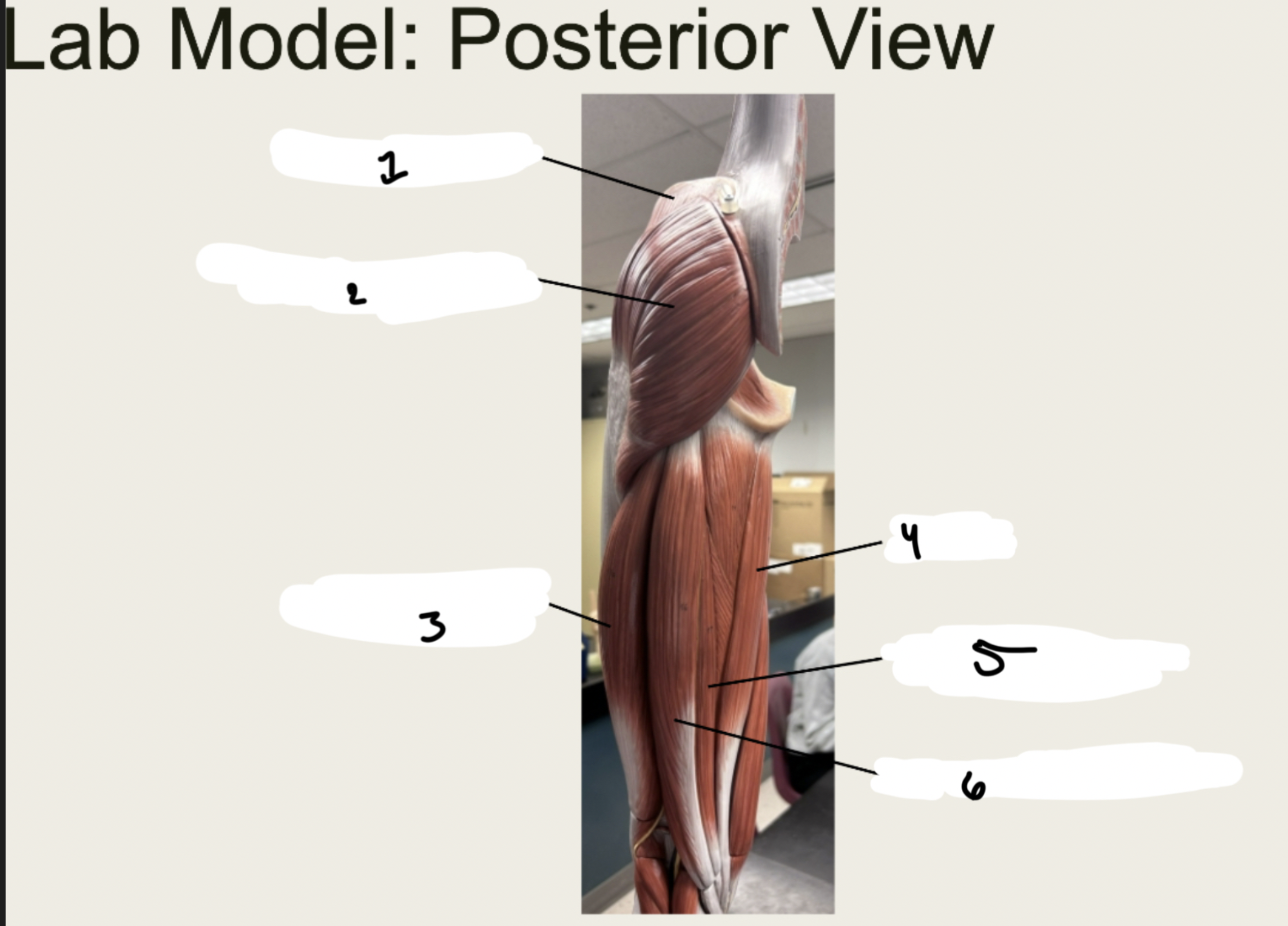
Muscles Acting on the Hip and Thigh:
Actions
■ XX: Flexes thigh and lumbar vertebrae
■ XX XX XX: Flexes, abducts, and medially rotates
■ XX XX: Extends and abducts thigh
■ XX XX: Abducts and medially rotates thigh
Muscles Acting on the Hip and Thigh:
Actions
■ Iliopsoas: Flexes thigh and lumbar vertebrae
■ Tensor Fasciae Latae: Flexes, abducts, and medially rotates
■ Gluteus Maximus: Extends and abducts thigh
■ Gluteus Medius: Abducts and medially rotates thigh
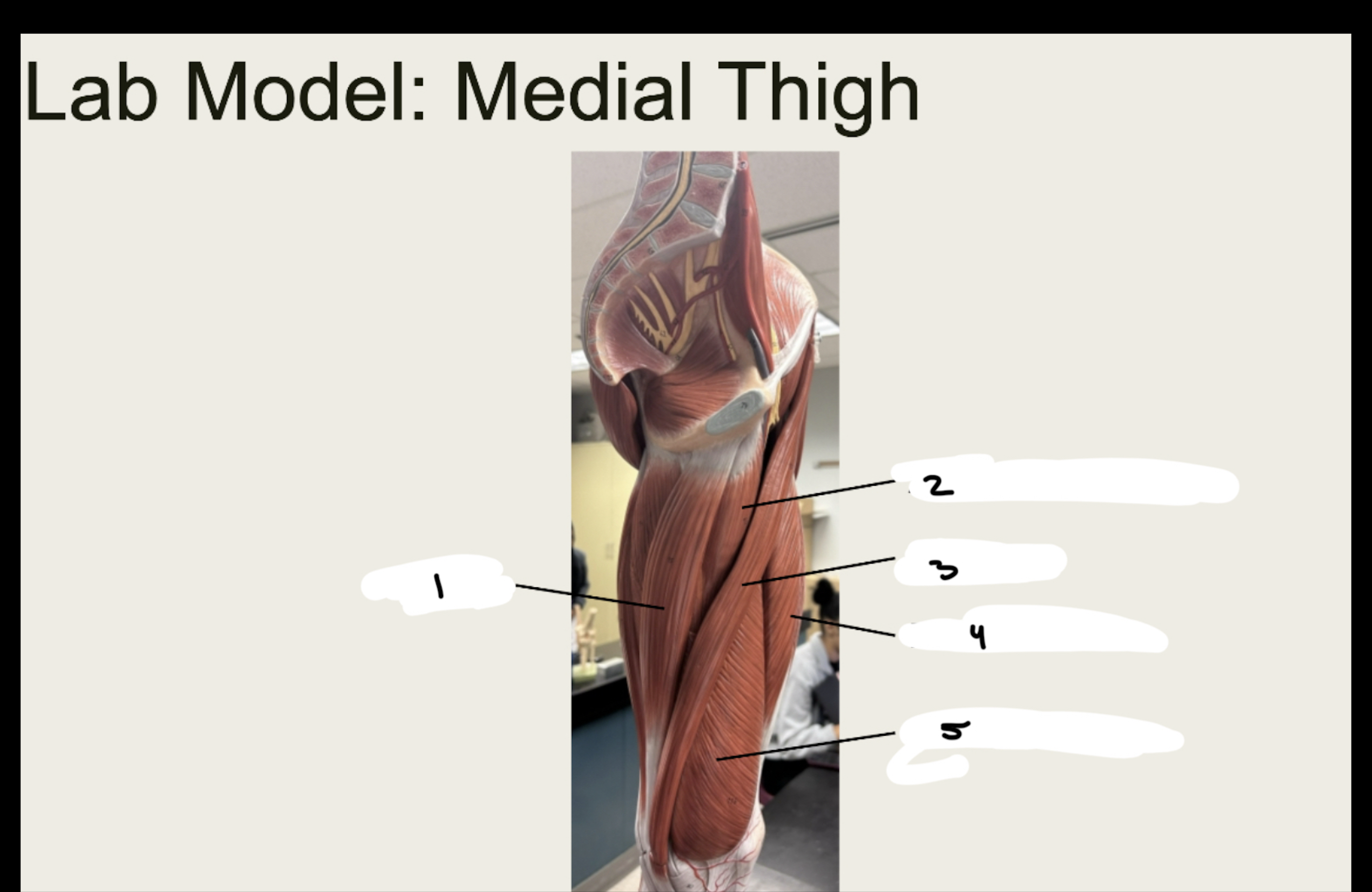
Medial Adductor Compartment of the
Thigh: Actions
■ XX XX: Adducts, flexes, and medially rotates thigh
■XX: Adducts, flexes and medially rotates leg,
■ XX: Adducts and flexes thigh
Medial Adductor Compartment of the
Thigh: Actions
■ Adductor Longus: Adducts, flexes, and medially rotates thigh
■ Gracilis: Adducts, flexes and medially rotates leg,
■ Pectineus: Adducts and flexes thigh
Muscles Acting on the Knee and Leg:
Actions
■ Quadriceps
– XX XX: Flexes thigh, extends knee
– XX XX: Extends knee
– XX XX: Extends knee
– XX XX: Extends knee
■ XX: Flexes and laterally rotates thigh, flexes knee
■ Hamstrings
– XX XX: Extends thigh, flexes knee
– XX: Extends thigh, flexes knee
– XX: Extends thigh, flexes knee
Muscles Acting on the Knee and Leg:
Actions
■ Quadriceps
– Rectus Femoris: Flexes thigh, extends knee
– Vastus Lateralis: Extends knee
– Vastus Medialis: Extends knee
– Vastus Intermedius: Extends knee
■ Sartorius: Flexes and laterally rotates thigh, flexes knee
■ Hamstrings
– Biceps Femoris: Extends thigh, flexes knee
– Semitendinosus: Extends thigh, flexes knee
– Semimembranosus: Extends thigh, flexes knee
Muscles Acting on the Foot: Actions
■ XX XX XX: Extends toes, dorsiflexes foot
■ XX XX: Dorsiflexes and inverts foot
■ XX: Flexes knee, plantar flexes foot
■ XX: Plantar flexes foot
■ XX XX XX: Flexes toes, plantar flexes, and inverts foot
■ XX XX: plantar flexes and everts foot
Muscles Acting on the Foot: Actions
■ Extensor Digitorum Longus: Extends toes, dorsiflexes foot
■ Tibialis Anterior: Dorsiflexes and inverts foot
■ Gastrocnemius: Flexes knee, plantar flexes foot
■ Soleus: Plantar flexes foot
■ Flexor Digitorum Longus: Flexes toes, plantar flexes, and inverts foot
■ Fibularis Longus: plantar flexes and everts foot
Muscles Acting on the Foot: Anterior
1.XX
2.XX
3.XX
4.XX
5.XX
6.XX
7.XX
8.XX
9.XX
1.PSOAS MAJOR
2.PECTINEUS
3.ADDUCTOR LONGUS
4.SARTORIUS
5.VASTUS MEDIALIS
6.ILIACUS
7.TENSOR FASCIA LATAE
8.RECTUS FEMORIS
9.VASTUS LATERALIS
1.XX
2.XX
3.XX
4.XX
5.XX
6.XX
1.GLUTEUS MEDIUS
2.GLUTEUS MAXIMUS
3.BICEPS FEMORIS
4.GRACILIS
5.SEMITENDINOSUS
6.SEMIMEMBRANOSUS
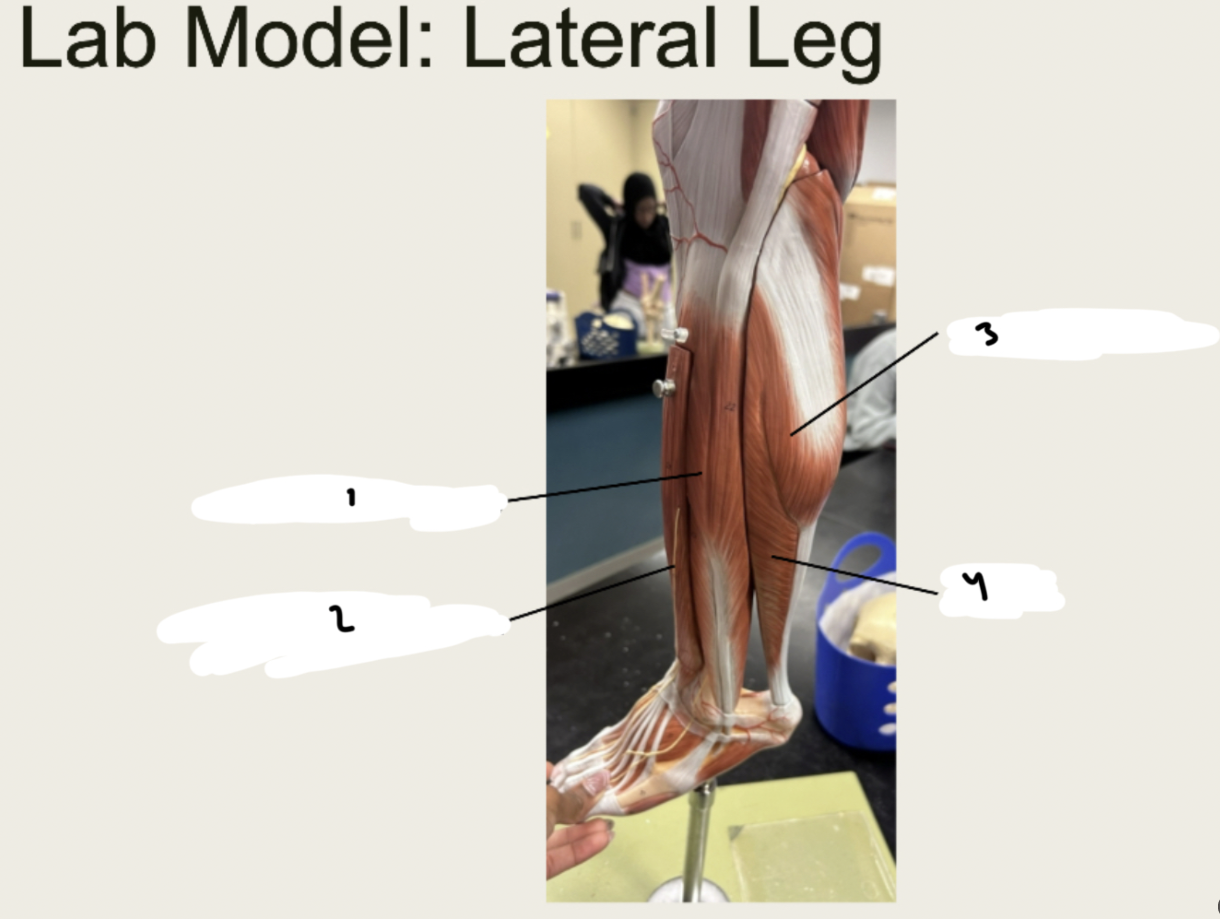
1.XX
2.XX
3.XX
4.XX
5.XX
1.GRACILIS
2.ADDUCTOR LONGUS
3.SARTORIUS
4.RECTUS FEMORIS
5.VASTUS LATERALIS
1.XX
2.XX
3.XX
4.XX
5.XX
6.XX
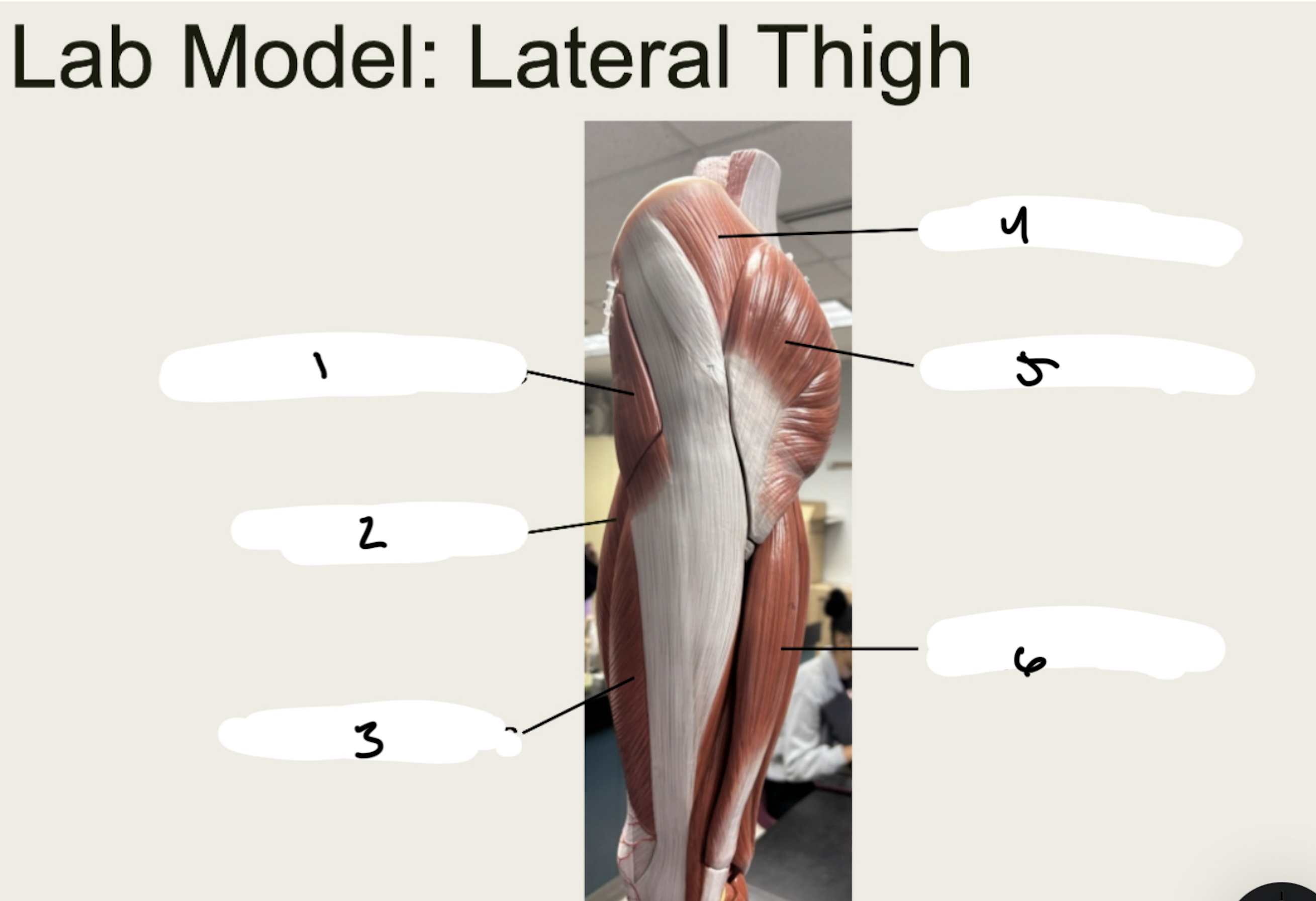
1.TENSOR FASCIA LATAE
2.RECTUS FEMORIS
3.VASTUS LATERALIS
4.GLUTEUS MEDIUS
5.GLUTEUS MAXIMUS
6.BICEPS FEMORIS
1.XX
2.XX
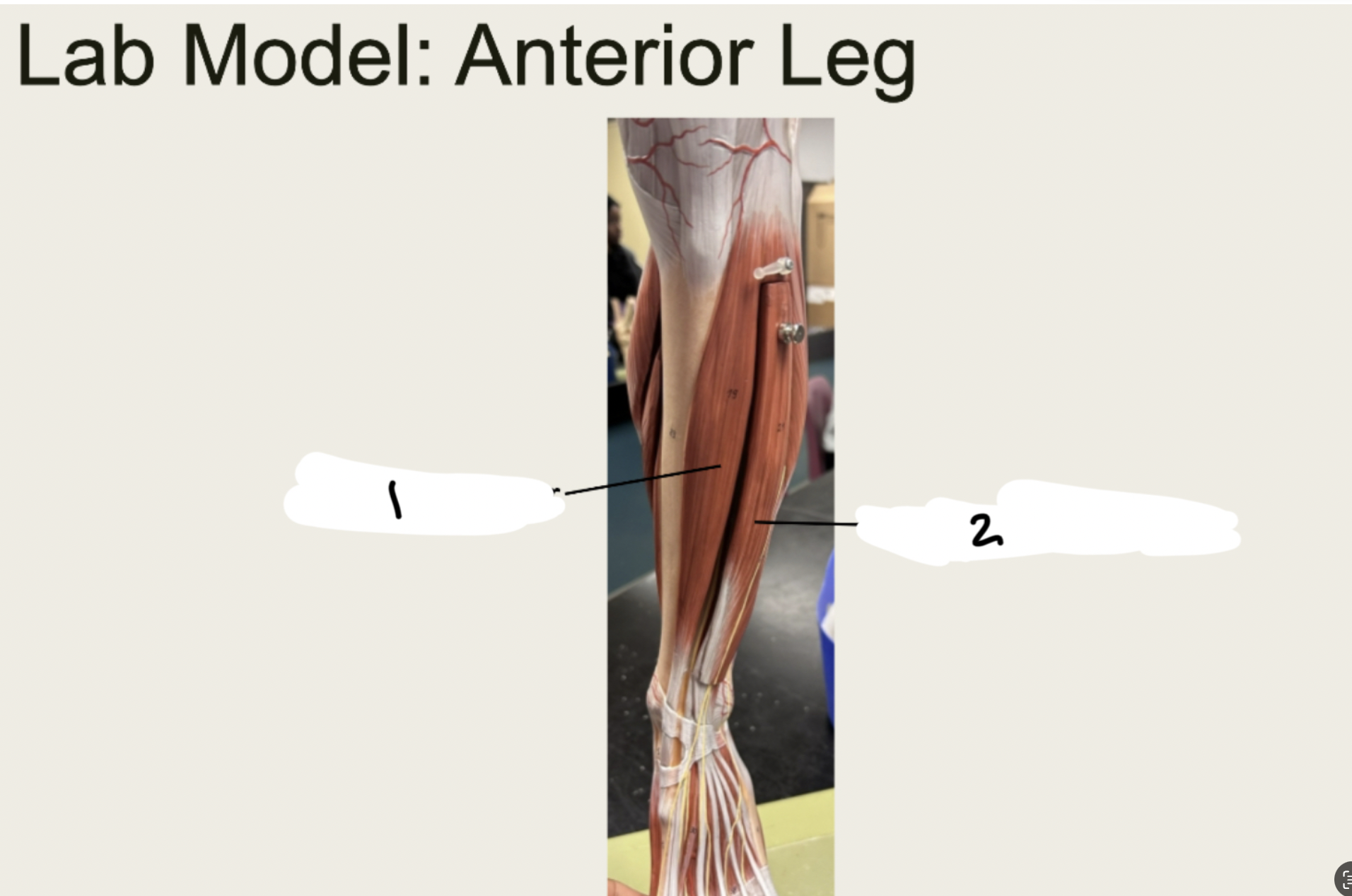
1.TIBIALIS ANTERIOR
2.EXTERNSOR DIGITORUM
1.XX
2.XX
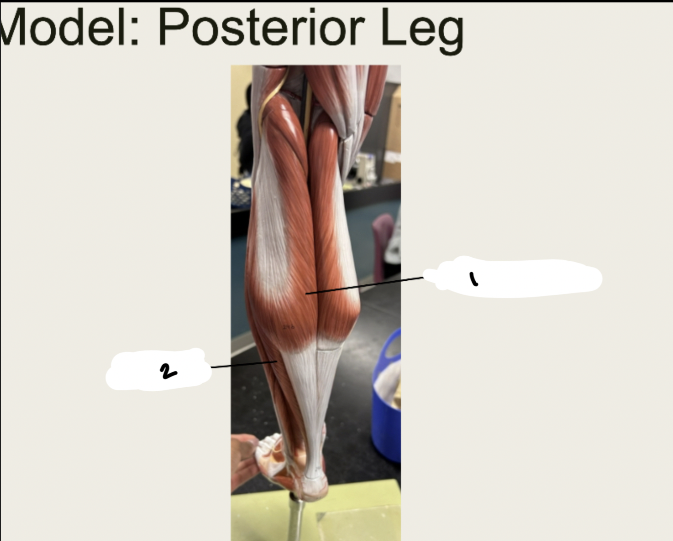
1.Gastrocnemius
2.SOLEUS
1.XX
2.XX
3.XX
4.XX
5.XX
1.GASTROCNEMIUS
2.SOLEUS
3.CALCANEAL TENDON
4.TIBIALIS ANTERIOR
5.FLEXOR DIGITORUM LONGUS
1.XX
2.XX
3.XX
4.XX
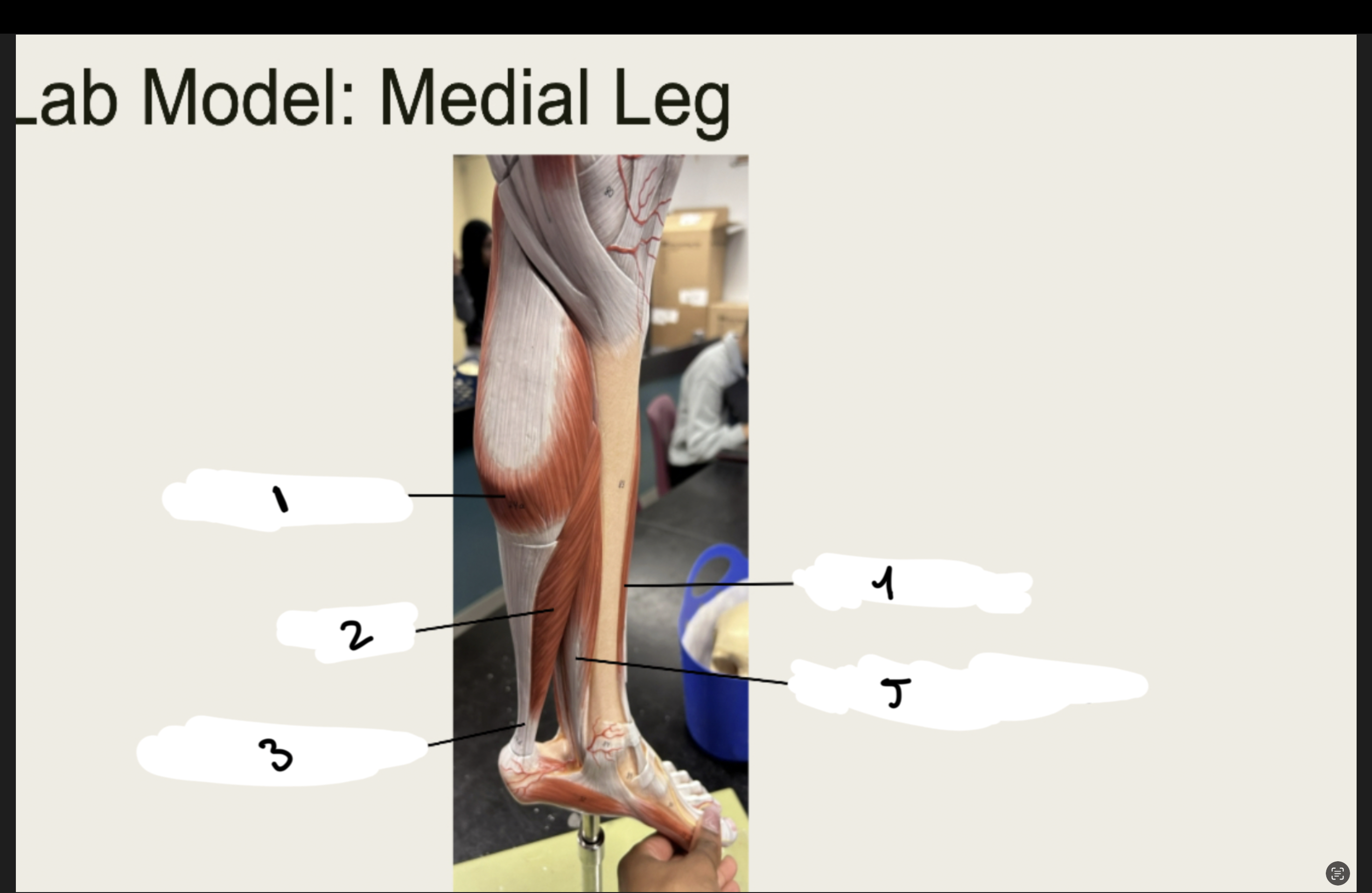
1.FIBULARIS LONGUS
2.EXTENSOR DIGITORUM
3.GASTROCNEMIUS
4.SOLEUS
Divisions of the Nervous System
■ Central Nervous System
– XX and XX XX
■ Peripheral Nervous System
– XX XX
– XX XX
– XX
– XX XX
Divisions of the Nervous System
■ Central Nervous System
– Brain and Spinal Cord
■ Peripheral Nervous System
– Spinal Nerves
– Cranial Nerves
– Ganglia
– Somatic Nerves
Peripheral Nervous System
■ Autonomic
– Controls XX that occur XX
– Sympathetic Nervous System: XX or XX
– Parasympathetic Nervous System: XX and XX
■ Somatic
– XX that occur under XX XX
Peripheral Nervous System
■ Autonomic
– Controls actions that occur automatically
– Sympathetic Nervous System: fight or flight
– Parasympathetic Nervous System: rest and digest
■ Somatic
– Actions that occur under conscious control
1.XX
2.XX
3.XX
4.XX
5.XX
6.XX
7.XX
8.XX
1.NISSL BODIES
2.DENDRITES
3.CELL BODY
4.NUCLEUS
5.CYTOPLASM
6.AXON HILLOCK
7.AXON
8.SCHWANN CELL
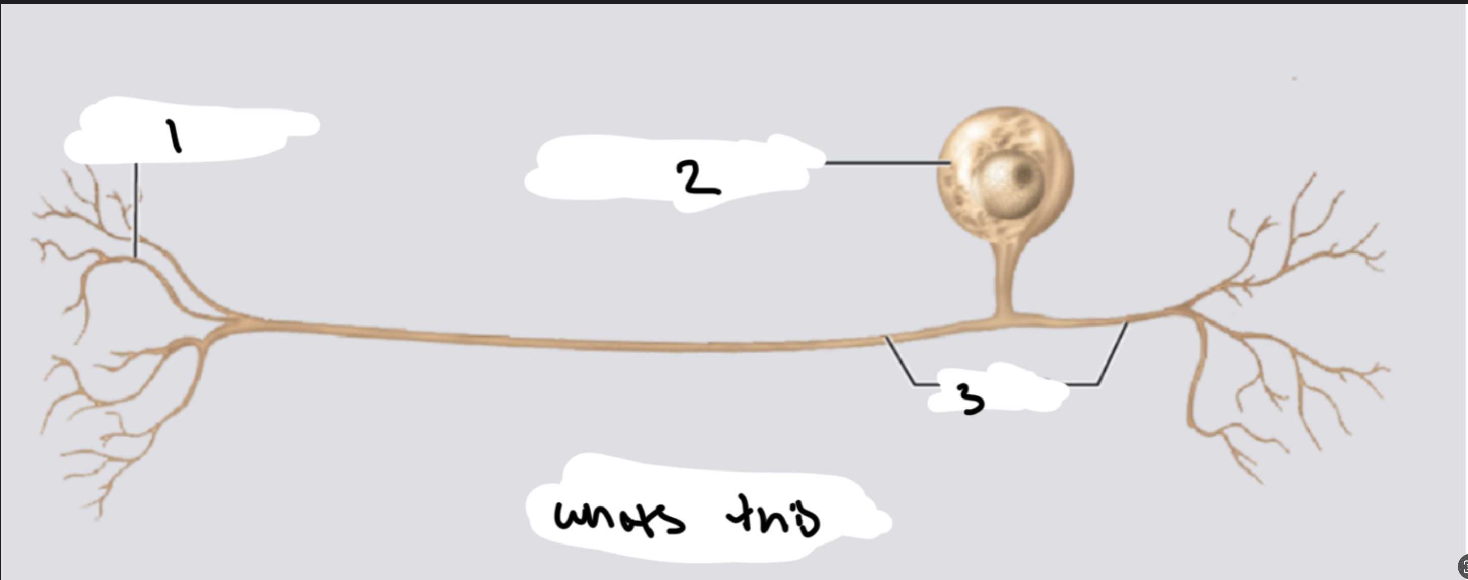
WHATS THIS:XX XX
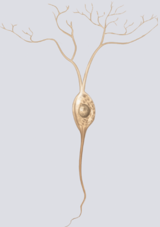
1.XX
2.XX
3.XX
MULTIPOLAR NEURON
1.DENDRITES
2.DENDRITIC SPINES
3.AXON
WHATS THIS: XX XX
1.XX
2.XX
3.XX
BIPOLAR NEURON
1.DENDRITES
2.NEUROSOMA
3.AXON
WHATS THIS: XX XX
1.XX
2.XX
3.XX
UNIPOLAR NEURON
1.DENDRITES
2.NEUROSOMA
3.AXON
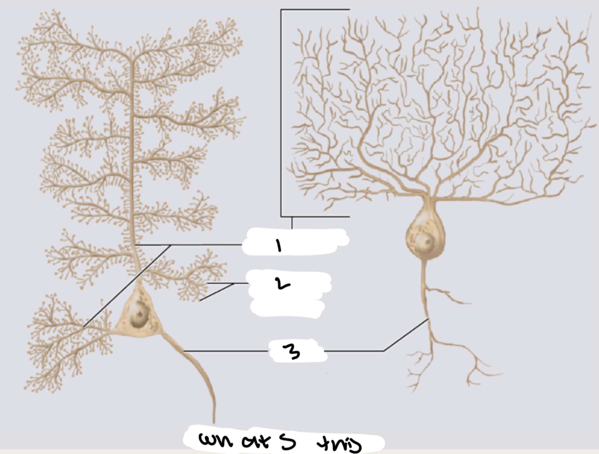
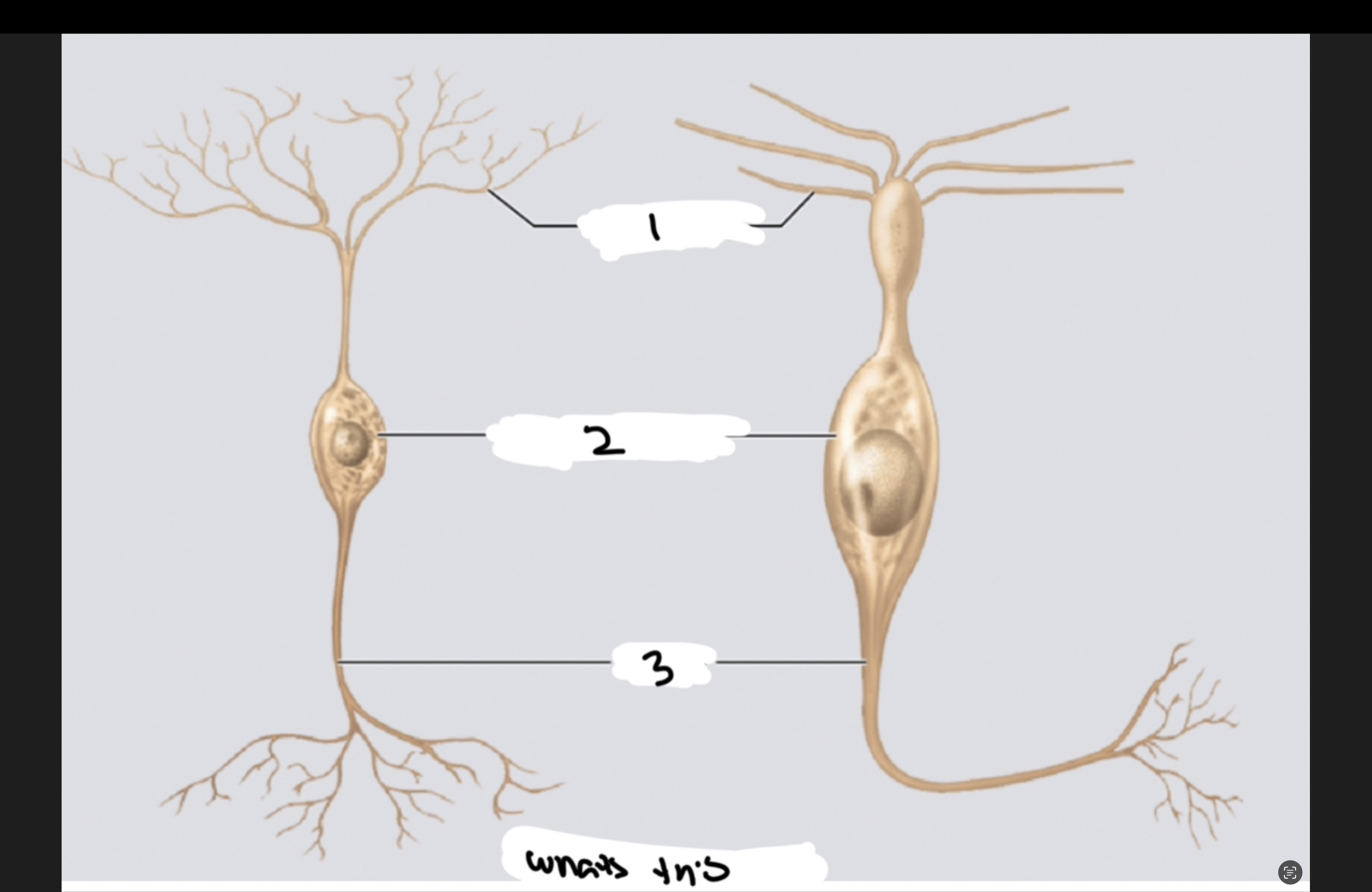
Specialized
Neurons: Pyramidal Cells
XX cell body
■ Found in the XX XX, XX,
and XX
■ Plays a role in XX XX
Triangular cell body
■ Found in the cerebral cortex, amygdala,
and hippocampus
■ Plays a role in cognitive functions
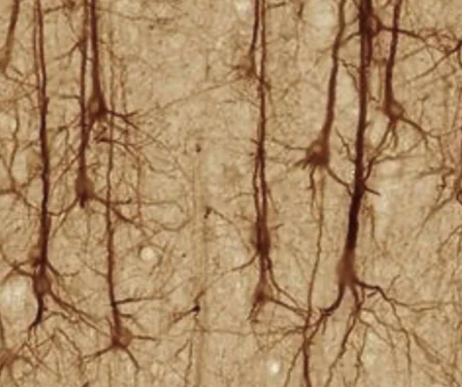
WHATS THIS?
A PYRAMIDAL CELL
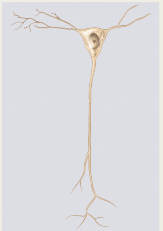
WHATS THIS
A PYRAMIDAL CELL
Specialized
Neurons: Purkinje Cells
■ Some of the XX XX in the brain
■ Functions in XX XX
■ Found in the XX XX of the
XX
Specialized
Neurons: Purkinje
Cells
■ Some of the largest neurons in the brain
■ Functions in motor coordination
■ Found in the gray matter of the
cerebellum
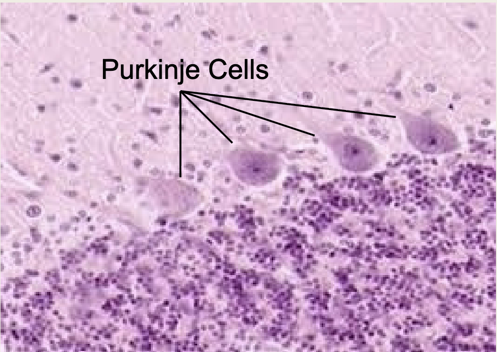
WHATS THIS
PURKINJE CELLS
WHATS THIS
PURKINJE CELL
Longitudinal Aspect of the Spinal Cord
■ Medullary Cone/Conus Medullaris
– The XX of the XX XX
– Occurs at about XX
■ Cauda Equina
– XX of XX XX branching off the
end of the XX XX
– Resembles a XX XX
Longitudinal Aspect of
the Spinal Cord
■ Medullary Cone/Conus Medullaris
– The end of the spinal cord
– Occurs at about L1
■ Cauda Equina
– Bundle of nerve fibers branching off the
end of the spinal cord
– Resembles a horse’s tail
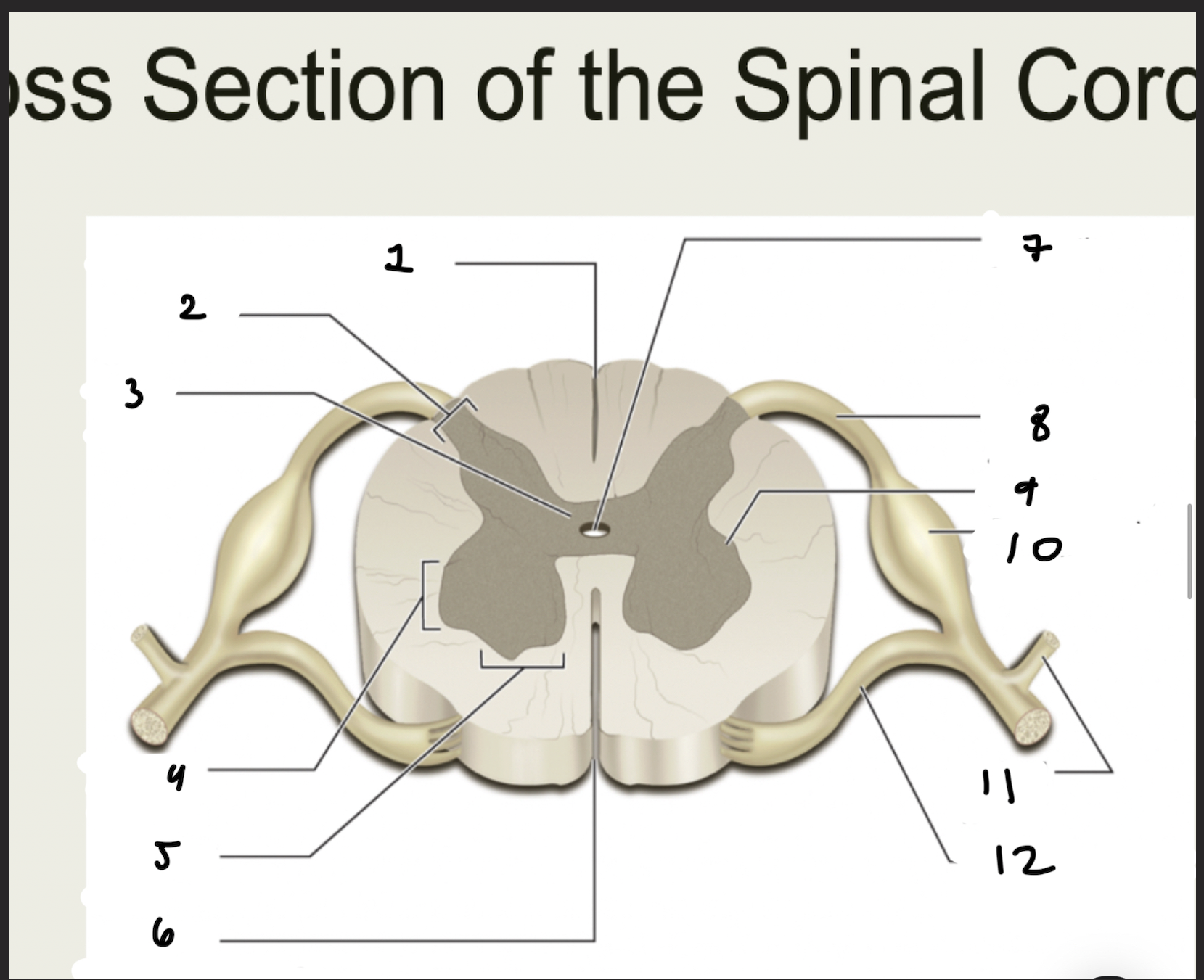
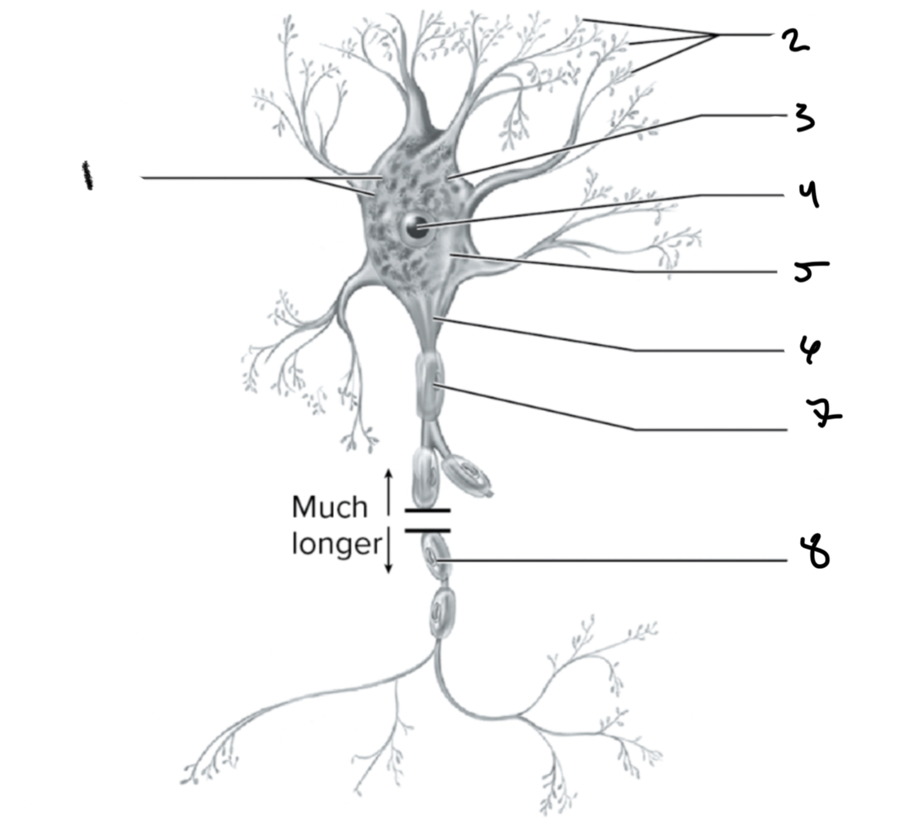
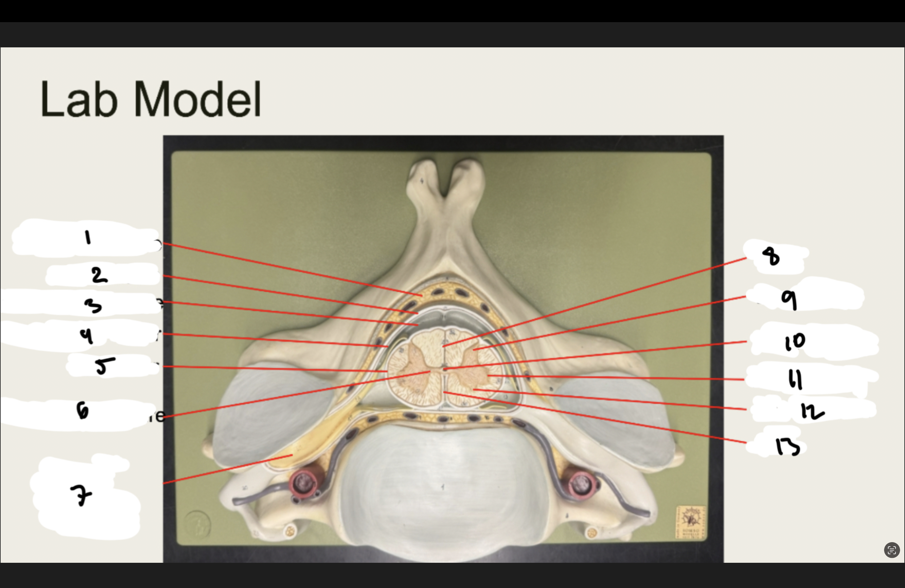
1.XX
2.XX
3.XX
4.XX
5.XX
6.XX
7.XX
8.XX
9.XX
10.XX
11.XX
12.XX
1.POSTERIOR MEDIAN SULCUS
2.POSTERIOR HORN
3.GRAY COMMISURE
4.LATERAL HORN
5.ANTERIOR HORN
6.ANTERIOR MEDIAN FISSURE
7.CENTRAL CANAL
8.DORSAL ROOT OF SPINAL NERVE
9.LATERAL HORN
10.DORSAL ROOT GANGLION
11.SPINAL NERVE
12.VENTRAL ROOT OF SPINAL NERVE
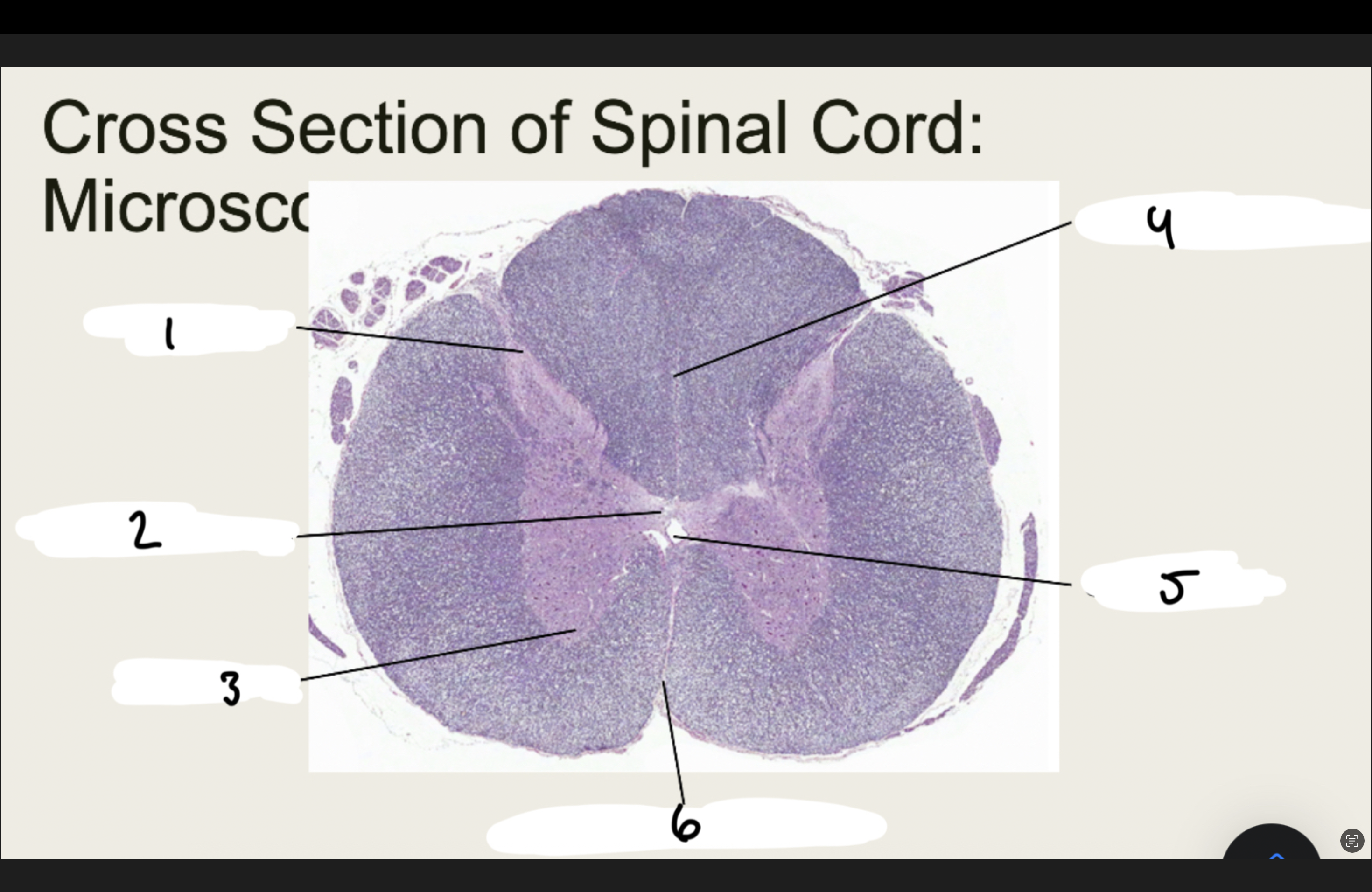
1.XX
2.XX
3.XX
4.XX
5.XX
6.XX
1.DORSAL HORN
2.GRAY COMMISURE
3. VENTRAL HORN
4.POSTERIOR MEDIAN SULCUS
5.CENTRAL CANAL
6.ANTERIOR MEDIAN FISSURE
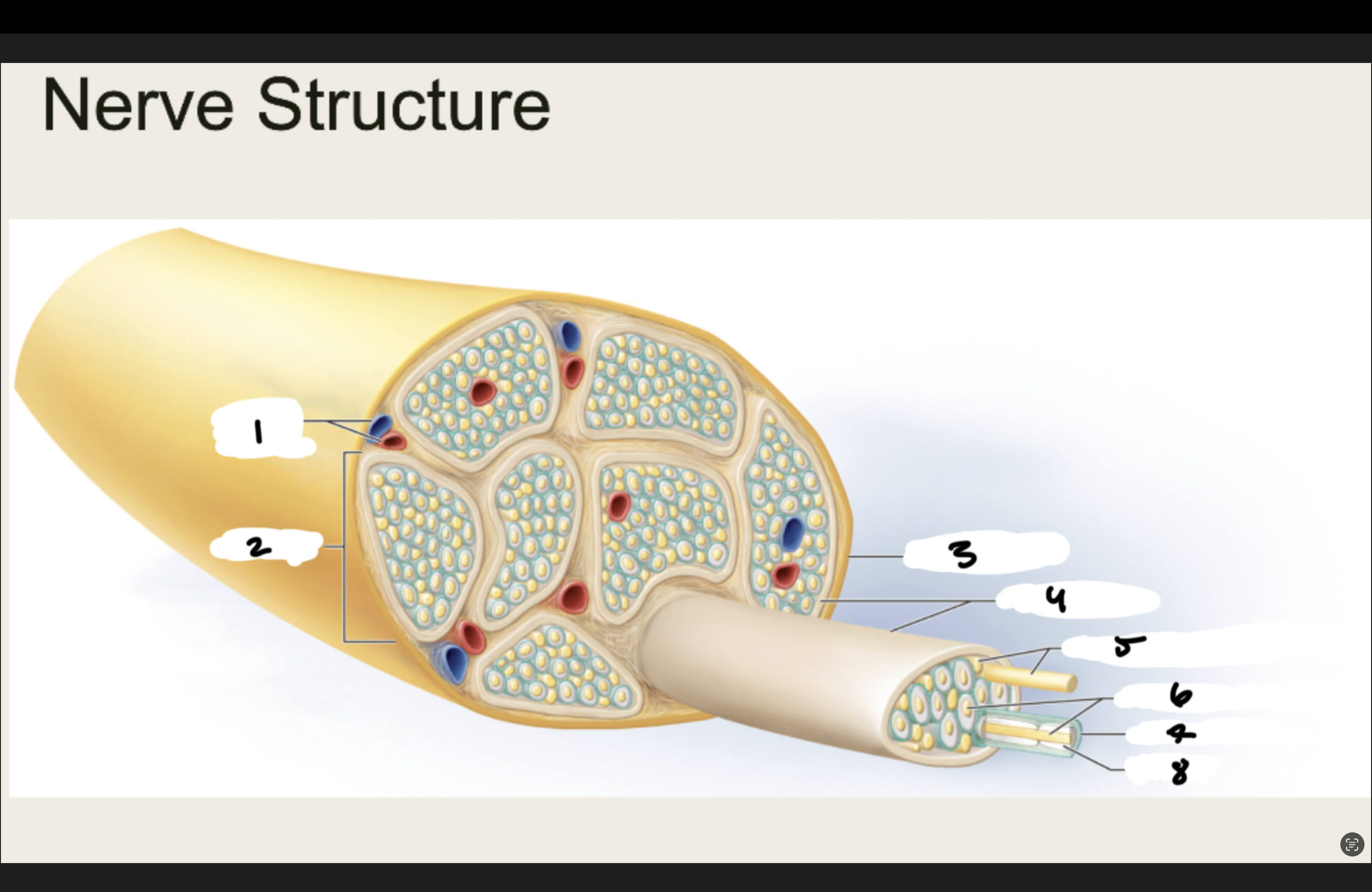
1.XX
2.XX
3.XX
4.XX
5.XX
6.XX
7.XX
8.XX
1.BLOOD VESSELS
2.FASICLE
3.EPINEURIUM
4.PERINEURIUM
5.UNMYELINATED NERVE FIBERS
6.MYELINATED NERVE FIBERS
7.ENDONEURIUM
8.MYELIN
1.XX
2.XX
3.XX
4.XX
5.XX
6.XX
7.XX
8.XX
9.XX
10.XX
11.XX
12.XX
13.XX
1.EPIDURAL SPACE
2.DURA MATER
3.SUBARACHNOID SPACE
4.ARACHNOID MATER
5.PIA MATER
6.GRAY COMMISURE
7.DORSAL ROOT GANGLION
8.POSTERIOR MEDIAN SULCUS
9.DORSAL HORN
10.CENTRAL CANAL
11.LATERAL HORN
12.ANTERIOR HORN
13.ANTERIOR MEDIAN FISSURE
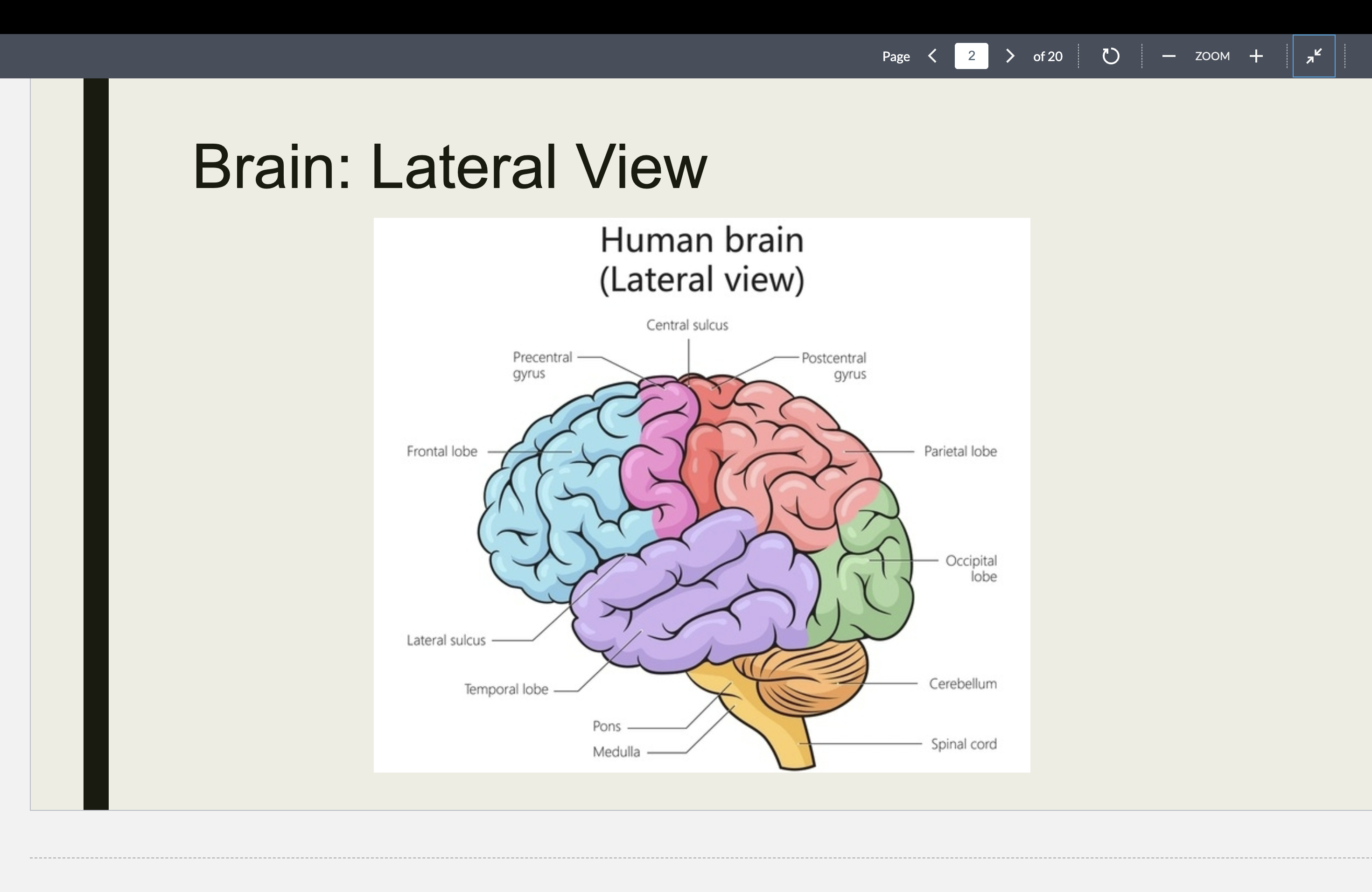
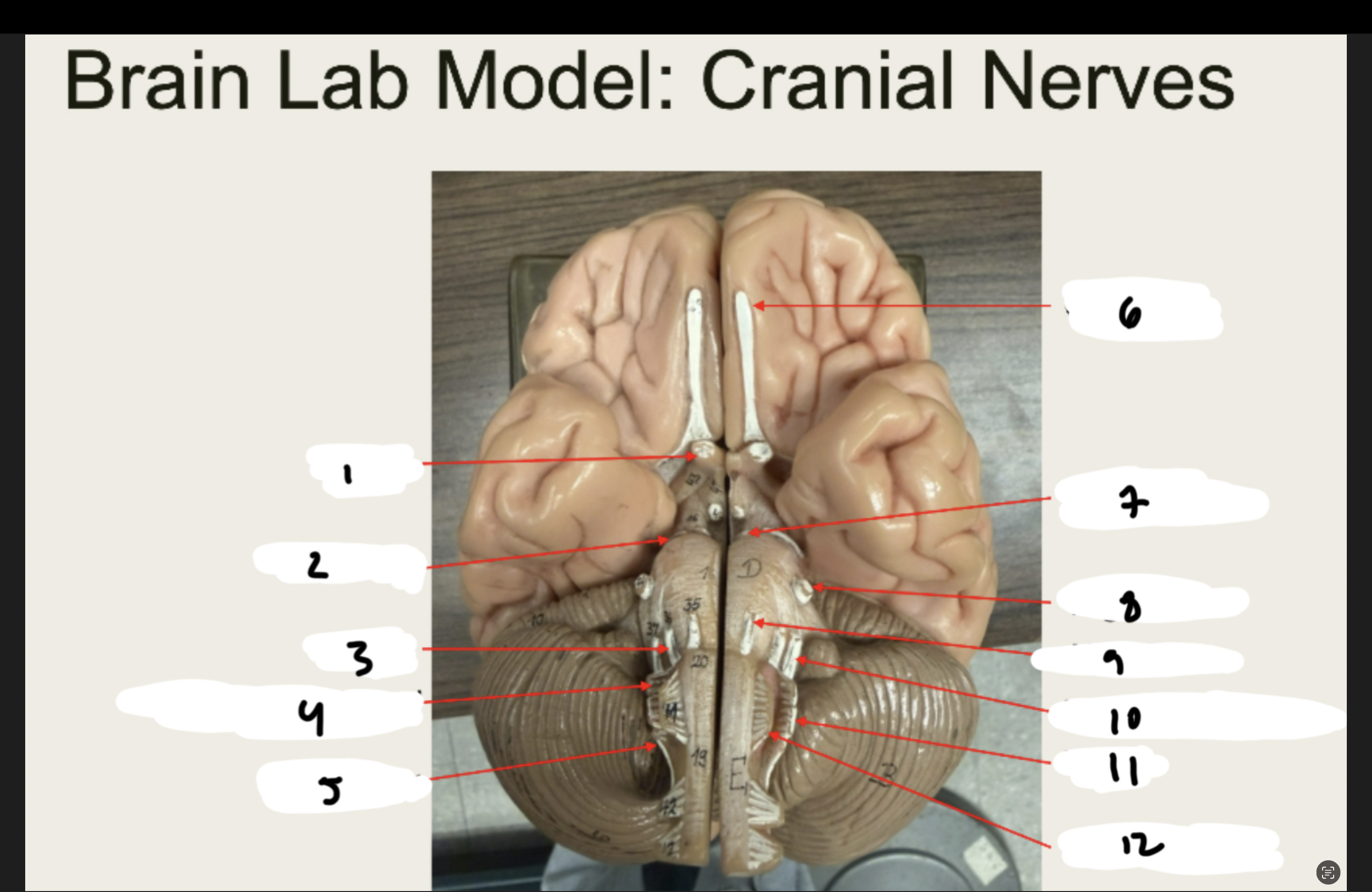
LATERAL VIEW OF BRAIN
LATERAL VIEW OF BRAIN
1.XX
2.XX
3.XX
4.XX
5.XX
6.XX
7.XX
8.XX
9.XX
10.XX
11.XX
12.XX
13.XX
14.XX
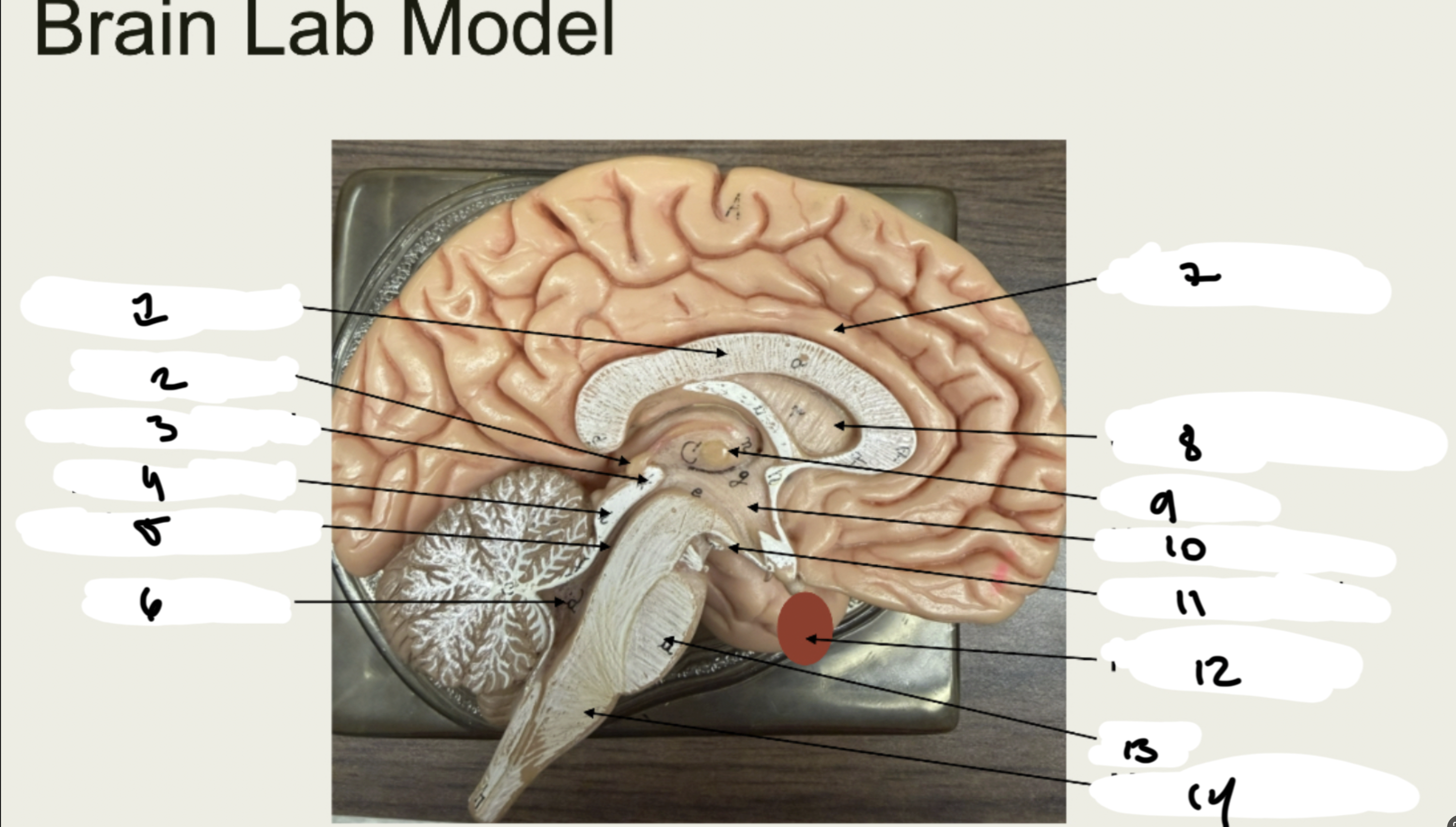
1.CORPUS CALLOSUM
2.PINEAL GLAND
3.SUPERIOR COLLUCLI
4.INFERIOR COLLUCULI
5.CEREBRAL AQUEDUCT
6.4TH VENTRICLE
7.CINGULATE GYRUS
8.SEPTUM PELLUCIDUM
9.THALAMUS
10.HYPOTHALAMUS
11.MAMMILLARY BODY
12.PITUITARY GLAND
13.PONS
14.MEDULLA OBLONGATA
NERVES
NERVES
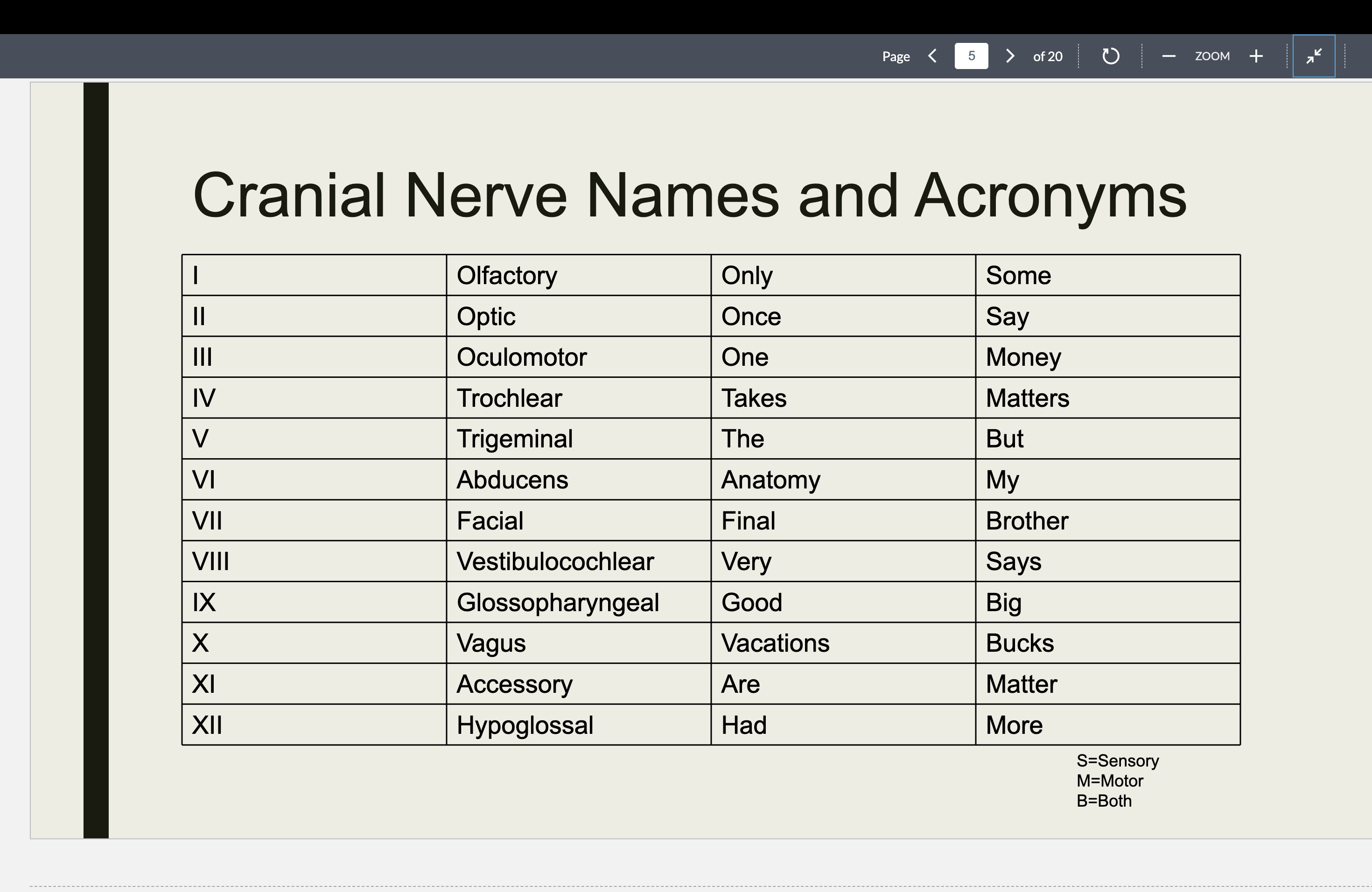
Olfactory
Nerve (I)
■ Function: XX of
XX (XX)
■ Passes through
XX XX of XX XX
Olfactory
Nerve (I)
■ Function: sense of
smell (sensory)
■ Passes through
cribriform plate of
ethmoid bone
Optic Nerve
(II)
■ Function: XX
(XX)
■ Passes through the
XX XX
Optic Nerve
(II)
■ Function: sight
(sensory)
■ Passes through the
optic canal
Oculomotor
Nerve (III)
■ Function: XX XX (XX)
■ Passes through
XX XX XX
Oculomotor
Nerve (III)
■ Function: Eye
movement (motor)
■ Passes through
superior orbital fissure

Trochlear
Nerve (IV)
■ Function:XX XX (motor)
■ Passes through XX XX XX
Trochlear
Nerve (IV)
■ Function: eye
movements (motor)
■ Passes through
superior orbital fissure
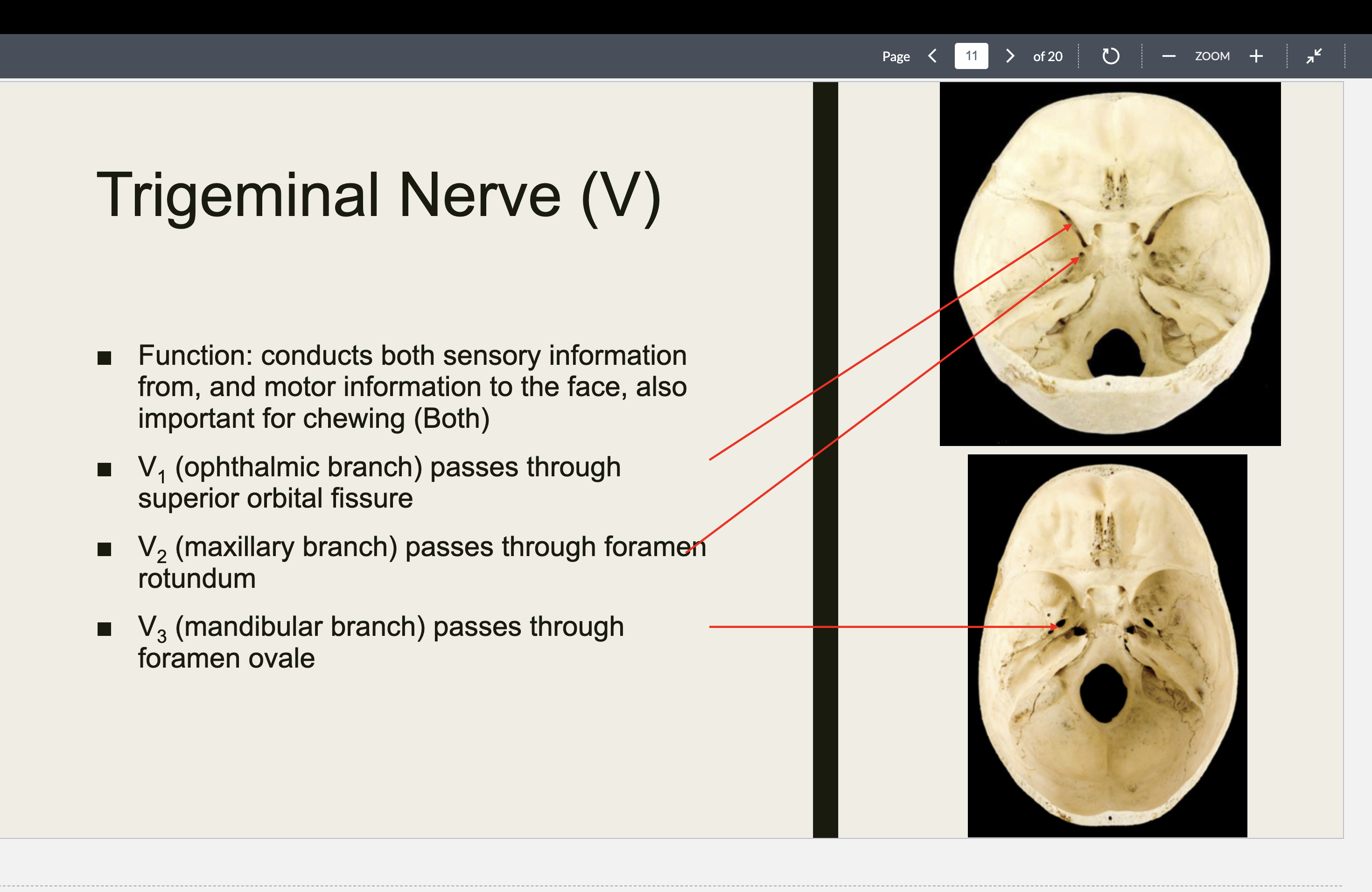
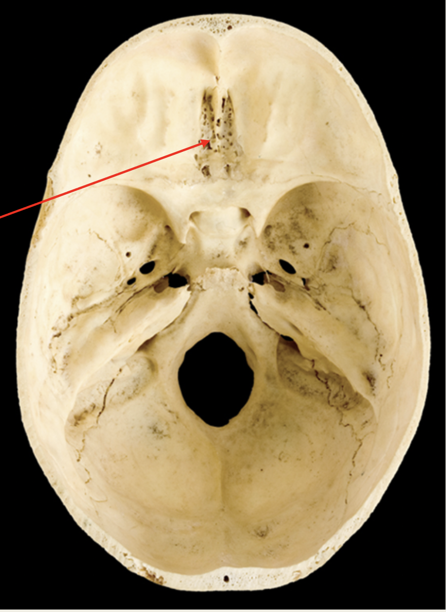
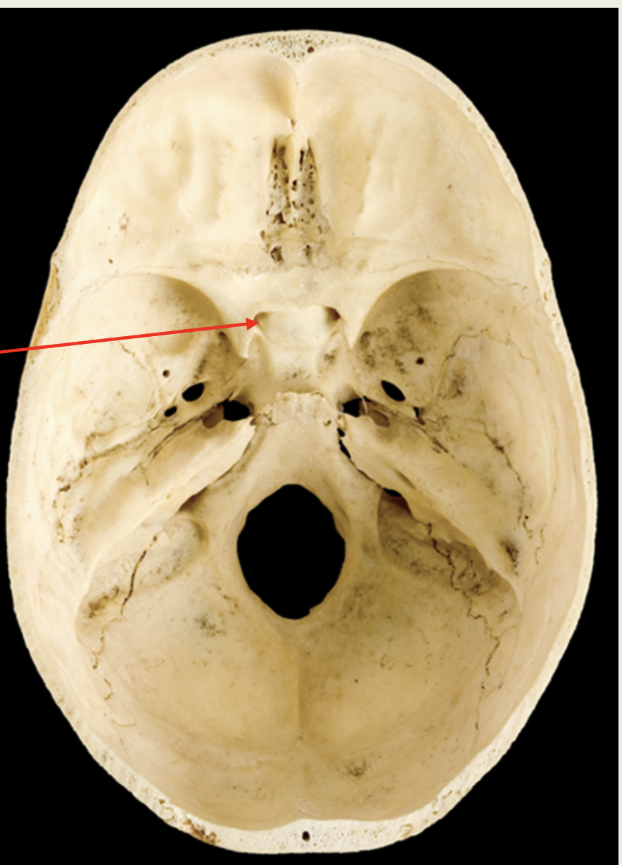
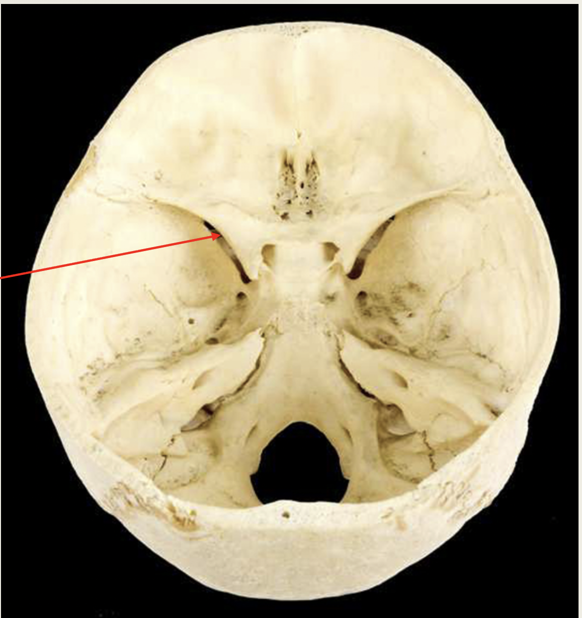
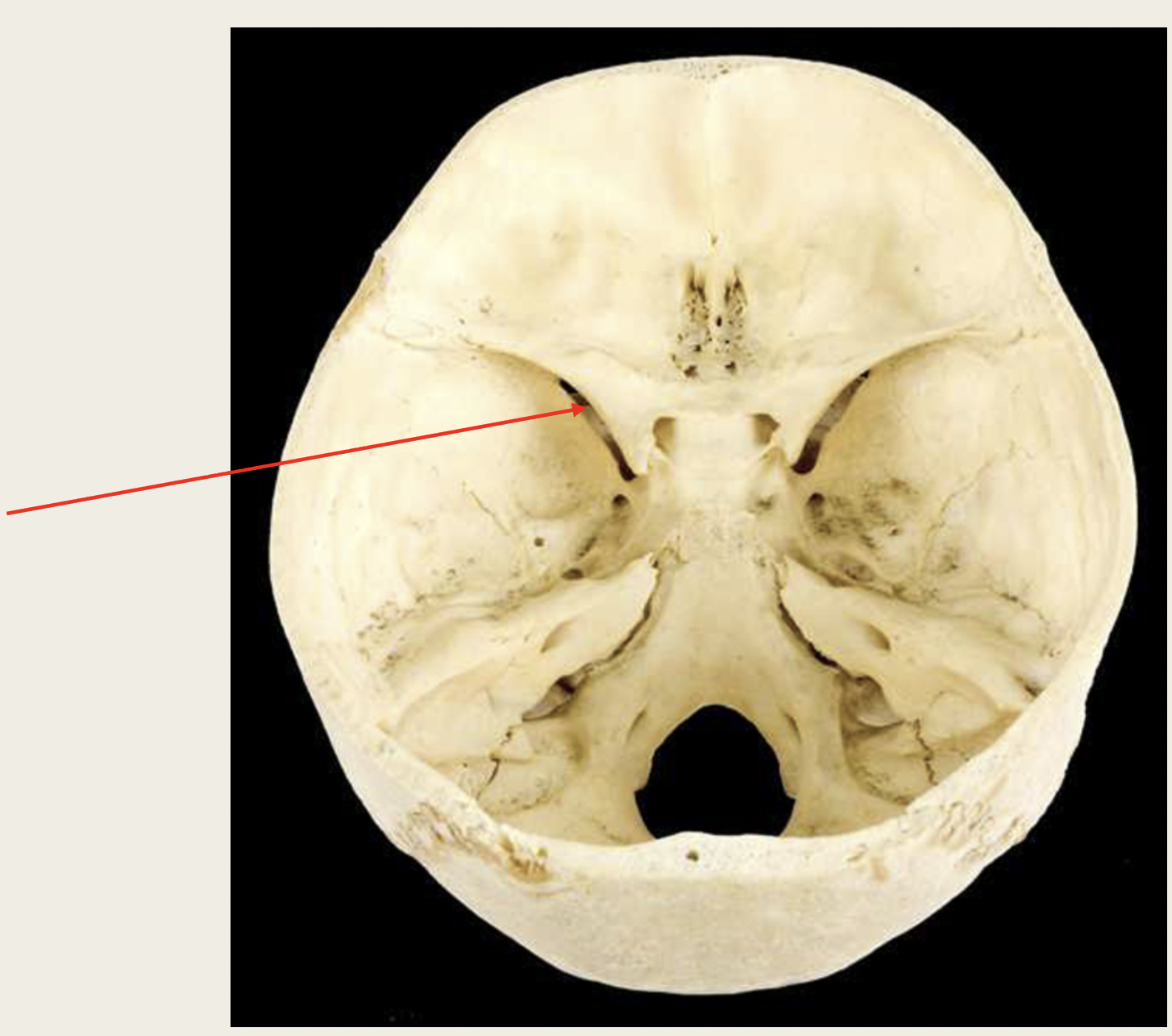
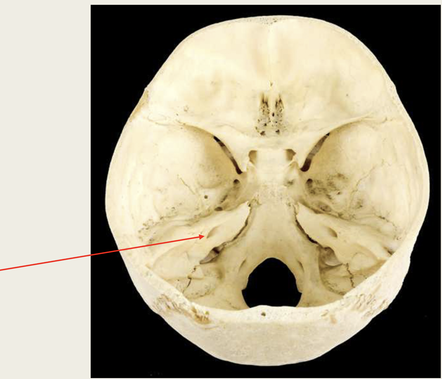
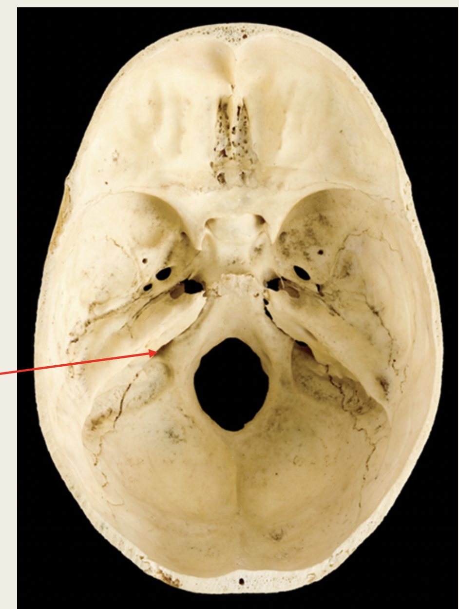
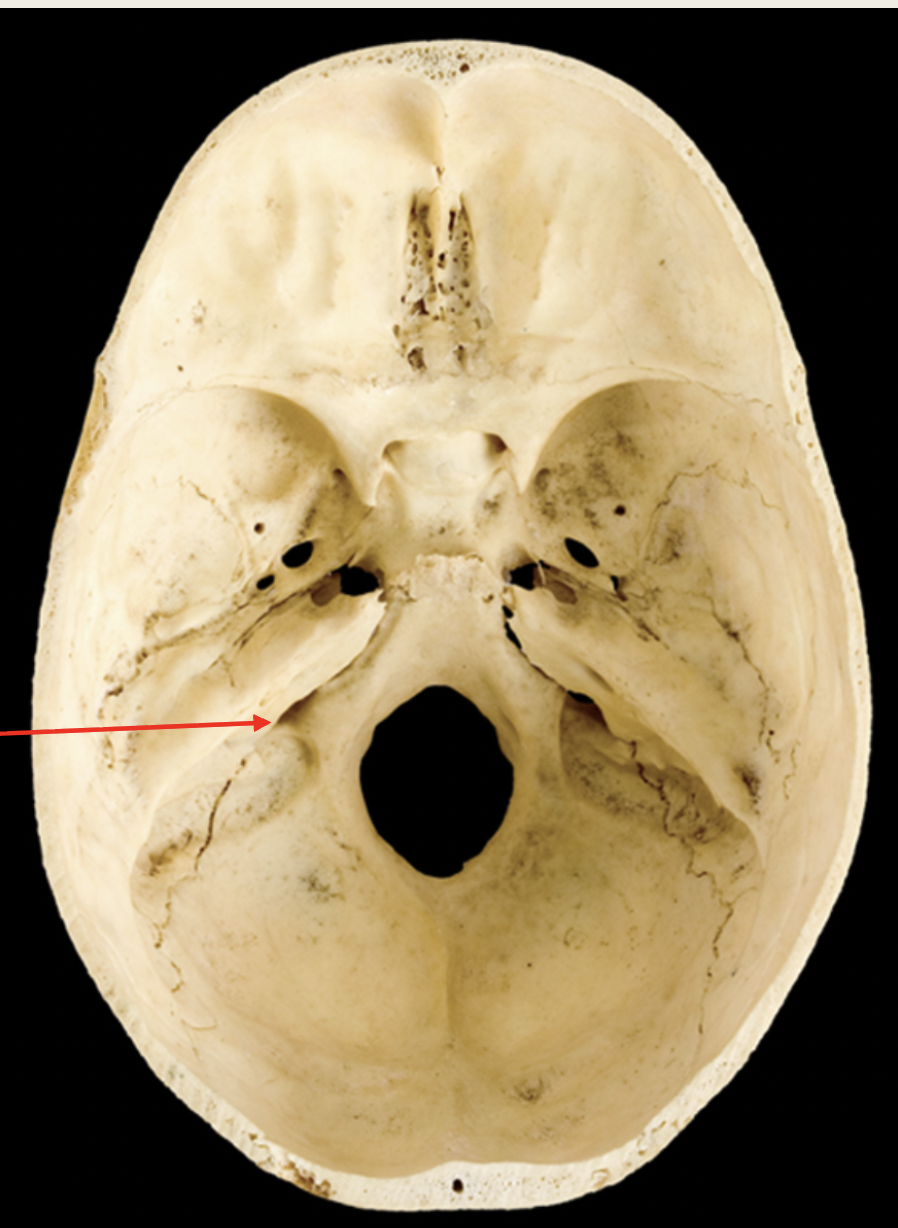
Trigeminal Nerve (V)
■ Function: conducts both XX information
from, and XX XX to the face, also
important for XX (XX)
■ V1 (XX branch) passes through XX XX XX
■ V2 (XX branch) passes through XX XX
■ V3 (XX branch) passes through XX XX
Trigeminal Nerve (V)
■ Function: conducts both sensory information
from, and motor information to the face, also
important for chewing (Both)
■ V1 (ophthalmic branch) passes through
superior orbital fissure
■ V2 (maxillary branch) passes through foramen
rotundum
■ V3 (mandibular branch) passes through
foramen ovale
Abducens
Nerve (VI)
■ Function: XX XX (XX)
■ Passes through XX XX XX
Abducens
Nerve (VI)
■ Function: Eye
movements (motor)
■ Passes through
superior orbital fissure

Facial
Nerve (VII)
■ Function: Receives
XX information
from XX and controls
XX XX for XX XX (both
XX & XX)
■ Passes through XX XX XX
Facial
Nerve (VII)
■ Function: Receives
sensory information
from taste and controls
facial muscles for facial
expression (both
sensory & motor)
■ Passes through internal
acoustic meatus
Vestibulocochlear
Nerve (VIII)
■ Function: XX and
XX (XX)
■ Passes through XX XX XX
Vestibulocochlear
Nerve (VIII)
■ Function: hearing and
equilibrium (sensory)
■ Passes through internal
acoustic meatus
Glossopharyngea
l Nerve (IX)
■ Receives XX XX for XX
and has XX XX (XX)
■ Passes through XX XX
Glossopharyngea
l Nerve (IX)
■ Receives sensory
information for taste
and has motor functions
(Both)
■ Passes through jugular
foramen
Vagus
Nerve (X)
■ Function: Receives
XX information
from XX organs
and controls XX
in XX XX (XX)
■ Passes through XX XX
Vagus
Nerve (X)
■ Function: Receives
sensory information
from visceral organs
and controls functions
in various organs (Both)
■ Passes through jugular
foramen
Accessory
Nerve (XII)
■ Function: innervates
muscles of the XX
and XX, important for
XX (Motor)
■ Passes through XX XX
Accessory
Nerve (XII)
■ Function: innervates
muscles of the neck
and back, important for
swallowing (Motor)
■ Passes through jugular
foramen
Hypoglossal
Nerve (XII)
■ Function: XX XX (XX)
■ Passes through XX XX
Hypoglossal
Nerve (XII)
■ Function: Tongue
movement (Motor)
■ Passes through
hypoglossal canal
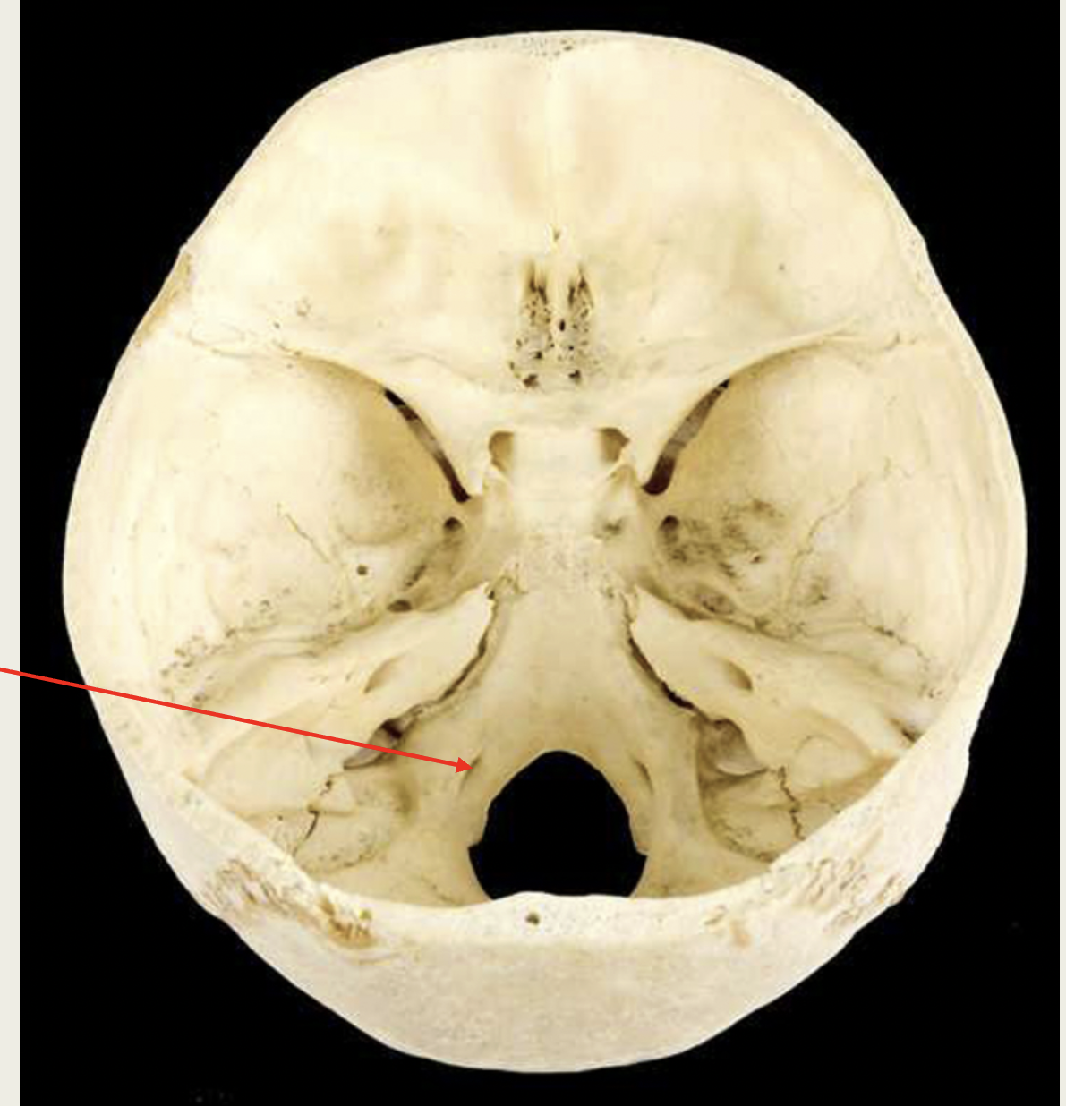
1.XX
2.XX
3.XX
4.XX
5.XX
6.XX
7.XX
8.XX
9.XX
10.XX
11.XX
12.XX
1.OPTIC
2.TROCHLEAR
3.FACIAL
4.GLOSSOPHARYNGEAL
5.ACCESSORY
6.OLFACTORY
7.OCULOMOTOR
8.TRIGEMINAL
9.ABDUCENS
10.VESTIBULOCHLEAR
11.VAGUS
12.HYPOGLOSSAL
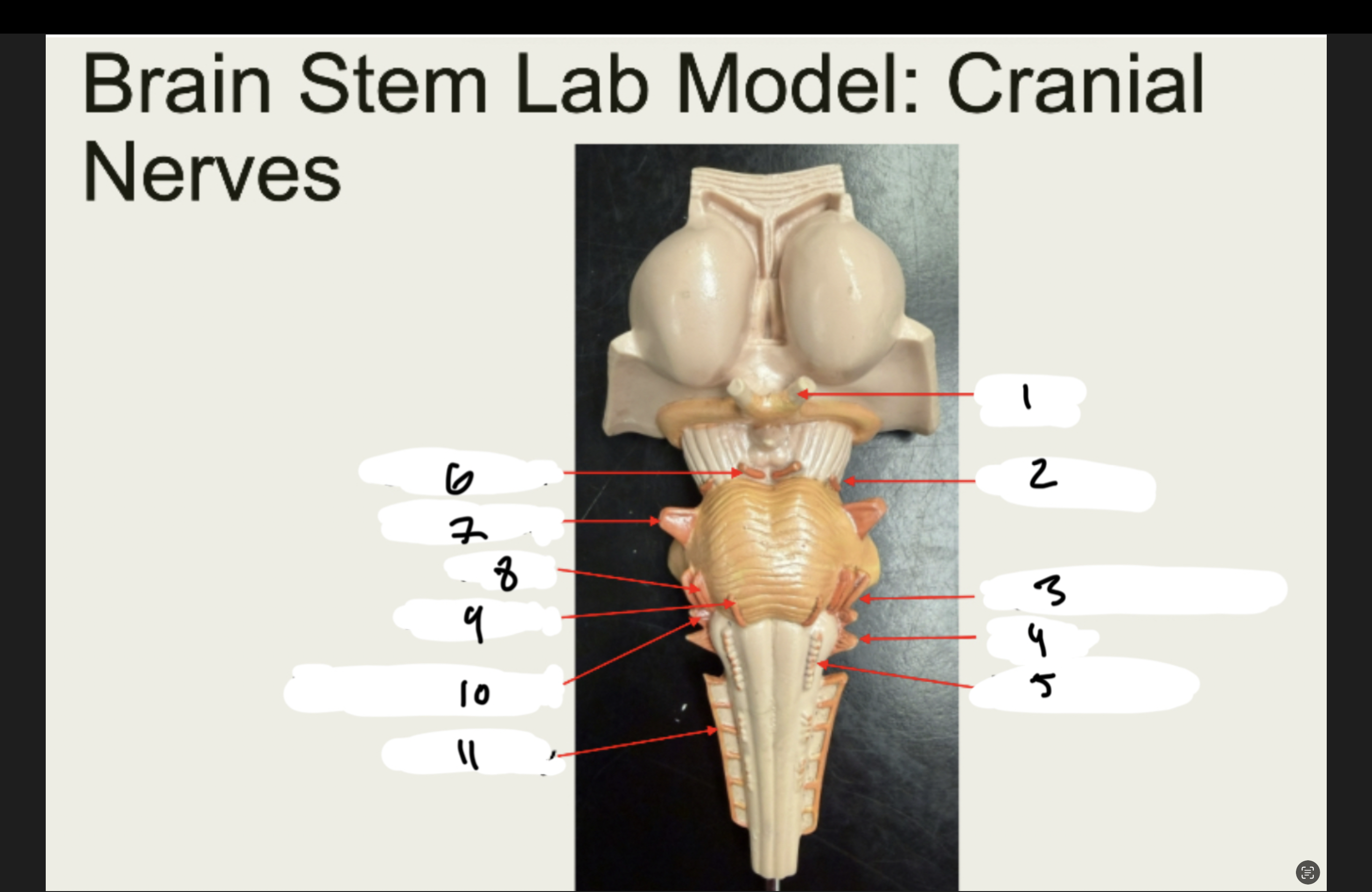
1.XX
2.XX
3.XX
4.XX
5.XX
6.XX
7.XX
8.XX
9.XX
10.XX
11.XX
1.OPTIC
2.TROCHLEAR
3.VESTIBULOCHLEAR
4.VAGUS
5.HYPOGLOSSAL
6.OCULOMOTOR
7.TRIGEMINAL
8.FACIAL
9.Glossopharyngeal
10.ACCESSORY
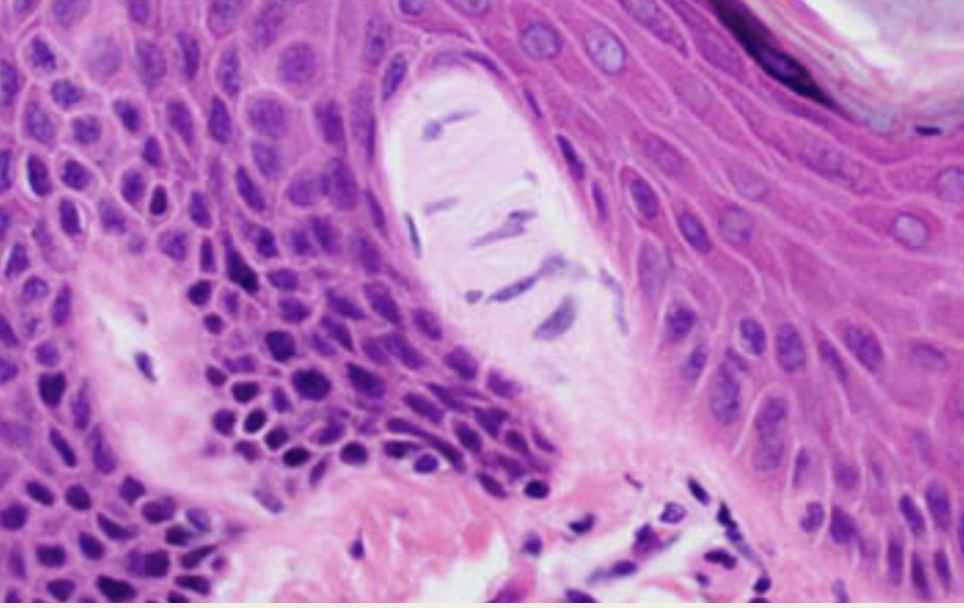
LIGHT TOUCH
Tactile/Meissner’s Corpuscles
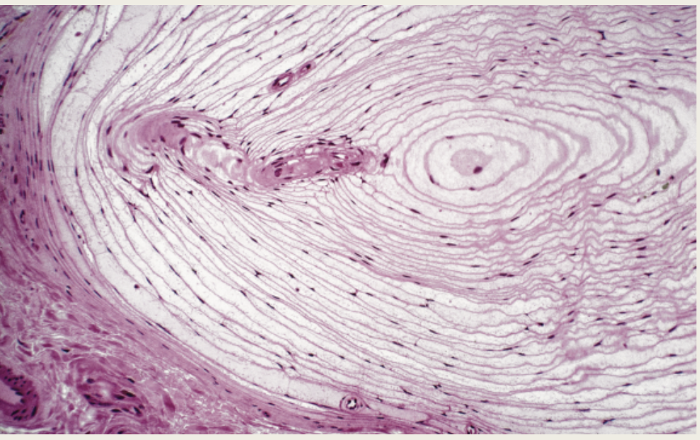
Deep Touch
Lamellar/Pacinian Corpuscle
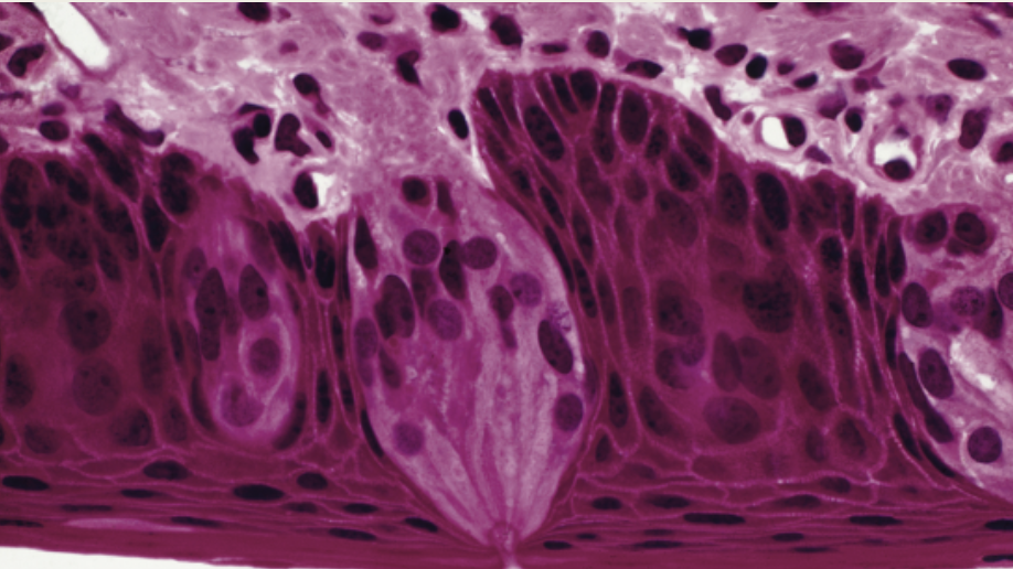
Found on the sides of vallate papillae
TASTE BUDS
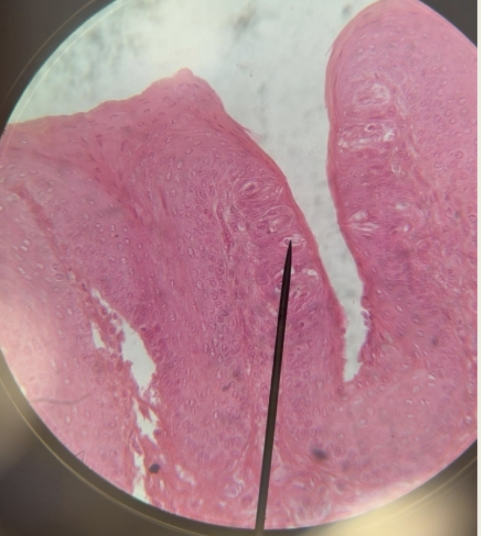
WHATS THIS
TASTE BUDS