(2) Fundamental Principles of Imaging Modalities
1/27
There's no tags or description
Looks like no tags are added yet.
Name | Mastery | Learn | Test | Matching | Spaced | Call with Kai |
|---|
No analytics yet
Send a link to your students to track their progress
28 Terms
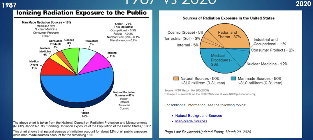
ionizing radiation exposure
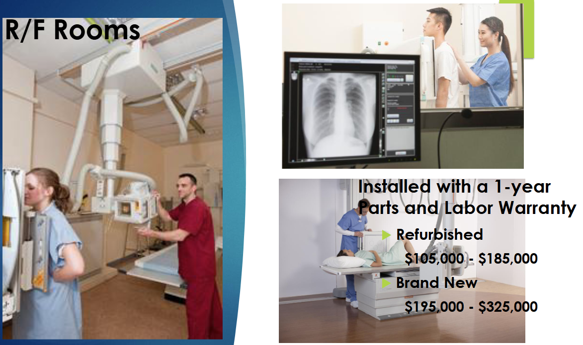
what is an R&F room?
radiography and fluoroscopy room
rooms for diagnostic imaging made form still x-ray images and real time moving images (fluoroscopy)
how does an xray tube work?
hot cathode (-) emits electrons that are attracted to tungsten anode (+)
cathode : generates electrons
anode : stops electrons
when fast moving electrons hit the metal target (anode) what happens to kinetic energy
99% → heat
1% → exits as electron beam (goes to target or is scattered)
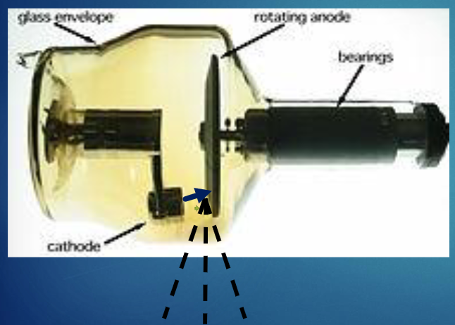

what is the primary function of the envelope?
provide support and electrical insulation for anode and cathode
maintain vacuum in the tube
what do x rays penetrate?
less dense matter
skin
body tissue
NOT BONE
how does bone appear on xray?
bone is dense and blocks xrays
appears white
how does soft tissue appear on xray?
grey
how does air filled areas appear on xray?
black
how do metallic objects appear on xray?
stark white → high contrast
how does blood appear on xray?
grey
where does scatted from xray come from?
1% backscatter from walls
3% leaks from tube
96% from patient
what is a film badge dosimeter?
used for monitoring cumulative radiation dose due to ionizing radiation
photographic film
holder
should be work in front of body (between shoulders and waist)
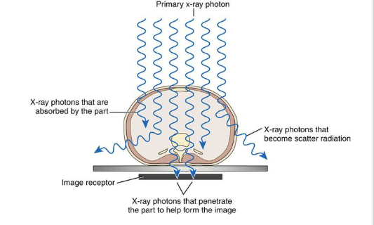
what is primary radiation? remnant? scatter? attenuation?
primary : beam of photons before it interacts with patient
remnant : resulting beam that is able to exit from patient
scatter : radiation that interacts with matter and continues in a different direction
attenuation : primary radiation that is partially absorbed as it travels through the pt
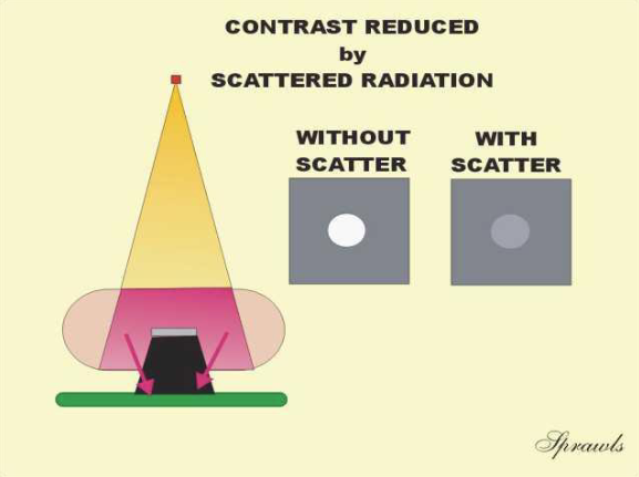
scatter reduces
contrast
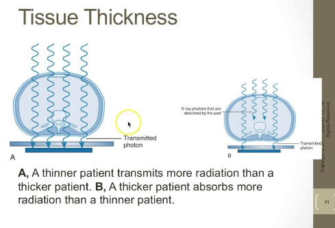
which patient transmits more radiation? thinner or thicker
which patient absorbs more radiation? thinner of thicker
transmits : thinner
absorbs : thicker
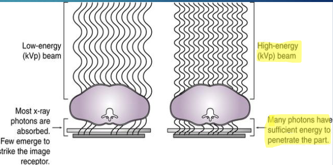
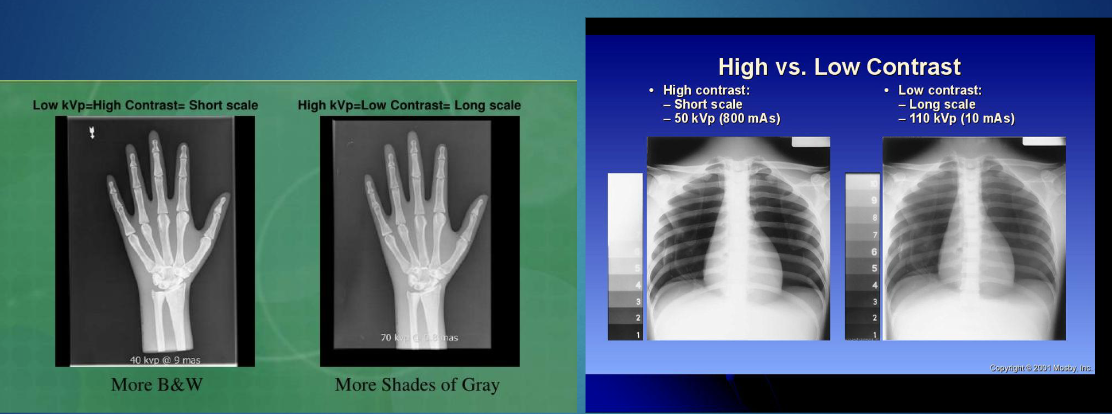
what determines the quality/contrast of the beam?
kVp = energy of xrays
more kVp improves image quality but reduced contrast
what is contrast?
difference in densiy of adjacent structures on the image
high contrast & low kVp : more B&W
this is because lower energy photons are more easily absorbed by denser tissue rather than just passing through them
creates greater difference in attenuation
low contrast & high kVp: more greys
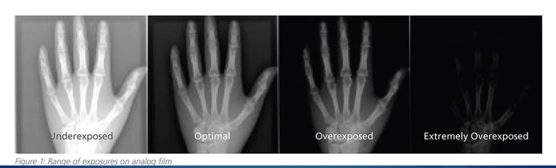
what is Mas?
milliampere-seconds
quantity of xray produced
tube current (mA) x exposure time (s)
*directly controls number of xray photons → determines image brightness
in what position of xray source/film would increased organ size be seen?
xray source is closer to the organ than xray film
*OID (object to image receptor distance)
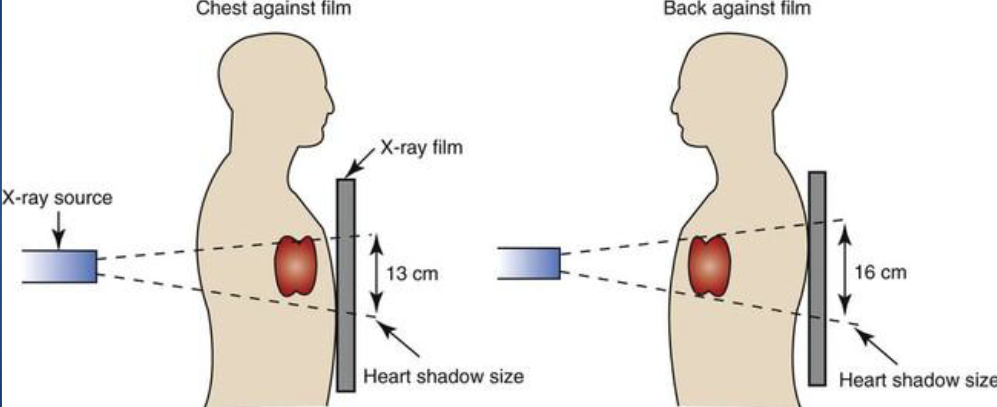
know
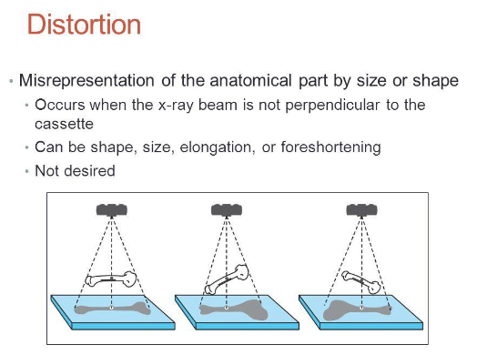
what is distortion?
misrepresentation of anatomical part by size/shape
occurs when x ray beam is not perpendicular to cassette/film
shape, size, elongation,foreshortening
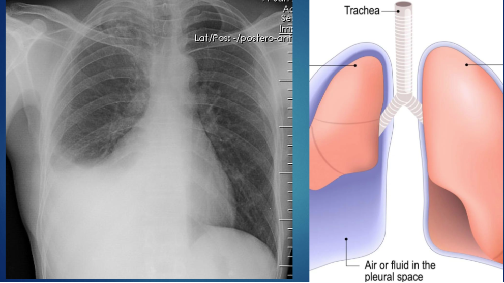
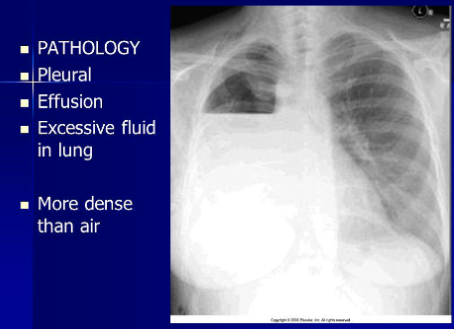
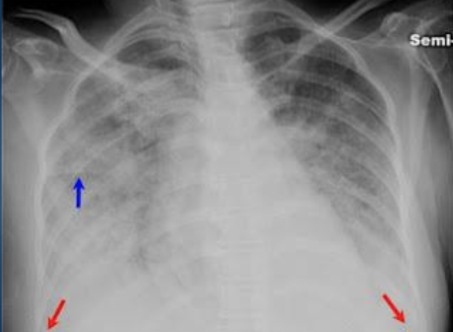
blunted angles → pleural effusion
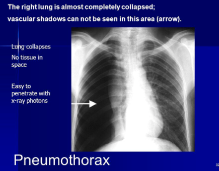
collapsed lung
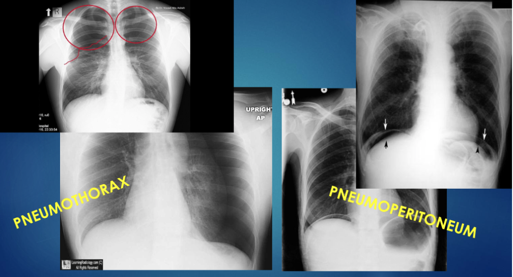
opacities indicate
fluid
infectious infiltrate
aspiration
blood
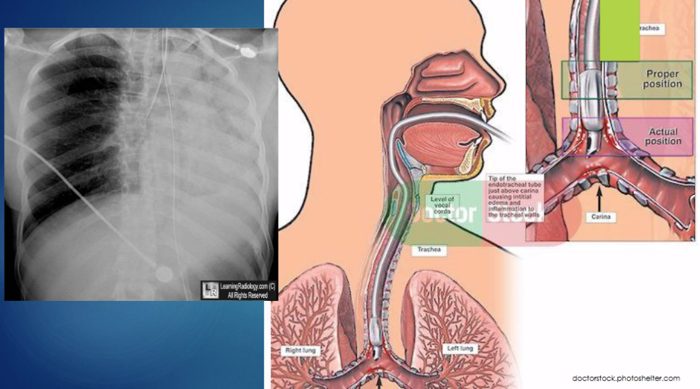
what is this
endotrachial tube can be used to treat collapsed lung