BMS Quiz 6
1/49
Earn XP
Description and Tags
Chapter 12: 12.6, 12.7, 12.8, 12.9, and 12.10 Chapter 14: 14.1, 14.2, 14.3, and 14.5 (only the first bullet point plus it's 2 sub-bullets)
Name | Mastery | Learn | Test | Matching | Spaced | Call with Kai |
|---|
No analytics yet
Send a link to your students to track their progress
50 Terms
Distinguish between a pump and a channel
Channels:
Allow substances to move down their concentration gradient
Do not require cellular energy
Pumps:
Maintain concentration gradients by moving substances against their gradient
Require cellular energy
Leak channels
Always open
Allow for continuous diffusion of one type of ion
Ex: K+ leak channel
Chemically-gated channels
Ligand-gated channels
Closed at rest
Open briefly in response to Neurotransmitter binding
Allow for diffusion of one type of ion
Ex: Chemically-gated cation channel
Voltage-gated channels
Closed at rest
Open briefly in response to changes in electrical charge across the membrane
Allow diffusion of one type of ion
Ex: Voltage-gated Na+ channel
Sodium/potassium pumps
Maintain the resting membrane potential
Account for 2/3 of neurons energy expenditure
Move 3 Na+ to ECF and 2 K+ to ICF
Calcium pumps
Establish a concentration gradient for Ca2+ in the axon terminal
important for synaptic transmission
Moves Ca2+ to the ECF
Can be used for later work
List the four functional neuron segements
Receptor segment
Initial segment
Conductive segment
Transmissive segment
What is in the Receptive segment and the 3 channels
Dendrites
Soma
Receives signals
Chemically gated cation channel
Chemically gated K+ channels
Chemically gated Cl- channels
What is in the Initial segment and the 2 channels
Axon hillock
Generates initial action potentials
Voltage-gated Na+ channels
Voltage-gated K+ channels
What is in the conductive segment and the 2 channels
Axon
Propagates action potentials
Voltage-gated Na+ channels
Voltage-gated K+ channels
What is in the transmissive segment?
Axon terminals
Releases neurotransmitters
Voltage-gated Ca2+ channels
Ca2+ pumps
Define the terms: electrical gradient, electrical potential, voltage, membrane potential
Electrical gradient: There is an unequal distribution of Ions across the plasma membrane
Electrical potential: An electrical gradient represents potential energy of electrical potential (think of a dam with a hole)
Membrane potential and voltage: Membrane potential refers to the difference in electric charge inside and outside of a cell, which can change in response to ion movement. Voltage is the measure of that electric potential difference.
A cell is polarized due to
The unequal distribution of ions across its membrane results in a difference in electrical charge inside and outside the cell. Like the North and South polls.
Resting Membrane Potential (RMP)
The potential difference across a cell’s plasma membrane when it is not being stimulated (at rest)
-70 mV
Explain how the RMP is established and maintained in neurons
Diffusion of K+ through K+ leak channels (primary)
down the electrochemical gradient
Diffusion of Na+ through Na+ leak channels
Na+/K+ pumps (always)
to maintain RMP
Describe the distribution of substances between the inside and the outside of a neuron
The outside of the cell (ICF):
More Na+ (sodium), Cl- (chloride), Ca2+ (calcium)
The inside of the cell (Cytosol):
More K+ (potassium)
Explain the roles of K+, Na+, and Na+/ K+ pumps in establishing and maintaining the RMP
K+ diffuses out of the cell through leak channels, making the inside negative.
Na+ diffuses in but less so due to fewer channels.
The Na+/K+ pump actively transports 3 Na+ out and 2 K+ in, maintaining the RMP at approximately -70 mV.
Depolarization
Gain of positive charge makes the cytosol less negative
Ex: Influx of Na+
What are the 2 definitions for Hyperpolarization
Loss of positive charge makes the cytosol more negative
Ex: Efflux of K+
Gain of negative charge makes the cytosol more negative
Ex: Influx of Cl-
Repolarization
The Na+/K+ pump returns the membrane potential to RMP/polarized state
Define a graded (postsynaptic) potential
A.K.A. Postsynaptic Potentials, Local Potentials
Occur along the RECEPTIVE SEGMENT
Result from the opening of chemically-gated channels
May cause depolarization or hyperpolarization
Size of the change in membrane potential varies
Travels only a short distance
Describe the events of a graded (postsynaptic) potential
Graded (postsynaptic) potentials are changes in membrane potential that occur in response to synaptic transmission. They result from neurotransmitter binding to receptors, leading to the opening of ion channels, which can cause local depolarization or hyperpolarization.
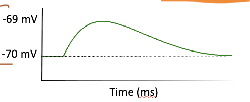
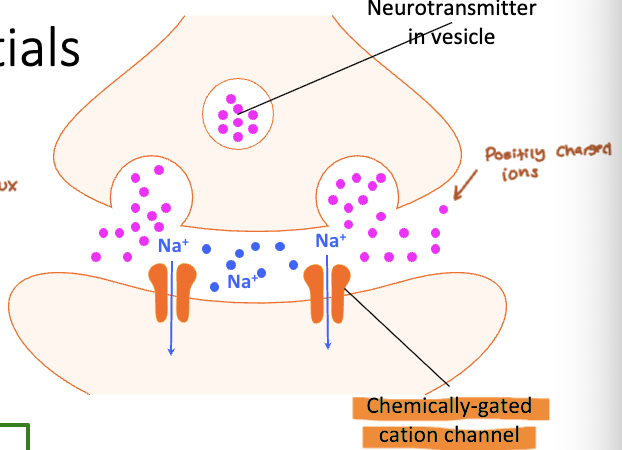
Chemically-gated cation channel graded potential
Neurotransmitter binds receptor/opens the channel
Na+ diffuses into the cell (influx)
Depolarization (less negative)
Excitatory Postsynaptic Potential (EPSP)
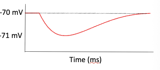
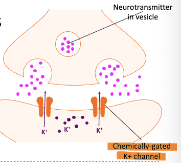
Chemically-gated K+ channel graded potential
Neurotransmitter binds receptor/opens channel
K+ diffuses out of the cell (Efflux)
Hyperpolarization (More negative)
Inhibitory Postsynaptic Potential (IPSP)
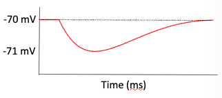
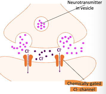
Chemically-gated Cl- channel graded potential
Neurotransmitter binds receptor/opens channel
Cl- diffuses into the cell (Influx)
Hyperpolarization (more negative)
IPSP
Excitatory
Increase the likelihood the axon will fire an axon potential
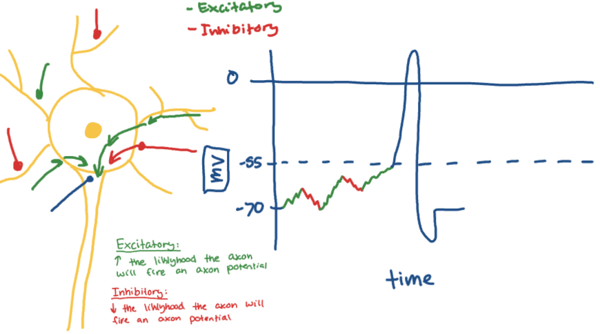
Inhibitory
Decrease the likelihood the axon will fire an axon potential

Define summation
The changes in membrane potential generated by all the graded potentials (EPSP + IPSP) are:
Added together at the initial segment
Explain how summation relates to threshold potential
An action potential will be generated of graded potentials arriving at the axon hillock move them membrane potential to:
Threshold potential → -55mV
What happens when threshold is reached?
Voltage-gated channels at the axon hillock open, initiating an ACTION POTENTIAL
Describe an action potential
Begins in the INITIAL SEGMENT
Occurs along the CONDUCTIVE SEGMENT
Results from the sequential opening and closing of voltage-gated channels
Causes a large, stereotypical change in the membrane potential
“All or none”
Describe what causes depolarization, repolarization, and hyperpolarization in an action potential
Depolarization
Na+ influx causes “rising phase”
Repolarization
K+ efflux causes “falling phase”
Hyperpolarization
Excess K+ efflux causes “refractory period”
Return to resting membrane potential
Depolarization phase
Mediates by voltage-gated Na+ channels
A. Na+ enters from adjacent areas, and membrane potential changes: -70mV to -55mV
Reaching threshold, VGNCs open & membrane potential depolarizes: -55mV to +30mV
VGNCs close
Repolarization, hyperpolarization, and return to RMP
Mediated by voltage-gated K+ channels
D. VGKCs open slowly, to coincide with peak depolarization, and K+ exits the cell & the membrane repolarizes: +30mV to -70mV
VGKCs remain open for longer time than needed, & the membrane hyperpolarizes: -70 mV to -80mV
VGKCs close and RMP is reestablished by Na+/K+ pumps (-70mV)
Refactory period
Brief time period after an action potential (AP) when it is impossible or difficult to fire another AP, during which a neuron cannot fire another action potential due to inactivated sodium channels and increased potassium permeability.
Continuous conduction
Unmyelinated axons
Sequential opening of VGNCs along the entire length of the axon
A method of action potential propagation in which action potentials are generated at each segment of the axon, allowing impulses to travel continuously down the membrane.
Saltatory conduction
Myelinated axons
Action potentials jump between nodes of Ranvier
A method of action potential propagation that increases the speed and efficiency of impulse transmission.
Requires less energy
Action potential: Velocity
The velocity of an AP is influenced by
Myelination axons = faster velocity
Axon diameter = Bigger = Faster velocity
Synaptic transmission
Occurs in the TRANSMISSIVE SEGMENT
Initiated by the arrival of an action potential at theaxon terminal
Causes release of neurotransmitter from thesynaptic knob
Depends on a concentration gradient for Ca2+
What are the steps os synaptic transmissions?
Arrival of an action potential in the synaptic knob opens voltage-gated Ca2+ channels (VGCCs)
Ca2+ enter the synaptic knob & binds synaptic vesicles
Synaptic vesicles fuse with the plasma membrane &neurotransmitter is released by vesicular exocytosis
Neurotransmitter diffuses across the synaptic cleft and binds receptors on the plasma membrane of the postsynaptic neuron
Action potential: Frequency
Amplitude of an AP: is always the same
Frequency of an AP: depends on stimulus strength
Stronger stimulus = more frequent action potentials
Identify the four classes of neurotransmitters based on structure
Structural classification = chemical structure
Acetylcholine (Ach)
Significantly different from the others
Biogenic amines (monoamines)
Modified amino acid
Melatonin
Amino acids
Neuropeptides
Chains of amino acids
opioids
Describe how neurotransmitters are classified based on function
Functional classification = effect on membrane potential
Excitatory: induce an EPSP
E.g. Glutamate
Inhibitory: Induce an IPSP
E.g. GABA (gamma-Aminobutyric acid)
What are the 5 subdivisions of the spinal cord and their nerves
Cervical
(C1-C8)
Thoracic
(T1-T12)
Lumbar
(L1-L5)
Sacral
(S1-S5)
Coccygeal
(Co1)
Describe the locations and function of the spinal cord meninges.
The meninges are three protective membranes surrounding the spinal cord that provide structural support, contain cerebrospinal fluid, and cushion the spinal cord.
Deepest = Pia Mater
Middle = Arachiod Mater
Superficial = Dura Mater
Distinguish the anatomical locations of gray and white matter in the spinal cord
Grey matter
Deep/middle and forms a butterfly
cell bodies, dendrites, unmyelinated axons
White matter
Outer part of the spinal cord
myelinated axons
Name each sub-region of gray and white matter
White matter:
Dorsal/Posterior funiculus
Lateral Funiculus
Ventral/Anterior funiculus
Grey matter:
Posterior horn
Lateral horn
Anterior horn
Differentiate the four functional groups (nuclei) found within each gray matter region
Sensory nuclei: receive stimuli
Somatic and visceral
In the posterior horn
Motor Nuclei: Send out signals to muscles and glands
Somatic and autonomic
In the lateral and anterior horns
Describe the components (roots) of a typical spinal nerve.
Formed by:
Posterior (Dorsal) root: Contains sensory axons
Cell bodies in the posterior root ganglion
Anterior (ventral) root: contains motor axons
Cell bodies in anterior & lateral horns of spinal cord
Classify spinal nerves based on function (sensory, motor, or mixed)
A spinal nerve is always a mixed nerve