5. cerebral cortex and higher cortical functions
1/64
There's no tags or description
Looks like no tags are added yet.
Name | Mastery | Learn | Test | Matching | Spaced |
|---|
No study sessions yet.
65 Terms
cerebral cortex
seat of intelligence
basic sensorimotor, visual and auditory processing
cognition
cognition
information-processing functions carried out by the brain
attention
memory
executive functions: planning, problem solving, self-monitoring, self-awareness, and metacognition
comprehension of language and formation of speech
calculation abilities
visual perception
praxis
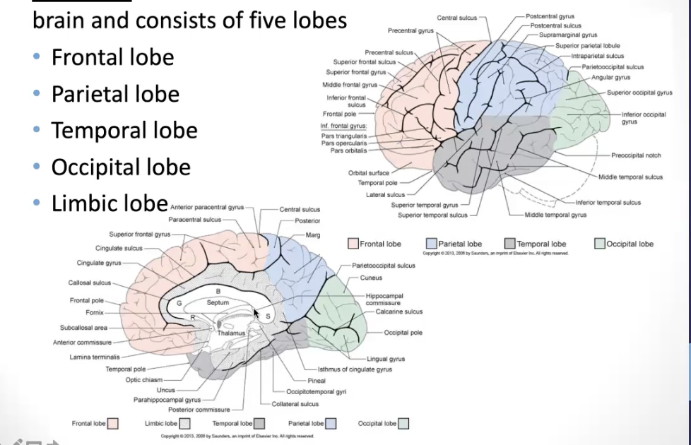
structure of cerebral cortex
entire surface of brain
five lobes
parietal lobe
frontal lobe
limbic lobe (sagittal section)
temporal lobe
occipital lobe
projection fibers
ascending and descending
fibers project form cortical areas to subcortical structures or from subcortical areas to the cortex
thalamocortical fibers: main source of input to the cortex originating from non-cortical structures
callosal/commissural fibers
these fibers connect areas of the cortex in one hemisphere with areas of cortex in the opposite hemisphere
association fibers
These fibers connect areas of cortex within the same hemisphere (short fibers connect gyri of the same lobe and long fibers connect gyri of adjacent lobes)
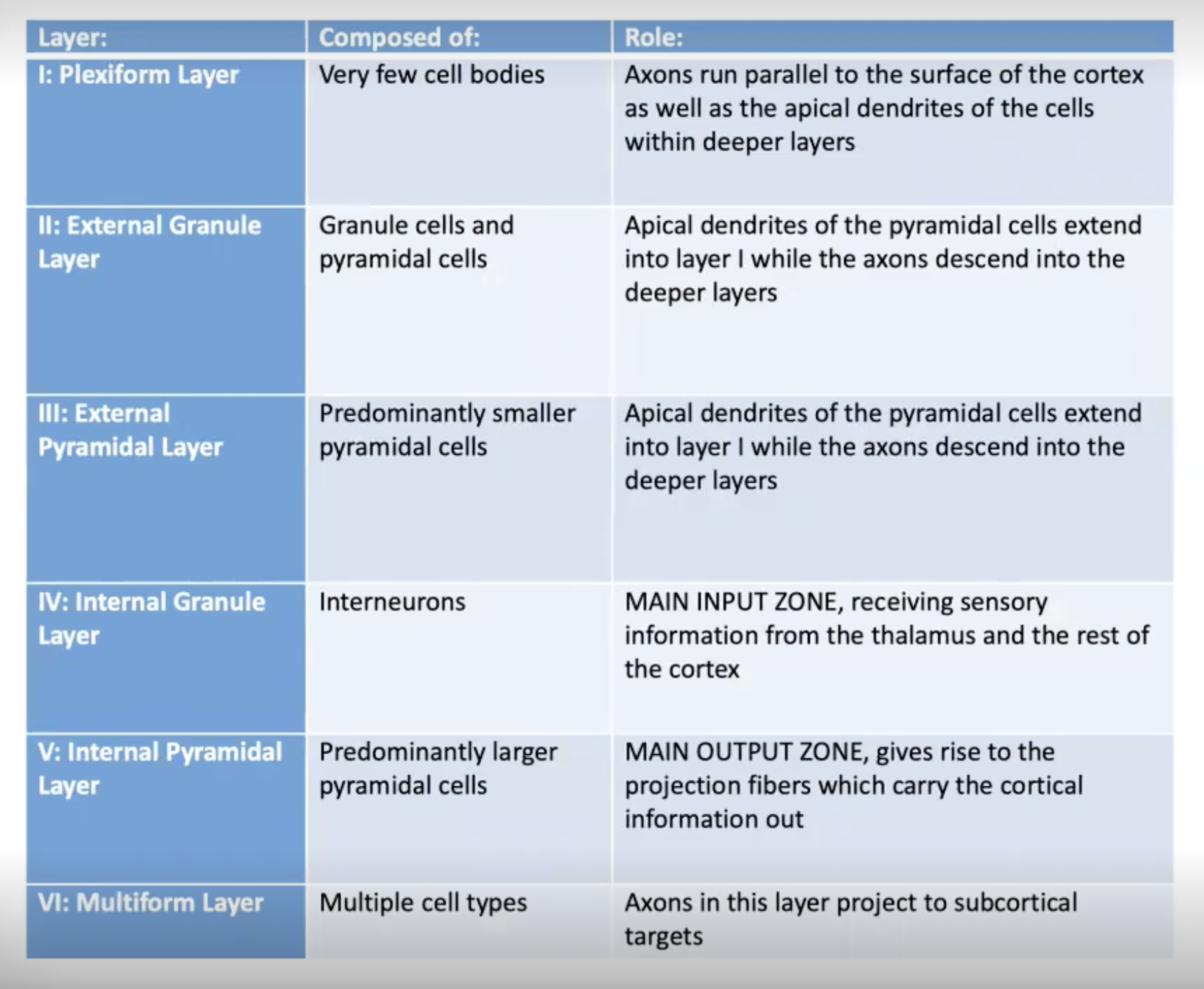
neocortex
six layers (I-VI)
Two major cell types
pyramidal neurons: one apical dendrite and multiple dendritic trees; project OUT of the cortex to other regions of the brain and to the spinal cord
granular neurons: smaller dendritic trees and shorter axons; REMAIN in the cortex - considered interneurons
neocortex
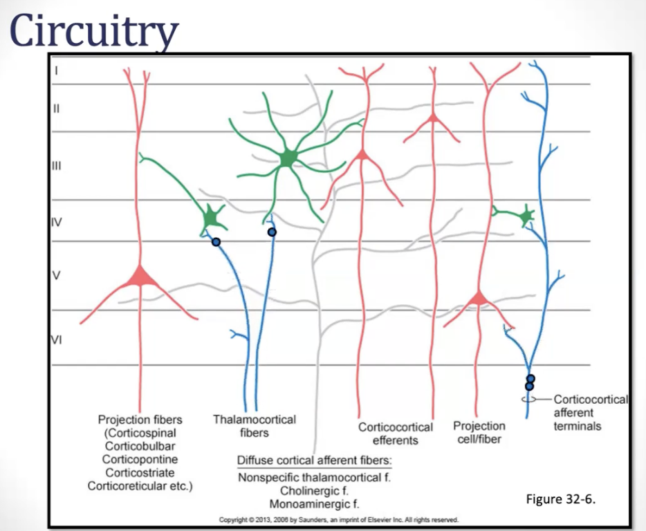
circuitry: intrinsic circuitry
high degree of convergence and divergence within the neural circuitry of the cerebral cortex - allowing for complex integration of information
afferent fibers
input to the cortex
thalamocortical fibers
cortico-cortical fibers
diffuse inputs
afferent fibers: thalamocortical fibers
input from thalamic nuclei
excitatory
afferent fibers: cortico-cortical fibers
excitatory or inhibitory
afferent fibers: diffuse inputs
e.g. ascending reticular activating system; raphe nuclei
excitatory
local circuit neurons
processing and integrating information (layer IV)
excitatory and inhibitory interneurons
efferent fibers
pyramidal cells - excitatory output from the cortex
cortico-cortical fibers (i.e. association or callosal - layers II and III)
projection fibers (layer V)
internal circuitry
Histological Organization
cytoarchitecture: not every of cortex has the same makeup
differs based on function
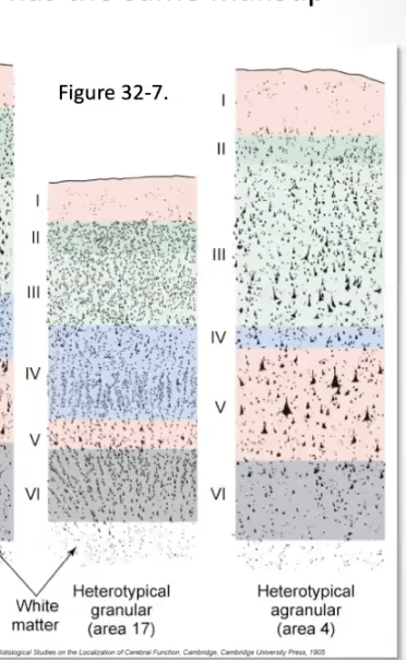
homotypical cytoarchitecture
all six layers are clearly represented in most areas of the cortex
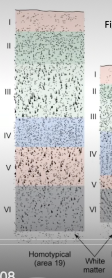
heterotypical cytoarchitecture (dont need to know difference between granular and granular cortex)
layers vary in thickness
granular cortex: layer IV is especially thick and layer V is thin - primary sensory cortex
agranular cortex: layer V is especially thick and layer IV is thin - primary motor cortex
b
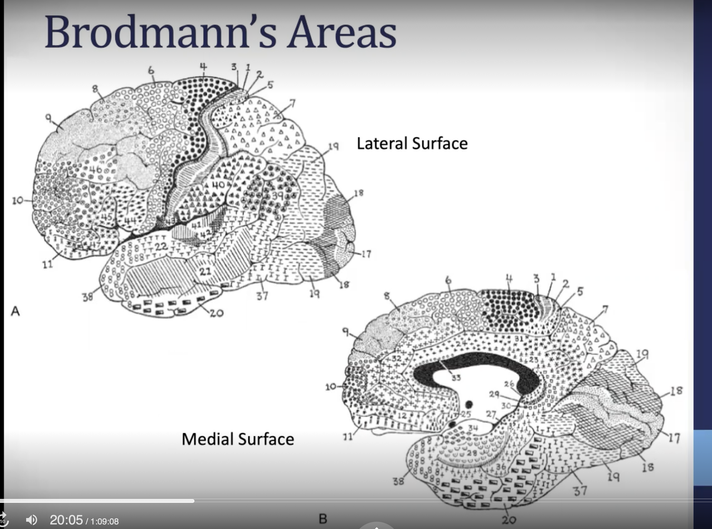
brodmann’s areas: frontal lobe (dont need to know numbers, know function)
areas 4, 6, 8-12, 32, and 44-47
primary motor cortex: area 4
premotor and SMA (Motor planning): area 6
frontal eye fields: area 6 and 8
broca’s area (speech): areas 44 and 45
prefrontal and orbitofrontal 8-12, 32, 45-47
parietal lobe brodmann’s areas(dont need to know numbers, know function)
primary somatosensory cortex
posterior parietal cortex: visuospatial functions
superior parietal cortex: somatosensory association cortex, dorsal stream of vision - “where” and “how” stream of vision)
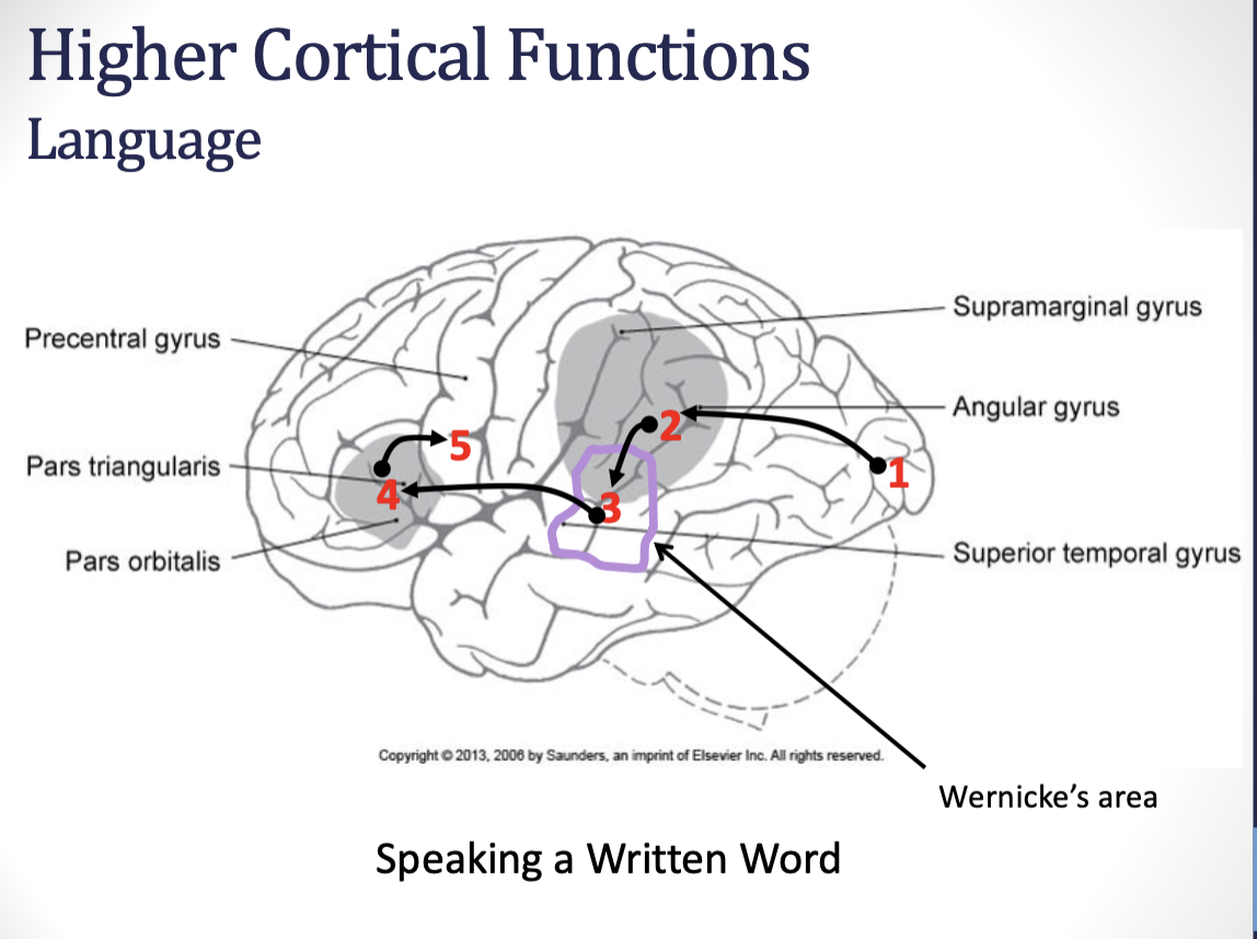
inferior parietal cortex: angular gyrus and supramarginal gyrus
temporal and occipital lobe areas (brodman’s areas)
primary auditory cortex
primary visual cortex
visual and auditory association cortices:
wernicke’s area
ventral stream of vision (what and who stream of vision)
limbic lobe
lateralization
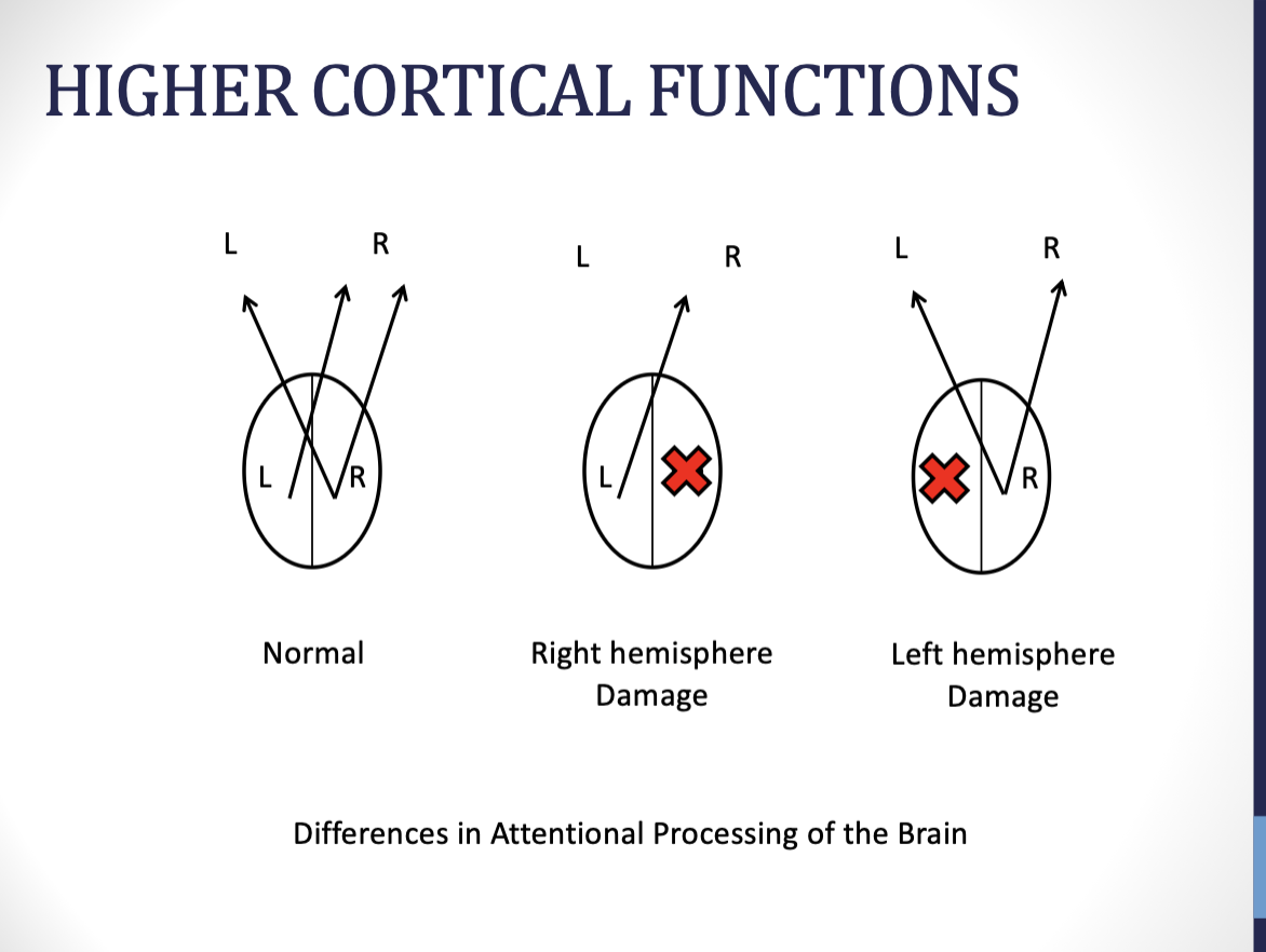
right brain: big picture, help explain context
left brain: detail orientated, help with routines but not full understanding
motor and sensory functions pertaining to one side of the body are controlled by the opposite hemispehre
information from one visual field is processed by the opposite hemisphere
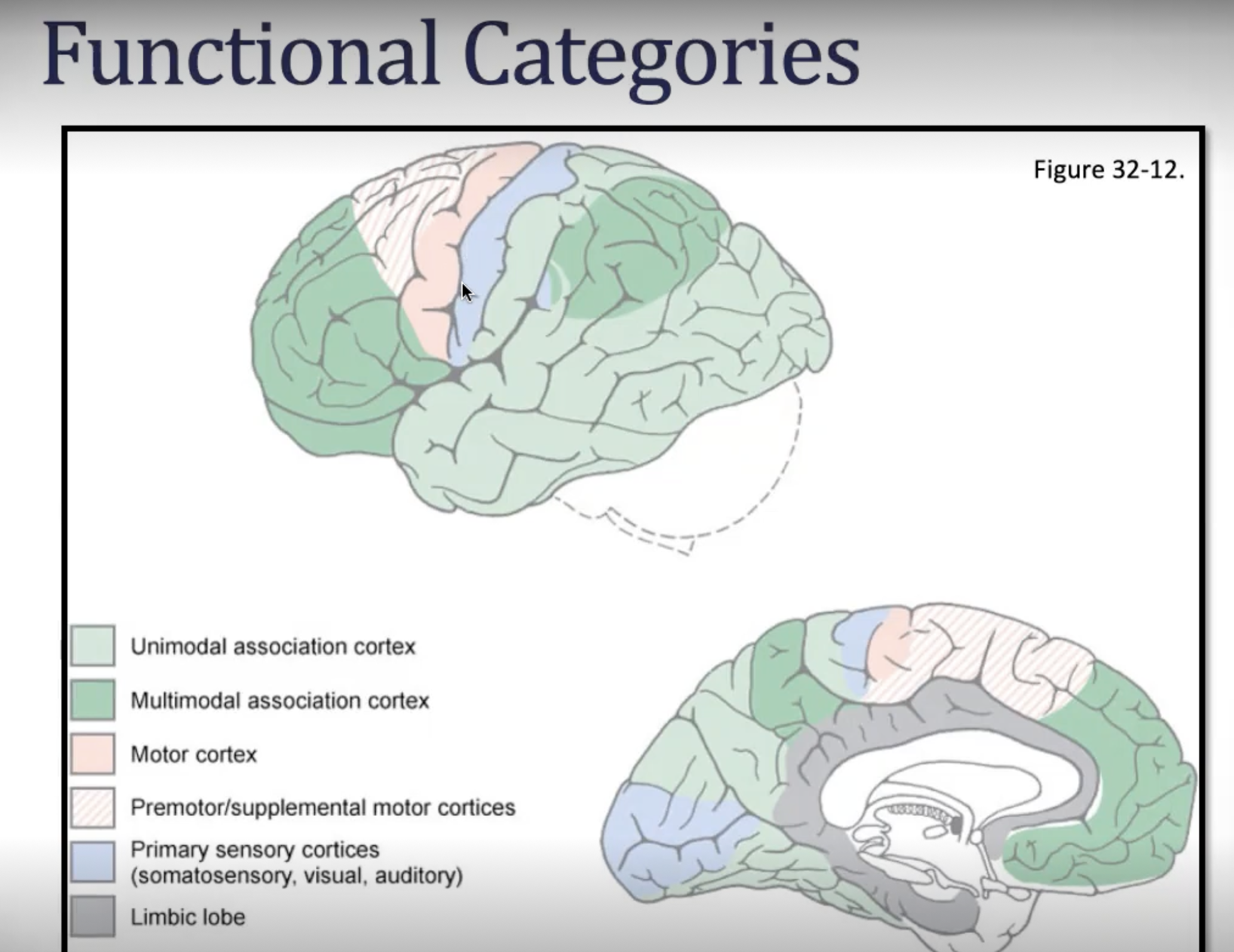
Functional categories: motor
primary motor cortex, supplementary motor area, premotor cortex
functional categories: primary sensory
primary somatosensory cortex, primary visual cortex, primary auditory cortex
functional categories: association areas
majority of cortex
unimodal association: areas that are adjacent and related to primary areas
multimodal association: there are large areas of cortex which receive information from several different sensory modalities and create a broader, more complete understanding of ourselves and our environment
primary motor cortex
localized to the pre central and anterior paracentral gyri of frontal lobe
somatotopic organization: motor homunculus
sends motor output to the opposite side of the body (corticospinal and corticonuclear tracts)
lesions of this area result in upper motor neuron signs contralaterallypr
primary somatosensory cortex (PSC)
localized to the post central and posterior paracentral gyri of parietal lobe
somatotopic organization: sensory homunculus
receives sensory input from the opposite side of the body (PCMLS and ALS)
lesions of this area result in decreased awareness of sensory stimuli and/or poor localization of sensory stimuli - contralaterally high
higher cortical functions: arousal and attention
arousal/alertness is first step
starts with the reticular activating system
if person is not alert enough, there can be no attention
if a person is over-aroused/hyper-alert, attention will be affected
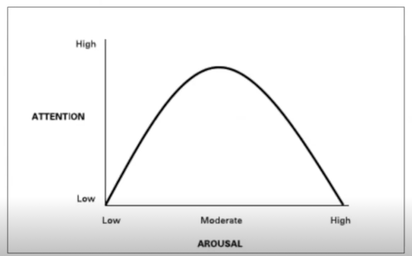
higher cortical functions: arousal and attention
attention (Frontal and parietal association areas)
attentional capture: automatic or reflexive orienting to a stimulus
selective attention: goal-directed focus of attention with the ability to ignore irrelevant stimuli
sustained attention: ability to maintain vigilance over time
divided attention: ability to perform two or more tasks at the same time, or process two or more sources of information at the same time
alternating attention/task-switching: rapidly switching between different skills/tasks/cognitive sets
higher cortical functions: language
frontal and temporal association areas
language is the faculty of communication to describe things, events, and to express ideas - language is a function of multimodal sensory and motor processing areas and includes speaking, reading, and writing
hemisphere that controls language is considered the dominant hemisphere
almost all right-handers and 50-70 percent of left-handers are left-cerebral dominant
in brain lesions which impact language, about 95% of those cases are left hemisphere involvement
right hemisphere involved in language in regards to prosody of speech (rhythm, tone, stress, etc.)
aphasia: disturbance in the understanding or formulation of language not due to hearing/vision/motor impairments
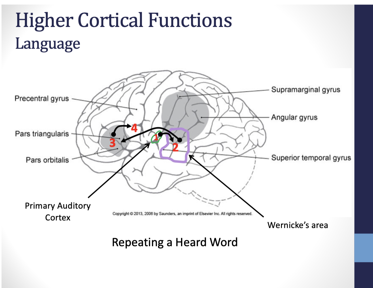
speaking a written word
aphasia
any disturbance in language, affecting the production of speech, the comprehension of speech, ability to read, ability to write
usually due to a lesion of the left hemisphere
fluent aphasia: can still produce speech, but meaning is impaired (usually a wernicke’s aphasia)
non-fluent aphasia: difficulty communicating orally and with written words, as it is the production of speech, but comprehension is usually intact (usually Broca’s aphasia)
broca’s aphasia
expressive aphasia
lesions in the left inferior frontal gyrus, aka Broca’s area or brodmann’s area 44 and 45
frontal lobe tumors and occlusions of the frontal branches of the middle cerebral artery
often with a contralateral hemiparesis
might still be able to get key words out, grammar is off
might have problems repeating words
might have agraphia (impairments in writing)
usually able to understand spoken and written words
aware of deficits usually- can understand they are not saying what they want to say
wernicke’s aphasia
receptive aphasia
lesions in left inferior parietal lobe, brodmann’s area 39 and 40, as well as posterior superior temporal gyrus, aka wernicke’s area, or brodmann’s area 22
occlusions of temporal and/or parietal branches of middle cerebral artery
might have agraphia (impairments in writing)
might have Alexia (inability to read)
usually present with clear speech, normal prosody, but content is unintelligible (paraphasic speech)
patients usually have impaired awareness of the deficits; cannot comprehend what they are saying; usually less frustrated
conduction aphasia
disruption of the arcuate fascicles, bundle of long association fibers that connect wernicke’s to Broca’s areas
type of fluent aphasia; spontaneous expression is intact and comprehension is functional; patients will have trouble repeating phrases or translating/interpreting what they have heard into an appropriate reply
global aphasia
damage to both Broca’s and wernicke’s areas, usually due to occlusion of the internal carotid artery; results in very gross left hemisphereic damage
loss of virtually all language
may be able to use some gestures
motor control
motor planning (motor and parietal association areas)
praxis: conception and planning of a new action in response to environmental demand
apraxia: inability to execute learned purposeful movements despite having the desire to perform them as well as the motor/sensory capabilities required to execute them
secondary to lessons of the left hemisphere (premotor cortex, supplementary motor area, and/or parietal association cortex)
forms of apraxia
ideomotor: most common; impaired motor performance in response to a verbal cue despite intact sensory/motor/language, usually with tool use or gesturing
ideational: inability to coordinate activities with multiple, sequential steps or movements
agnosia
inability to know, name, identify, or extract meaning from visual, auditory, or tactile information - parietal and temporal association areas
visual agnosia: inability to visually recognize objects, although sensory function is generally normal (parietal and temporal areas)
ventral stream of vision (who/what path)
social-emotional agnosia: inability to correctly perceive or comprehend social-emotional information conveyed by voice, gesture, or facial expression
prosopagnosia: inability to recognize faces
executive functions
prefrontal cortex
organization: attention, planning, sequencing, problem solving, cognitive flexibility, abstract thinking, etc.
regulation: initiation of actions, self-control, emotional regulation, moral-reasoning, decision-making, etc.
damage to the prefrontal cortex
deficits in attention
planning deficits: inability to anticipate or predict future events based on current information
disorganized
perseveration
lack of judgment
socially inappropriate behavior
mirror neuron system (MNS)
distributed network of neurons located throughout the frontal (e.g. ventral premotor cortex, inferior frontal gyrus), parietal (e.g. inferior parietal lobe), temporal (e.g. superior temporal gyrus), cingulate and insular cortices
network is active when we perform a particular action and when we observe another individual perform an action
distributed network is thought to allow us to understand the actions performed by another person and predict the consequences of their actions by using mental simulation (simulation theory)
plays a role in motor learning
plays a role in empathy
deficits in the functioning of this network have been implicated in autism spectrum disorders and schizophrenia
theory of mind
theory-theory
how we develop theories about peoples minds; cognitive capability to consider the wants, needs, knowledge, and mental state of another
neural network
medial prefrontal; precuneus (superior parietal lobule)
temporal/parietal junction (right)
cingulate cortex/amygdala
default mode network
consists of discrete cortical areas (bilateral and symmetrical)
medial prefrontal cortices
posterior cingulate cortices
medial and lateral parietal cortices
medial and lateral temporal cortices
typically more active when wer aren’t engaged in attention demanding goal directed tasks
thought to allow emotional processing, self referential mental activity, and the recollection of prior experiences
salience network
anterior cingulate cortices
anterior insulation subcortical nodes involved in affect and reward processing
integrates internal and external stimuli
plays a key role in identifying the most relevant stimuli in order to rot guide attention and behavior for adaptive purposes - important for switching between default mode and central executive networksce
central executive network
dorsolateral prefrontal cortex
anterior cingulate
orbitofrontal cortex
lateral posterior parietal cortex
operates during all goal-directed activities and filters out distractions that might interfere with performance
plays a key role in cognitive processing, reasoning, task flexibility, and decision making
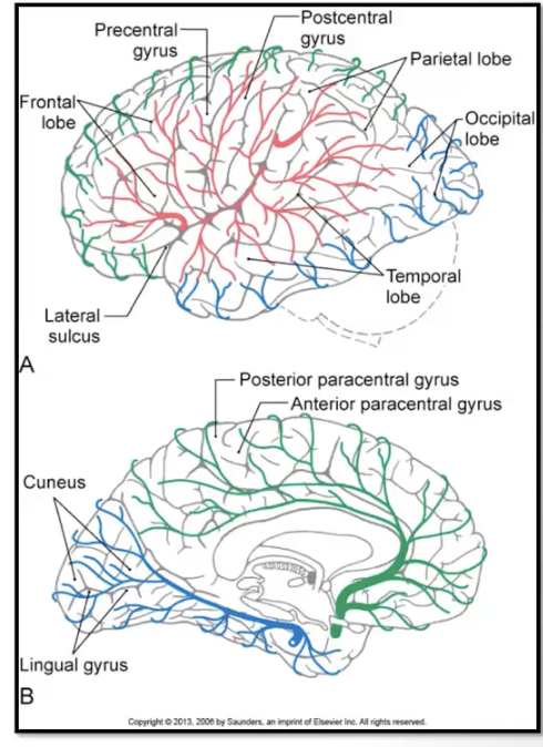
blood supply to the cerebral cortex
anterior cerebral: serves majority of medial surface of the hemispheres
middle cerebral: serves majority of the lateral surface of the hemispheres
posterior cerebral: serves the inferior temporal lobe and medial occipital lobe
circle of willis and border zones!
anterior cerebral artery
motor cortex for LE
primary somatosensory cortex for LE
motor planning areas in medial frontal lobe (dominant hemisphere)
prefrontal cortex
posterior cerebral artery
supplies following areas
optic radiations and primary and visual association cortices
hippocampal formation and fornix
middle cerebral artery
primary motor and somatosensory cortex for face, trunk, arm/hand
deep white matter (corticobulbar and corticospinal)
broca’s area: superior cortical brancha nd wernicke’s area (inferior cortical branch) in language dominant (L)
frontal eye fields
parts of frontal and parietal lobed important for lateralized attention, visuospatial analysis and emotional expression (body language/tone of voice) (R hemisphere)
optic radiations (inf cortical branch)
MCA stroke
main stem: key deficits
contralateral hemiplegia; contralatteral somatosensory loss; contralateral homonymous hemianopia; head and eye deviation toward side of lesion (acutely)
right hemisphere lesion: denial, neglect, disturbed spatial perception, emotional flatness possible
left hemisphere lesion: global aphasia
MCA superior cortical branch deficits
superior cortical branch
contralateral hemiparesis impacting the face and UE; possible contralateral somatosensory loss impacting the face and UE; head and eyes deviated toward side of lesion (acutely)
right hemisphere lesion: neglect and disturbances ins spatial perception
left hemisphere lesion: Broca’s aphasia
MCA inferior cortical branch deficits
possible mild weakness of contralateral face and arm possible contralateral somatosensory loss; contralateral superior Quadrantanopia
right hemisphere lesion: denial, neglect, disturbed spatial perception
left hemisphere lesion: wernicke’s aphasia
anterior cerebral artery
deficits
contralateral hemiparesis involving the leg; contralateral somatosensory loss involving the leg; frontal lobe behavioral abnormalities
ACA-MCA cortical border zone deficits
weakness of proximal limb girdles of arm, leg, bothPCA
PCA
penetrating branch supplying thalamus: may produce loss of all somatic sensation in the contralateral face and body; this initial hemianesthesia may later develop into thalamic pain syndrome
unilateral cortical branches of PCA: contralateral homonymous hemianopia
bilateral cortical branches of PCA: inability to form new semantic or episodic memories; cortical blindness
lacunar strokes (aka small vessel disease)
these strokes are consequences of blockage of blood flow to a single small deep penetrating vessel supplying the subcortical white matter region, basal ganglia, internal capsule, corona radiate, thalamus, or paramedian pons
blood vessels responsible
lenticulostriate arteries of the MCA
thalamogeniculate arteries of the PCA
paramedian perforating arteries of the basilar artery
common lacunar stroke syndromes: pure motor hemiparesis
pure motor hemiparesis
most common lacunar syndrome (33-50%) usually involved the internal capsule, corona radiate, or basis pontis; presents clinically with contralateral hemiparesis
common lacunar stroke syndromes: pure sensory
typically involves the thalamus (VPL), internal capsule or corona radiate; presents clinically with contralateral sensory loss
common lacunar stroke syndromes: sensory-motor stroke
typically involves the thalamus and posterior limb of internal capsule; presents clinically with a combination of contralateral motor and sensory lossa
common lacunar stroke syndromes: ataxic hemiparesis
typically involves the internal capsule, pons, or corona radiate; presents clinically as a combination of cerebellar and motor symptoms, including weakness and clumsiness, on the ipsilateral side of the body; typically leg > arm
common lacunar stroke syndromes: dysarthria-clumsy hand
usually involves the pons, anterior limb, or gene of internal capsule; presents clinically with dysarthria and contralateral paresis/clumsiness of the arm and hand