Stifle Joint & Thigh Muscles
1/32
There's no tags or description
Looks like no tags are added yet.
Name | Mastery | Learn | Test | Matching | Spaced |
|---|
No study sessions yet.
33 Terms
What type of bones are the tibia and fibula?
• Type = long bones
What are the main roles of the tibia and fibula?
Does rotation occur within these two bones?
• Tibia:
main weight bearing
Prioritize stabilizing
• Fibula:
much reduced but present in dog
Lateral aspect of limb, along length
Absent in larger species
• No rotation
Describe key features of the distal end of the tibia/fibula.
• Distal end
• Medial malleolus, dorsal surface of tibia
Attachment of medial collateral ligament of hock
• Lateral malleolus in fibula
Attachment of lateral collateral ligament of hock (or tarsus)
• Both palpable
Describe key features of the proximal end of the tibia/fibula.
Proximal End
• Tibial plateau / plate
Medial condyle (c)
Lateral condyle (c)
Covered by smooth bone surface - articular cartilage
Articulate with medial & lateral condyles of femur
• Intercondylar ridge (r)
Non-articular rough bone surface
ligament attachment
• Tibial crest
• Tibial tuberosity
• insertion of patellar ligament (Which is a tendon = a fake ligament)
Is palpable in life
Describe the centers of ossification in the tibia.
• Tibia:
• Proximal end (epiphisys)
• Tibial tuberosity
• Body (diaphysis)
• Distal end (epiphysis)
Total = 4
Describe the centers of ossification in the fibula.
• Fibula: maximum = 3 (carnivores)
Proximal and distal epiphysis + diaphysis
How does the tibia / fibula look in:
Carnivores + Pig
Ruminant + Horse
• Carnivores + pig: present as two separate long bones
• Ruminant & horse:
• Incomplete & fused
• Horse - present in proximal aspect, fused to body of tibia, lateral mallelolus incorporated into tibia
• Ruminants - tear shaped tuberosity fused to lat. condyle of tibia (proximal) + isolated maleollar bone (distal)
What comprises the stifle joint?
1/ Femorotibial joint
2/ Femoropatellar joint
3/ Proximal tibiofibular (where fibula present)
4/ Femur + paired sesamoids (carnivores)
What are the articular surfaces, meniscus/menisci of femorotibial joint.
1/ Femorotibial - incongruent surface
Articular surfaces :
Lateral & medial femoral condyles
Lateral & medial tibial condyles
Meniscus / Menisci: produce a congruent surface
C shaped wedges of cartilage
Attached to tibial condyles (medial and lateral)
Thick to thin from outer to inner
Movement occurs between femoral condyles & menisci
What gives the femorotibial joint stability?
Stability:
1. Medial & lateral collateral ligaments
Prevent lateral and medial movement
2. Cranial & Caudal Cruciate ligaments:
Intercondylar eminence - intercondylar fossa
Named for attachment to tibia
CRANIAL CRUCIATE - attaches to cranial intercondylar ridge of tibia and runs to caudal intercondylar fossa of femur
CAUDAL - attaches to caudal intercondylar ridge of tibia and runs to cranial intercondylar fossa of femur
• Maintain femur on mensici
• Resist rotation, keeps bones firmly attached together, preventing excessive cranial and caudal movement
What is the clinical significance of a cranial cruciate rupture?
• Cranial cruciate * rupture:
• Joint instability
'Cranial draw' sign
Tibia moves cranial relative to femur
What are the components of the femoropatellar joint?
• Components, articulation of the femur and patella
• Trochlear groove (femur)
• Patella
• Patella is embedded in patellar ligament (which is a TENDON!!)
• Parapatellar (fibro)cartilages -
What provides the femoropatellar joint with stability?
• Stability: No lateral movement:
Medial and lateral trochlear ridges of the femur
Lateral & medial (collateral) femoro-patellar ligaments
Insert onto the femur itself in large species
Insert onto famellae sesamoids in domestics
Tend to be mineralized in canine species
Fascia / retinaculum
How does proximal and distal glide of the patella occur?
What movements will this result in?
• Proximal & distal glide only:
Patella pulled proximally by contraction of quadriceps
Produces stifle extension
Patella pulled distally by the caudal muscles (biceps femoris)
Allows stifle flexion
Describe the different types of sesamoid bones found within the stifle joint.
Note any species differences as well.
• Sesamoid bones:
• Patella (x1)
Visible all species
More pointed in cat than dog
• Fabellae (sesamoids) (x2):
In Gastrocnemius muscle
Not visible in larger species
Medial often not mineralised in cat
• Popliteal sesamoid (x1):
Usually visible dog & cat
Not found in large animal species
Dog & cat: maximum total = 4
Large species: total = 1
Describe the joint capsule of the stifle joint.
How does communication between joint compartments vary between species.
• Typical synovial joint:
• Extensive joint capsule
• Forms 3 compartments:
Femoro-patellar
Lateral femoro-tibial
Medial femoro-tibial
• Communication between compartments varies between species
• Dog - good
• Horse - poor / none
Describe radiographic appearance of the stifle joint.
• Radiography:
Soft tissue components not visible
Retropatellar fat pad
Describe the key features of the equine stifle joint.
• Medial trochlear ridge:
larger than lateral
tubercle at proximal end, allowing for ligaments to “hook”
• Trochlear groove
2 parts to articular surface
Vertical = portion for gliding
Horizontal = portion for resting
• Patella:
Large medial cartilage projection
3 patellar ligaments:
Medial / middle /lateral
All insert - tibial tuberosity
Patella & medial patellar ligament form hook
What is the patellar locking mechanism.
The patellar locking mechanism in horses, part of the stay apparatus, allows them to sleep standing by locking the patella (kneecap) over a ridge on the femur (thigh bone) using the medial patellar ligament.
Patella pulled proximally as stifle extended, by contracting quadriceps
Patella rotated medially due to contraction
Engages or hooks horizontal / resting surface
Tubercle of medial trochlear ridge now sits between middle & medial patellar ligaments
Locks joint in extension
Describe how the equine patellar locking mechanism contributes to the stay apparatus.
How does it “unlock”?
• Patella pulled proximally then back to midline to unlock
• Contributes to stay apparatus:
• Locks leg in extension
• Can rest other hindlimb / sleep standing up
• Clinical significance:
• Upward fixation / 'locked' patella
Summarize the stifle joint components.
• 1/ Femur - tibia components:
• Medial & lateral menisci
• Collateral ligaments
• Cranial & caudal cruciate ligaments
• 2/ Femur - patella components:
• Medial and lateral trochlear ridges
• Medial & lateral femoro-patellar ligaments
• Retinaculum
• Patella & patellar ligament - palpable
• Locate joint space
• Further proximal than you would think!
What are the nerves supplying the hindlimb?
• Spinal nerves
• L5, L6, L7, S1, S2.
• Dorsal branches - dorsal structures
• Ventral branches - lumbosacral plexus
• Emerging peripheral nerves from the lumbosacral plexus to hindlimb:
Gluteal
Obturator
Femoral
Sciatic - tibial
fibular / peroneal
Describe the differences in flexor angles in the hindlimb.
At hip - flexor angle is a cranial aspect
Hip joint - first mobile joint
Protraction = flexion
Retraction = extension
What is the pivotal point for the hind limb?
What structures provide the following movement?
Abduction
Adduction
Protraction
Retraction
• Hip joint = Pivotal point
Ball & socket joint
Movement in all directions
1. Abduction (carnivores)
Dorsal thigh muscles
2. Adduction
• Medial thigh muscles
3. Protraction
• Hip flexion
• Cranial thigh muscles
Retraction
• Hip extension
• Caudal thigh muscles
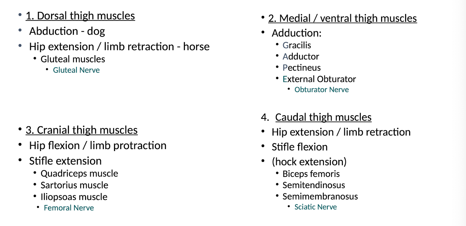
Describe the following for the gluteal muscles.
Origin
Insertion
Function
Nerve Supply
Gluteal muscles:
• 3 parts
• O - sacrum & pelvis
• I - greater trochanter of femur
acts as lever
Function:
• Hindlimb abductors - dog
• Hindlimb retractor / hip extensors - horse
Since mostly flexion and extension in these species, angle at which muscles inserts restricts movement to flexion and extension
Nerve Supply;
• Gluteal nerve
Describe the following for the medial/ventral thigh muscles. (The GAPE muscles)
Origin
Insertion
Function
Nerve Supply
Medial /ventral thigh muscles:
• O - Ventral surface pelvis
• I - medial aspect limb
• 'GAPE' muscles =
• Gracilis (+)
• I = tibia
• I = calcaneus (tarsus)
(via common calcanean tendon)
• Adductor (*)
• Pectineus (#)
• Both | = femur
• (External obturator)
• Function - Adductors
• Nerve Supply - Obturator Nerve
Describe the following for the Quadriceps muscle. (Cranial Muscle)
Origin
Insertion
Function
Nerve Supply
• Quadriceps muscle
Rectus femoris: O-ilium
Vastus lateralis: O - lateral femur
Vastus medialis: O - medial femur
Vastus intermedius: O - cranial femur
• I - tibial tuberosity
(via patellar ligament)
• Function:
• Stifle extensor
• (Hip flexor / limb protractor)
• Nerve Supply:
• Femoral Nerve
Describe the following for sartorius. (Cranial Thigh muscle)
Origin
Insertion
Function
Nerve Supply
• Sartorius (blue)
• O - ilium
• | - femur
• I - tibial tuberosity
(via patellar ligament) fusion
• Function:
• Stifle extensor
• Hip flexor / limb protractor
Nerve supply: Femoral nerve
Describe the following for iliopsoas.
Origin
Insertion
Function
Nerve Supply
• Iliopsoas muscle
• O - lumbar vertebrae & ilium
• I - medial proximal femur
• Function:
• Hip flexor / limb protractor
Nerve supply: Femoral nerve
Describe the following for the biceps femoris.
Origin
Insertion
Function
Nerve Supply
• O - tuber ishium
• | - fascia latae
• I - calcaneus (tarsus)
(via common calcanean tendon)
• Functions:
• Hip extensor / hindlimb retractor
• Stifle flexor
• Hock extensor
• Nerve supply
• Sciatic nerve
Describe the following for the semitendinosus and semimembranosus.
Origin
Insertion
Function
Nerve Supply
• Semitendinosus (blue)
• O - Tuber Ishium
• I - Tibia
• I - calcaneus (tarsus)
(via common calcanean tendon)
• Semimembranosus (pink)
O - Tuber ischium
I - femur & tibia
• Function:
Hip extensor / hindlimb retractor
Stifle flexor
(Hock extensor)
Nerve supply:
Sciatic Nerve
Summarize the muscles (and their functions), of those located in the hindlimb.
What are some notable comparative differences considering the thigh muscles in the ruminant and horse?
• Ruminants - angular
• Horse - rounded rump / croup
• Gluteal muscles:
Very well developed
• Caudal thigh muscles:
Biceps femoris
Semitendinosus
Semimembranosus
O- tuber ischium (same as other species)
Extra O- lumbosacral fascia
• Very powerful limb retraction / hip extension
• Allows horse to gallop, rear, buck / kick and jump better than cows!