Bioenvironmental Microbiology
1/105
There's no tags or description
Looks like no tags are added yet.
Name | Mastery | Learn | Test | Matching | Spaced |
|---|
No study sessions yet.
106 Terms
Bacteria (Prokaryotes) and Archaea Ribosomes
70S ribosomes = 30S + 50S S = Sveberg = s edimentation coefficient
30S= 16S + 21 proteins
50S = 23S + 5S + 32 proteins
5S (~120 nucleotides)
16S (~1500 nucleotides)
23S (~3000nucleotides)
16S and 23S have many conserved sequences.
Eukarya ( Eukaryotes) Ribosomes
80S ribosomes = 40S + 60S
40S = 18S (2000 nucleotides) + 30 proteins
60S =1 EA; 5S (120 nucleotides), 5.8S, 28S (5000 nucleotides) + 50 proteins
Why is 16S used in taxonomy
A "universal" 16S rRNA primers can be used to identify different species of bacteria because
bacteria have segments of the 16S rRNA sequence that are identical.
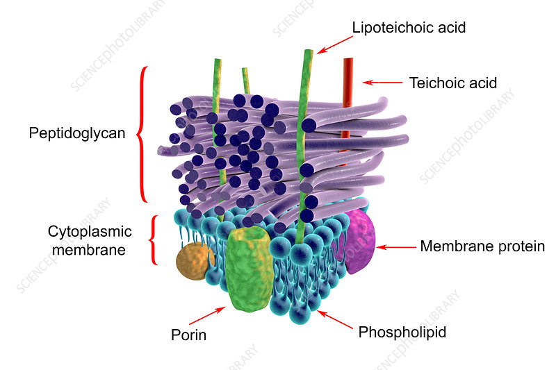
Gram Positive envelope
Thick ell wall with multiple peptidoglycan layers. No outer membrane, periplasmic space, or lipopolysaccarides, They possess long chain teichoic acids intertwined among peptodoglycan

Teichoic Acid
Teichoic acids are only found in gram positive bacteria. THey are polymers in which glycerol or ribitol residues alternate with phosphate groups. Some teichoic acids are attached to peptidoglycan and extend throughout the cell wall other are covalently attached to lipids in the cytoplasmic membrane call lipoteichoic acid
Peptidoglycan
Long chains polysaccharide consisting of alternate residues of N-acetyl-glucoamine NAG and N-acetyl-muramic acid NAM are cross-linked by short chains of amino acids
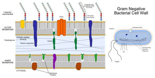
Gram Negative Envelope
Single thin peptidoglycan layer but no teichoic acids. Possess an outer membrane containing lipopolysaccharide and periplasmic space between the outer and inner membrane

Outer Membrane
Found in gram-negative only. Provides outer diffusion barrier to molecules greater than 700 to 1500 molecular weight and protects cell against antibiotics, toxic metals and other noxious chemicals
Lipopolysaccharide
Typically consist of hydrophobic domain known as lipid A an endotoxin, a non-repeating core oligosaccaride, and a distal polysaccharide. Lipid A is the hydrophobic anchor of lipopolysaccharide is glucosamine based phospholipid that maked up the outer monolayer of the outer membrane
Enbotoxin
Activates mediators of inflammation are pyrogens, cause fever, and diarrhea, In the bloodstream it cause hypertension, reduces oxygen delivery, and respiratory failure
Periplasmic Space
It’s between the ouer membrane and the inner membrane of gram negative cells. This space contains variuos degradative enzymes and binding proteins involved in the uptake of amino acids or sugar ect. When gram-negative cells
contain plasmids specifying antibiotic resistance the enzymes which break down antibiotics, e.g.,
beta-lactamase, are sometimes located in the periplasmic space.
Inner (cytoplasmic) Membrane
Comprised of one third lipid and two thirds protein. In addition, the surface of the lipid layer is covered by 2 to 3 layers of "extrinsic" protein molecules. The lipid bilayer behaves as a 2-dimensional liquid allowing membrane proteins to drift laterally. The IM controls entry into the cytoplasm and acts as an electrical insulator for the electron transport chain.
Capsule
Layer found loosely attached to the outside of many bacteria. Not essential and usually consist of simple or complex carbohydrates rarely protein. Capsules are protective and keep both bacteriophages and macrophages at a distance and desiccation
Pilus
Sex pili and common pili are both composed of protein monomers arranged helically. Sex pili are involved in binding of the donor and recipient cells together for subsequent transfer of DNA. Only one or two sex pili per cell are made. Common pili (also called fimbriae; singular = fimbria) are made by both sexes and appear in higher numbers per cell. They are involved in adhesion to suitable surfaces or in floating- bacteria lacking common pili clump together and sediment.
Nanowire
Electrically conductive appendage. Geobacter nanowires are composed of stacked cytochromes. Nanowires can transfer electrons to extracellular electron acceptors. Can reduce U (VI) to U (IV).
Flagellum
Prokaryotic flagella are quite different from those of eukaryotes bacteral flagella consist of a single filament made of helical protein subunits. THe basal structure in th inner membrane act as a motor and the whole shaft of the flagella rotates it is powered by the proton motive force in bacteria in eukarya it uses ATP
Lipids
The chemical can be use the differintiat Archaea and Eubacteria. Bacteria and eukaryotes synthesize membrane lipids with a backbone of fatty acids linked by an ester bond to a molecule of glycerol. The linkage to glycerol is ester based in Bacteria id Eukarya the makes a linear chain. Eukarya have cholestrol fungal membranes contain ergosterol
Drugs that target ergosterol
Drugs like the
azoles (Fluconazole or Itraconazole) and polyenes classes (i.e. amphotericin B) disrupt
ergosterol synthesis or function. Echinocandins prevent the synthesis of (1,3)‐β‐D‐glucan, a
key component of the fungal cell wall.
Archaeal Lipids
Consist of ether linked molecules and are long-chain branched hydrocarbon either of phytanyl or biophytanyl type are present and are bond by ether linkage to glycerol molecules
Viroids
Small circular single stranded RNAs which are not complexed with any protein and contain no protein-encoding genes.Viriod’s replication mechanism uses RNA polymerase II and host cell enzymes
Viruses
are separated into groups based on the type and form of nucleic acid genome and the
size, shape, structure, and mode of replication of the virus particle. There are viruses for
eukaryotes and prokaryotes. Viruses for microorganisms play an important role in the life cycle
of microorganisms.
Bacteriophages
The most numerous organism on earth and through their impact on bacteria they play a crucial role in shaping the biogeochemical cycle.
phage biologists expect that phage population densities will exceed bacterial densities by a ratio of 10-to-1 or more.
Archaeal viruses
Biruses that infect archaea are less studied virus group. All known archaeal viruses have DNA genomes. The cellular receptor to which zrchaeal viruses bind is not identified \
Many archaeal viruses can bind to extracellular structure such as pili
Mycoviruses
ubiquitous throughout all major taxonomic
groups of fungi, and persistently infect their hosts, usually without any discernable phenotypic
changes. The primary focus of mycoviral research has been on mycoviruses that infect plant
pathogenic fungi, due to the ability of some to reduce the virulence of their host and thus act as
potential biocontrol against these fungi.
Cryphonectria parasitica
Asia resulted in the destruction of billions of
mature American and European chestnut trees during the first half of the last century. Myco virus
infection makes the pathogen hypo virulent
Autotroph
Organisms whose growth and reproduction are independent of external sources of organic compounds.
"Self-feeding" ; obtain needed carbon by the reduction of CO2 and needed
energy from conversion of light to ATP or the oxidation of inorganic compounds to provide the
free energy for the formation of ATP.
Heterotroph
organisms requiring organic compounds for growth and reproduction the organic compounds serve as a source of carbon and energy
Chemotroph: Lithotroph
Energy from the oxidation of inorganic matter. ATP synthesis is coupled to the oxidation of the electron donor
Chemolithtroph source of energy by chemical oxidation and the use of inorganic
compounds as electron donors; carbon obtained by reduction of carbon dioxide.
aka- Chemoautotroph
Chemoorganothroph
energy from the oxidation of organic matter
organisms that obtain energy from the oxidation of organic compound and cellular carbon from preformed organic compouns
Photoautotroph
organisms whose source of energy is light and whose source of carbon is CO2 e.g. plants, algae, and some prokaryotes
Photoheterotroph
organism that obtains energy from light but requires organic compounds for growth
Archaea
Separated based on analysis of their 16S ribosomal RNA. They appear to be be primitive bacteria.
Found in extreme habitata many have not been cultivited or are very hard to
Four Archaeal Superphyla
A. TACK- Thaumarchaeota, Aigarchaeota, Crenarchaeote and Korarchaeota
B. Euryarchaeota
C. DPANN - Diapherotrites, Parvarchaeota, Aenigmarchaeota, Nanohaloarchaeota and
Nanoarchaeota
D. Asgard-only one isolate cultured to date. Asgard genomes encode many more of the
diverse eukaryotic signature proteins than do other archaea (Liu et al. 2021)
TACK- Thaumarchaeota
Thaumarchaeota are chemolithotroph found in marine, freshwater, and terrestrial environments
mesoplilic temperatures are
from 22–72 °C and pH 4.0–7.5 aerobically.
Most not cultured
TACK- Aigarchaeota
Widely distributed in terrestrial and subsurface geothermal systems and marine hydeothermal environments.
Physiology and ecology poorly understood
May be involved in dissimilatory sulfite reduction and possibly carbon monoxide oxidation
TACK- Crenarchaeota recently renamed Thermoproteata
Extreme thermophiles sulfur dependent anaerobic organotrophs or lithotrophs that are extreme thermophiles. All have been isolated from geothermally heated soils or
waters containing sulfur and most of the species metabolize sulfur. This ground is anaerobic with two exceptions Sulfolobus acidocaldarius and Acidianus infernus
TACK- Korarchaeota
Hyperthermophilic archaea most not culture first identified in hot springs in yellowstone
Euryarchaeota
Includes organisms that produce methane as a metabolic by-product and the extreme halophiles, organisms that survive extreme concentrations of salt and some extremely thermophilic organisms Some can colonize humans
Methanogens
Obligate anaerobes use CO2 as terminal electron acceptor in anaerobic respiration. They are autotrophic when CO2 serves as the source of carbon and the terminal electron acceptor
three classes of methanogens based on the
substrate they are able to utilize for release of free energy for the production of ATP.
1) CO2- type
CO2 + 4H2 CH4 + 2H20 4CO +
2H20 CH4 + 3CO2
2) Methyl group
4CH3OH 3CH4 + CO2 + 2H20
3) Acetoclastic- Acetate substrate
CH3COOH CH4 + CO2
Extreme Halophiles
Obilgate aerobes Halophiles require at least 1.5M most 3-4M NaCl. All halophilic Archaea stain Gram negatively, reproduce by binary fission, and do not form resting stages or spores. They contain extremely large plasmids (25-35% of total cellular DNA). The sodium ion cannot be replaced by the K ion. The
Na ion is required to maintain cell wall integrity.
Bacteriorhodopsin acts as a
proton pump, i.e., it captures light energy and uses it to move protons across the membrane
out of the cell. The resulting proton gradient is subsequently converted into chemical
energy (ATP). Phototroph
DPANN
Known as nanoarchaea or ultra-small archaea due to their
smaller size (nanometric) compared to other archaea. Members all have small genome (many are
below 1 Mbp and with an estimated maxima of ∼1.5 Mbp ) they have limited but sufficient
catabolic capacities to lead a free life, although many are thought to be episymbionts that depend on
a symbiotic or parasitic association with other organisms. Lack biosynthetic pathways for amino
acids, nucleotides, vitamins, and cofactors, which aligns with a host-dependent lifestyle.
DPANN- Diapherotrites
Found by phylogentic analysis of genomes recovered from the groundwater filtration of a gold mine
Asgard
First identified in deep sea hydrothermal vent in Artic ocean Archaea in this superphylum are prokaryotes that contain many proteins and gentic sequences that are otherwise found only in eukaryotes
Archaea Cell Walls
Archaea contain no peptidoglycan some have pseudopeptidoglycan which consist of polymer chains of glycan cross linked by short peptide connections. N-acetylmuramic acid is replaced by N- acetyltalosaminuronic acid and two sugars are bonded with a beta 1-3 glycosidic linkage instead of a beta 1-4 also the peptides are L-amino acids
A second cell wall types is composed out of polysaccharides
A third type consist of glyoprotein and occurs in hyperthermophiles and some methanogens
The fourth type is composed only of surface layer proteins
Bacteria
There are 30 major phylogenetic lineages (phyla), and some are not culturable. Over 90% of
the culturable bacteria belong to four phyla Actinobacteria, Firmicutes, Proteobacteria and
Bacteroidetes.
Aquifex/Hydrogenobacter group
Hyperthermophilic chemolithotrophs that oxidize H2 or reduce sulfur compounds. Aquifex grows up to 95 C and is the closet know relative to the universal ancestor of all bacteria
Thermotoga
Found in geothermal marine sediments it is a hyperthermoplhile and a chemoorganotroph. It produces unique lipids containing long chain fatty acids and its cell wall contains peptidoglycan. Cell is wrapped in a unique sheath like outer membrane call a toga. They are strict anaerobes
Phototrophic Bacteria- Green non-Sulfur Bacteria
dilamentous thermophiles that can grow organotrophically in the dark under aerobic conditions or photoautotrophically using H2S or H2 as electron donors and contain chlorosomes. Anoxygenic metabolism utilizes organic
carbon. Example: Chloroflexus (Phototroph) The green non-sulfur bacteria only tolerates low
concentrations of H2S.
Chlorosomes
Cigar shaped structures that are bonded by a membrane that is attached to but not continuous with the cytoplasmic membrane and contain bacteriochlorophyll C or D
Phototrophic Bacteria- Green Sulfur Bacteria
Phototrophic green sulfur bacterium strict anaerobes and obligate photorophic that are non-motile and contain chlorosomes. Anoxygenic photosynthetic metabolism. CO2 carbon source: most require reduced sulfur as electron donors as electron donors. Green bacteria contain bacteriochlorophyll c, d, or e. live in sulfur outflow and hot spring or ther benthic habitats and do not have gas vesicles
Green bacteria Photosystems
They have Type-I like phtosystems similar to PSI. cyanobacteria. PSI and Type I reaction centers are able to reduce ferredoxin, a strong reductant that can be used to fix CO2 and reduce NADP.
Difference between Green and Purple bacteria
Major differences between the purple bacteria and the green are the nature of their
photosynthetic membrane systems and the bacteriochlorophylls they contain. In green
bacteria the chlorosomes are attached but not continuous with the plasma membrane. Purple sulfur bacteria do not use chlorosomes for photosynthesis. Instead they use
intracellular membrane systems that contains pigments.
Purple Sulfur bacteria
Anoxygenic bacteria that can use elemental sulfur or sulfide as an electron donor in respiration. During autotropic growth H2S is oxidized to S^0 and deposited as granules that are stored inside the cell.
Purple bacteria photosynthesis
Purple bacteria photosynthetic pigment is part of the internal membrane system, connected to and produced from the plasma membrane and continuous with the plasma membrane-lamella (flat sheets) or vesicles each one binding molecules of bacteriochlorophyll and carotenoids non-covalently. Purple bacteria are anoxygenic phototrophs. They contain bacteriochlorophyll a or b The carotenoid gives them colors and absorbs light energy Purple bacteria use a Q-type photosystem, also known
as a Type II reaction center. The light-driven charge separation leads to the transfer of an electron
to a quinone molecule that transfer to the electron transport chain.
Purple Non-Sulfur Bacteria
Use organic electron donors instead or elemental hydrogen. Purple photosynthetic bacteria have given rise to non phototrophic relatives. They are oxygenic and play an important role evolution of life by producing an oxygenic atmosphere through their photosynthetic activity. Some cyanobacteria can produce cyanotoxins that can cause serious
illness or death in pets, livestock and wildlife. Some form herterocysts which are sites of nitrogen fixation.
Purple non-sulfur bacteria photosynthesis
Contain only chlorophyll a (430 nm-Blue and 680 nm-Red) and different phycobilins.
Phycocyanin- blue in color absorbs at ~620 nm (red-orange) and phycoerthrin- red in color
absorbs at ~550 nm (yellow-green). Many cyanobacteria contain gas vesicles. Cyanobacteria
have PS I and PSII.
Herterocysts
Some Cyanobacteria form herterocysts which are sites of nitrogen fixation
Deinococcus
Deinococcus unusual G+ cocci. Has an outer membrane usually
found only in G (-) and an atypical cell wall that contains ornithine instead of diaminopimelic
acid (DAP). Highly resistant to various environmental stresses, including nuclear radiation, extreme
temperatures, vacuum, oxidation, and desiccation. Deinococcu has an efficient mechanisms for
repairing damaged DNA. Most are red or pink due to carotenoids. Isolated from a can of meat
that had been irradiated at a dose 250-times higher than that used to kill E. coli .
Source of Taq polymerase used in PCR reaction.
Thermus aquaticus is a gram-negative organotrophic thermophile. T. aquaticus shows best growth
at 65 to 70 °C (149 °F to 158 °F) but can survive at temperatures of 50 °C to 80 °C (122 °F to
176 °F). It primarily scavenges for protein from its environment as is evidenced by the large
number of extracellular and intracellular proteases and peptidases as well as transport proteins
for amino acids and oligopeptides across its cell membrane.
Firmicutes
Gram Positive bacteria consist of two main classes Low GC group and High GC group
Actinomycetes- Streptomyces
Aerobic, multicellular, fliamentous bacteria that produce well developed vegetative hyphae and most produce spores.Soil bacteria that can degrade chitin and plant material. Produces earthy odor Streptomycetes produce over two-thirds of the clinically
useful antibiotics of natural origin
Actinomycetes- Actinomycese
– facultative anaerobes found in the soil and microbiota of animal. Filamentous
bacteria and produce spores. Degradation of chitin and plant material. Opportunistic pathogens
Nocardia
Filamentous soil saprophytes but also include pathoenic agents thay cause nocardiosis in animals in the lung, central nervous system, brain, and skin. They are opportunistic pathogen
Heliobacterium
Heliobacterium (contains Bch g (670, 788 nm) and forms spores)
Bacteriochlorophyll g is inactivated by the presence of oxygen, making them obligate
anaerobes. Heliobacteria (e.g., Heliobacterium mobilis and Heliobacterium modesticaldum)
common in the waterlogged soils of paddy fields and are the only known gram-positive
photosynthetic organisms. Strong nitrogen fixers; which is important in fertility of paddy fields
fertility.
Proteobacteria
Largest and most physiologically diverse group of all bacteria. Has anoxygenic metabolism. All proteobacteria are Gram-negative bacteria so they have an outer membrane around their cell wall. There are five major groups: alpha, beta, gamma, delta, epsilon based on phylogenetic
studies.
Proteobacteria- Alpha
The unifying characterstic of this class is that they are oligotrophs, organisms capable of living in low nutrient environment
Cell division in stalked or budding bacteria
Cell division in stalked or budding bacteria involves the
formation of a new daughter cell with the mother retaining the identity. The daughter
cell is immature and must form internal or budding structures before it can divide.
The majority of the budding and/or appendaged bacteria phylogenetically group in the alpha
purple bacteria.
Proteobacteria- Beta
Eutrophs needing substantial amounts of organic nutrients to grow. Some members are chemolithotrophs
Pathogenic species include e.g., Neiserria spp. (causing gonorrhea and
meningitis) and Bordetella pertussis (causing whooping cough). Burkholderia is a cross
domain causing plant disease and human infections. Ralstonia -plant pathogen.
Proteobacteria- Gamma
Enteric bacteria most diverse class of gram negative bacteria and exhibit a wide array of metabolic pathway
Proteobacteria- Delta
Bdellovibrio, Myxococcus and include sulfate-reducing bacteria (SRBs), (i.e.,
Desulfovibrio, Desulfobacter, Desulfococcus), so named because they use sulfate as the
final electron acceptor in the electron transport chain.
Also include ferric iron reducing Geobacter spp., the first organism identified with the
ability to oxidize organic compounds and metals, including iron, radioactive metals, and
petroleum compounds into carbon dioxide, while using iron oxide or other available metals
as electron acceptors. Can reduce the soluble, oxidized form of uranium U(vi) to the
insoluble form U(iv). The reduced form of U precipitates from contaminated groundwater
and prevents its further mobility. Uses nanowire electrically conductive appendages to
reduce metals.
Spirochetes
Wide spread in aquatic and humans. Flexuous helical rods. Resistant to rifampicin. Resistant to rifampicin
(binding in the polymerase subunit deep within the DNA/RNA channel, facilitating direct blocking
of the elongating RNA) therefore RNA polymerase is different from most bacteria.
Five genera based on habitat, pathogenicity, and morphological and physiological characteristics.
Critispira- digestive tract of mollusks, no known disease
Spirochaeta- aquatic free living; anaerobe or facultative aerobe
Leptospira- aerobic, free-living or parasite of humans or animals; leptospirosis
Borrelia - anaerobic, humans, animals or arthropods. B. burgdorferi is the causal
agent of lyme disease.
Treponema- anaerobic, parasitic in human and other animals, syphilis (T. pallidum)
Bacteridoales- Bacteroides
Obligate anaerobes and fermentative major part of human intestine flora. They are important for processing complex molecules into simple ones in the intestine
Bacteridoales- Flavobacteria
Non- spore forming strictly aerobic and yellow pigmented that are known to degrade complex organic compounds in freshwater. Marine, and soil. Move by gliding motility is the
ability of certain rod-shaped bacteria to translocate on surfaces without the aid of external
appendages such as flagella, cilia, or pili.
Bacteridoales- Cytophaga
Long slender rod able to digest cellulose and agar found in soil, marine, and freshwater sediment
Planctomyces
Bacteria found in brackish, marine, and freash water. Reproduce by budding organisms and lack peptidoglycan cell wall but contain a protein cell wall. Also called budding
and /or appendages bacteria. They are resistant to antibiotics that affect peptidoglycan synthesis
(eg. penicillin, cephalosporin, and cycloserine). .
Planctomycetes are the only free-living bacteria known to lack peptidoglycan in their cell
walls. In many cases their DNA is surrounded by a membrane similar to a eukaryotic nuclear
membrane.
Chlamydia
THis group has no ATP generating system and is obligatory intracellular parasite that is involved in diseases of both humans and animals. This group lives in a very restricted environment
EukaryoticMicroorganisms.
The most primitive evolutionary lineages in the Eukaryotic Domain are unicellular, anaerobic,
mesophilic organisms. Analyses of cell components such as cytochromes, ferrodoxins, and
rRNA indicate that mitochondria originated from proteobacteria (purple bacteria) and that
chloroplast originated from the cyanobacteria.
Superphyla Excavates- Diplomonads
Lack endoplasmic reticulum, mitochondria, and Golgi bodies and are generally endobionts in the gut of animals. They usually have two pairs of flagella associated with nulceus. Replicate asexually by binary fission
Giardia has two nuclei and eight flagella.
Superphyla Excavates- Parabasilia
Have parabasal structure associated with the flagellar base. ALck mitochindria often contain hydrogenosomes (organelle that contains enzymes that oxidize
pyruvate to hydrogen and CO2 under anaerobic conditions) and have well developed Golgi
bodies. Anaerobic flagellates that are mainly obligate symbionts or parasites of insects and
vertebrates
Superphyla Excavates- Kinetoplastids
Group of flagellated protozoans that are distinguished by the presence of DNA containing region call a kinetoplast in their single large mitochondrion
Superphyla Excavates- Euglenids
Large flagellates with two flagella although in many taxa inyl one flagellum free-living osmotroph or phagotroph capable of ingesting while eukaryotic cell. Euglena, acquired their chloroplasts, through secondary
endosymbiosis of a green alga. Non-pathogenic. Are phototrophic and chemotrophic.
SuperPhyla Aveolata
Have a pellicle, which is a flexible proteinaceous covering that provides form and flexibility
Characteristic is the presence of cortical (near the surface) alveoli (sacs). The
flattened vesicles (sacs) arranged as a layer just under the membrane and supporting it,
typically contributing to a flexible pellicle.
SuperPhyla Aveolata- Ciliates
Characterized by the presence of hair-like organelles called cilia. Commonly found almost anywhere in water. Most are heterotrophs and feedon smaller organisms like bacteria and algae
Eukaryotic nature (80S ribosomes and organelles),
SuperPhyla Aveolata- Dinoflagellates
Mostly marine phytoplanktonbut. Many are phtotsynthetic but a large fraction
of these are in fact mixotrophic, combining photosynthesis with ingestion of prey.
The chloroplasts in most photosynthetic dinoflagellates are bound by three membranes,
suggesting they were probably derived from some ingested algae. Most photosynthetic species
contain chlorophylls a and c2. Can cause red tides
Some have luciferin which when it is oxidized in produces blue colored light
Superphyla Stramenopiles
A clade of organisms distinguished by the presence of stiff tripartite external hairs. In most
species, the hairs are attached to flagella, in some they are attached to other areas of the cellular
surface, and in some they have been secondarily lost.
Many stramenopiles have plastids which enable them to photosynthesize. The plastids are colored
off-green, orange, golden or brown because of the presence of chlorophyll a, chlorophyll c,
and fucoxanthin. Most are single-celled, but some are multicellular including some large
seaweeds, the brown algae.
Diatoms
A nuclear envelope-bound cell nucleus that separates them from the prokaryotes archaea and bacteria.
Diatoms are a type of plankton alled phytoplankton. Diatoms are surrounded by a cell wall made of silica Major pigments of diatoms are chlorophylls a and c, beta-
carotene, fucoxanthin, diatoxanthin and diadinoxanthin
Diatoms, shells = diatomaceous earth- important industrial resources used for fine
polishing and liquid filtration.
Superphyla Stramenopiles- Oomycetes
Includes devasting plant pathogens. CAn cause late blight of potato, downy mildew, sudden oak death. THey produce zoospores in sporangia. THe cell wall is made of beta glucans and hydroxyproline. Can reproduce asexually by forming a structure called a sporangium or zoosporangium. Inside these sporangia, zoospores are produced, first the primary
zoospore and then the secondary zoospore, which is laterally flagellated. Their flagellum
allow the zoospores to move rapidly through water.
Golden Brown Algae or Golden algae
Known as chrysophytes found in both marine and fresh water the primary cell of chrysophytes cotains two specialized flagella. Chloroplast contain fucoxanthin which is brown. The contain chlorophyll c (447-452 nm
region). Like chlorophyll a and chlorophyll b, it helps the organism gather light and
passes a quanta of excitation energy through the light harvesting antennae to
the photosynthetic reaction center.
Rhizaria
Mostly heterotrophic unicellular eukaryotes including both amoeboid and flagellate forms. This supergroup includes many of
the amoebas, most of which have threadlike or needle-like pseudopodia.
Pseudopodia function to trap and engulf food particles and to direct movement in
rhizarian protists. These pseudopods project outward from anywhere on the cell
surface and can anchor to a substrate. The protist then transports its cytoplasm into
the pseudopod, thereby moving the entire cell. This type of motion, called
cytoplasmic streaming, is used by several diverse groups of protists as a means of
locomotion or as a method to distribute nutrients and oxygen.
Rhizaria- Chloroarachniophyte
Marine amoeba like phototrophs. Typically mixotrophic ingesting bacteria and small protist and conducting photosynthesis. Endosymbosised algal chloroplast.
Rhizaria- Foramoniferans
Marine microbes that form porous shells called test built from various organic materials and typically hardened with calcium carbonate. The tests may house photosynthetic algae, which the forams can harvest for nutrition.
Rhizaria- Radiolarians
Exhibit intricate exteriors of glass silica with radial or bilateral symmetry. Needle like pseudopobs supported by microtubules radiate outwrd from the cell bodies of these protist and function to catch food. are primarily heterotrophic, but many have
photosynthetic endosymbionts and are, therefore, considered mixotrophs.
Superphyla Amoebozoa
Amoebozoa are characterized by the presence of pseudopodia which are extensions that can be either tube-like or flat lobes and are used for mocomotion and feeding they primarily do phagocytosis. Species of Amoebozoa may be either shelled (testate) or naked, and cells may possess flagella.
Superphyla Amoebozoa- Slime molds
Non-phototrophic eukaryotes that have similarity to both fungi and protozoa. Divided into two groups: Cellular whoes vegetative forms are composed of single amoeba like cells and Acellular whose vegetative forms are a naked mass of protoplasm of indefinite size and shape called plasmodia. Slime molds live primarily on decaying plant material. Source of food is mainly other microorganisms, especially bacteria.
Superphyla Opisthokonta
A broad group of eukaryotes including both the animal and fungi One common characteristic of opisthokonts is that flagellate cells, such as most animal sperm and
chytrid spores, propel themselves with a single posterior flagellum. This gives the group its name. In
contrast, flagellate cells in other eukaryote groups propel themselves with one or more anterior
flagellae.
Superphyla Opisthokonta- Fungi
eukaryotic heterotrophic microorganisms that typically form reproductive spores; some
fungi are unicellular, but most form mycelia. Primary taxonomic groupings based on their sexual
structures (spores). All fungi are organotrophs and lack chlorophyll. Fungal cell walls are composed
of polysaccharides such as cellulose, chitin, mannans, glucans and galactans. Different species of
fungi contain a variety of these compound in varying degrees
Fungi- Ascomycota
Sac fungi, spetated hyphae, sexual spores are ascospore
Fungi- Basidiomycota
Club fungi, septated hyphae, sexual spore basidiospores
Fungi- Mucoromycota (Zygomycetes)
coenocytic hyphae, sexual spore is zygospores