BIOL20 - Ch 5 The Integumentary System
1/24
Earn XP
Description and Tags
Add together to study Unit 1 Lecture Exam
Name | Mastery | Learn | Test | Matching | Spaced | Call with Kai |
|---|
No analytics yet
Send a link to your students to track their progress
25 Terms
Organs include
skin
its accessory structures
hair
nails
glands
blood vessels
muscles
nerves
Functions of the skin
protection
thermoregulation: maintains temp
converts inactive vitamin D —> active
provides sensory info
maintains homeostasis
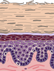
Structure: layers
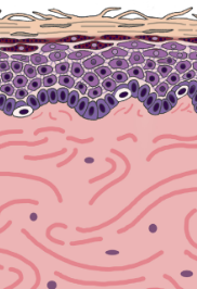
Epidermis - most superficial layer
Dermis - a layer deep to the epidermis
Hypodermis - subQ (subcutaneous layer), not a layer of skin, composed of areolar and adipose skin
Epidermis
4 major types of cells:
keratinocytes - waterproof skin
melanocytes - give pigment to absorb UV light
intraepidermal macrophages (aka langerhans cell) - eat big debris/germs
tactile epithelial cells (aka Merkel cell) - tell body when something is pushing through epidermis
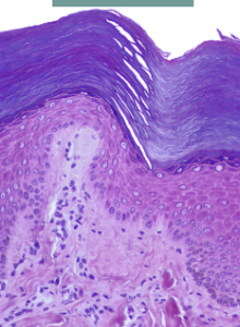
Layers of the skin - Thin skin
4 layers
Stratum corneum
Stratum granulosum
Stratum spinosum
Stratum basale
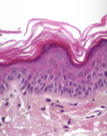
Layers of the epidermis - thick skin
Stratum corneum
Stratum lucidum
Stratum granulosum
Stratum spinosum
Stratum basale
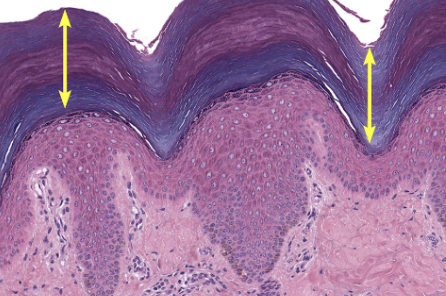
Stratum corneum
dead flat layer of keratinocytes
thick in thick skin, thin in thin skin
Stratum lucidum
ONLY in thick skin (eg. palms, feet)
doesn’t grow hair
layer lacks cell nuclei + organelles
dark line layer
Stratum granulosum
has lamellar granules
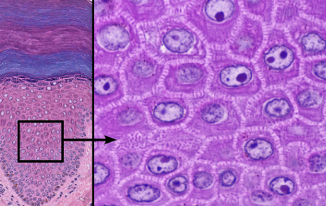
Stratum spinosum
3-10 layers of keratinocytes
cells have intermediate filaments w/ keratin
attached by desmosomes
largest epidermal layer
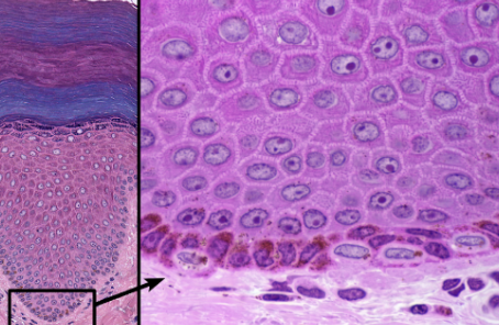
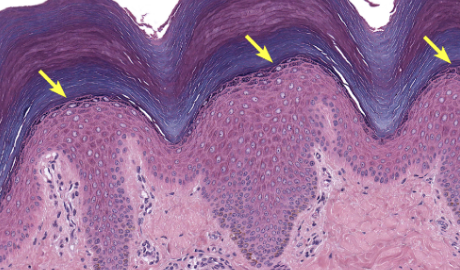
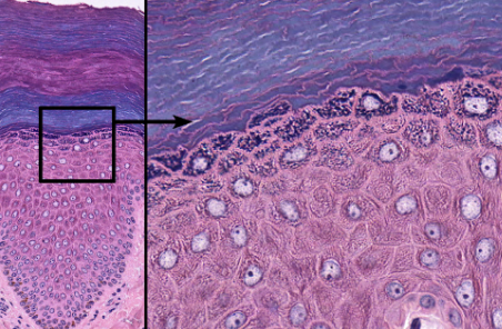
Stratum basale
deepest layer, rests on basal lamina
cell division primarily occurs here
formed daughter cells undergo keratinization + move up to superficial layer
Thick skin
covers the palms, palmar surfaces of digits and soles
hairless skin
Thin skin
covers all body regions excepts the palms, palmar surfaces of digits, and soles
hairy skin
Skin pigments
Melanin - produced by melanocytes in the stratum basale
Hemoglobin - red pigment in RBC
Carotene - yellow-orange pigment stored in corneum and adipose tissue

Dermis
composed of CT containing collagen + elastic fibers
contains blood vessels, lymph vessels, nerves, and other structures like hair follicles and sweat glands
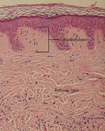
Layers of dermis
Papillary layer - areolar CT
reticular layer - dense irregular CT
Hypodermis
subQ layer
attaches skin —> underlying tissue + organs
consist of well-vascularized, loose, areolar CT, and adipose tissue
Hair + 3 types
composed of dead, keratinized epidermal cells
have sensory mechanisms
Lanugo - covers fetus
terminal - long, coarse, heavily pigmented hairs (normal hair)
vellus - short, fine, pale hairs (peach fuzz)
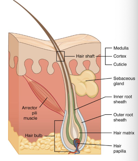
Hair structures
shaft - above skin surface
follicle - below level of skin
root - penetrates into dermis
epithelial root sheath
dermal root sheath
arrector pili muslce
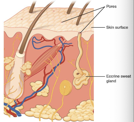
Skin glands - 4 types
Sebaceous (oil) gland
eccrine sweat glands
Apocrine sweat gland
cerumious glands
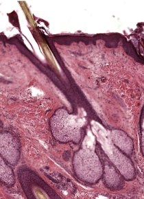
Sebaceous (oil) glands
connected to hair follicles
look like popcorn bubble
Eccrine v. Apocrine sweat glands
Eccrine - the most numerous, all over body
Apocrine - located mainly in hairy skin areas, stinky sweat
Ceruminous gland
modified sweat glands in ear canal
Nails
made of keratinized epidermal cells
extension of epidermis
protects furthest extensions of body (digits)
Integumentary system’s role in homeostasis
regulating body temperature through sweating and blood vessel dilation/constriction
protecting the body from environmental damage
preventing water loss
synthesizing vitamin D
providing sensory information to the nervous system to respond to external stimuli