Experiment 3: Bacterial Motility and Experiment 4: Bacterial Smear and Gram Staining
1/46
There's no tags or description
Looks like no tags are added yet.
Name | Mastery | Learn | Test | Matching | Spaced | Call with Kai |
|---|
No analytics yet
Send a link to your students to track their progress
47 Terms
motile
Organisms that possess the ability to propel themselves are said to be ___
Motility
an important characteristic used to identify organisms
flagella or axial filaments
Some bacteria possess ___ for movement
directly by using a flagella stain
indirectly by using wet mounts, hanging drop slides, or motility agar
Motility may be observed by:
hanging drop and wet mount techniques
allow for observation of living organisms
Wet mount
tend to dry out quickly under the heat of the microscope light;
it is simpler to perform than the hanging drop
it is useful for short-term observation only.
Hanging drop
is a more complex technique,
allows for longer-term observation and more reliable observation of motility
Brownian movement and true motility
On the wet mount or hanging drop slide, there are two types of movement:
Brownian movement
Movement caused by the molecules of the suspending liquid (water) colliding with the organism.
The movement is irregular or jerky or the cells appear to be vibrating.
This does not demonstrate true motility.
True motility
Movement in some consistent direction or path and they may be twisting and turning.
The cells exhibit independent movement over greater distances than in Brownian movement.
Flagellar motility
Spirochaetal motility
Gliding motility
Mechanisms of Bacterial Motility
Motion can be achieved by one of three mechanisms:
Flagellar Motility
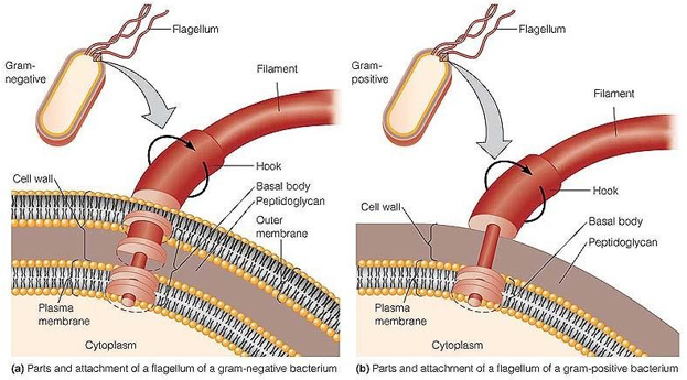
Flagella consist of a hollow, rigid cylinder composed of a protein called flagellin, which forms a filament anchored to the cell by a curved structure called the hook, which is attached to the basal body
Flagellum Structure
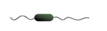
Polar/Monotrichous
Lophotrichous
Amphitrichous
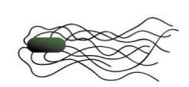
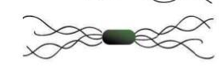
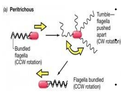
Peritrichous
Amphilophotrichous
What are the types of flagellar arrangement?
Polar/Monotrichous
single flagellum at one pole

Lophotrichous
tuft of flagella at one pole

Amphitrichous
flagella at both poles
Peritrichous
flagella all over
Amphilophotrichous
tuft of flagella at both ends
tumble
The direction of rotation determines the movement of the cell.
Periodically the direction of rotation is briefly reversed, causing what is known as a “___”, and results in reorientation of the cell.
When counterclockwise rotation is resumed, the cell moves off in a new direction.
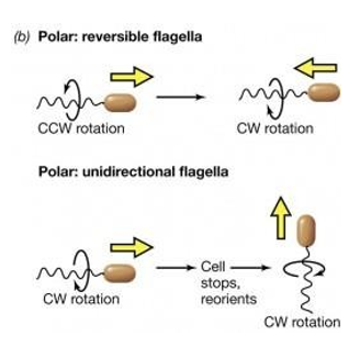
chemotaxis
Bacteria can sense nutrients and move towards them – a process is known as ___.
Additionally, they can also move away from harmful substances such as waste products and in response to temperature, light, gravity, etc.
This apparently intelligent behavior is achieved by changes in the frequency of tumbles.
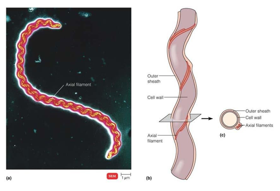
Spirochaetal Motility
The second type of motility is shown by Spirochaetes, helical bacteria which have a specialized internal structure known as the axial filament which is responsible for rotation of the cell in a spiral fashion and consequent locomotion.
Axial Filaments
Gliding Motility
is the movement of cells over surfaces without the aid of flagella, a trait common to many bacteria
copious slime
Gliding bacteria all secrete ___, but the exact mechanism which propels the cells is not known
true
[true or false] The gliding motility apparatus which propels the cells involves a complex of proteins, yet the full nature of this “motor” and how the components interact is not understood
cell wall structure
Most bacteria can be differentiated by the gram reactions due to difference in their ___
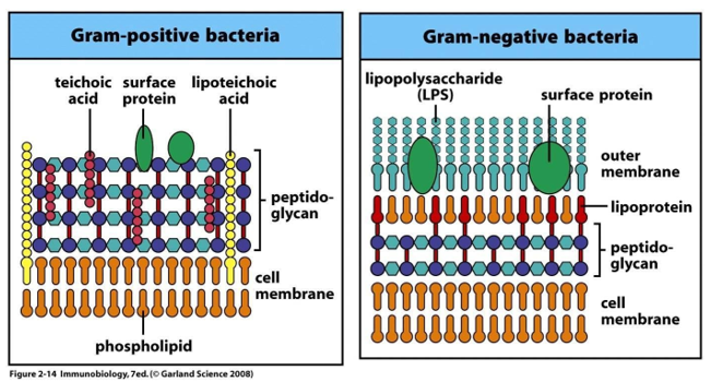
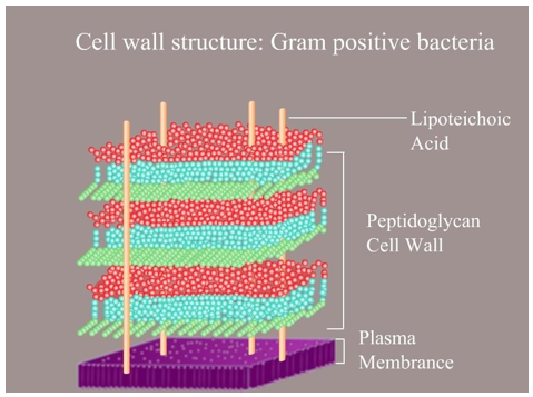
Gram positive bacteria
Cell wall has a large amount of peptidoglycan.
These are organisms that retain purple color with crystal violet and are not decolorized by acetone & iodine
Gram negative bacteria
They have less amount of peptidoglycan in their cell wall. They have lipopolysaccharide containing a compound known as lipid A or endotoxin.
The organisms that loose their color of crystal violet after being treated with acetone & iodine
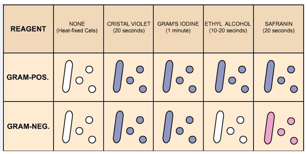
Application of the primary stain Crystal Violet (CV) to a heat-fixed smear of bacterial culture
Addition of Gram’s Iodine
Decolorization with 95% ethyl alcohol or acetone
Counterstain with Safranin
The four basic steps of the Gram Stain
Crystal Violet (CV)
___ dissociates in aqueous solutions into CV+ and Cl – ions. These two ions then penetrate through the cell wall and cell membrane of both Gram- positive and Gram-negative cells. The CV+ ions later interacts with negatively charged bacterial components and stains the bacterial cells purple.
Iodine
(I – or I3 –) acts as a mordant and as a trapping agent
mordant
is a substance that increases the affinity of the cell wall for a stain by binding to the primary stain, thus forming an insoluble complex which gets trapped in the cell wall. In the Gram stain reaction, the crystal violet and iodine form an insoluble complex (CV-I) which serves to turn the smear a dark purple color. At this stage, all cells will turn purple.
true
[true or false] Alcohol or acetone dissolves the lipid outer membrane of Gram negative bacteria, thus leaving the peptidoglycan layer exposed and increases the porosity of the cell wall. The CV-I complex is then washed away from the thin peptidoglycan layer, leaving Gram negative bacteria colorless.
On the other hand, alcohol has a dehydrating effect on the cell walls of Gram positive bacteria which causes the pores of the cell wall to shrink. The CV-I complex gets tightly bound into the multi-layered, highly cross-linked Gram positive cell wall thus staining the cells purple.
The decolorization step must be performed carefully, otherwise over-decolorization may occur. This step is critical and must be timed correctly otherwise the crystal violet stain will be removed from the Gram-positive cells. If the decolorizing agent is applied on the cell for too long time , the Gram-positive organisms to appear Gram-negative. Under-decolorization occurs when the alcohol is not left on long enough to wash out the CV-I complex from the Gram-negative cells, resulting in Gram-negative bacteria to appear Gram-positive.
Basic fuschin
The decolorized Gram negative cells can be rendered visible with a suitable counterstain, which is usually positively charged safranin, which stains them pink. Pink colour which adheres to the Gram positive bacteria is masked by the purple of the crystal violet (___ is sometimes used instead of safranin in rare situations

Gram Stain Mechanism
Gram-positive bacteria
have a thick mesh-like cell wall which is made up of peptidoglycan (50-90% of cell wall), which stains purple.
Peptidoglycan
mainly a polysaccharide composed of two subunits called N-acetyl glucosamine and N-acetyl muramic acid
transpeptidase enzyme
As adjacent layers of peptidoglycan are formed, they are cross linked by short chains of peptides by means of a ___, resulting in the shape and rigidity of the cell wall. The thick peptidoglycan layer of Gram-positive organisms allows these organisms to retain the crystal violet-iodine complex and stains the cells as purple.
Lipoteichoic acid (LTA)
another major constituent of the cell wall of Gram-positive bacteria which is embedded in the peptidoglycan layer. It consists of teichoic acids which are long chains of ribitol phosphate anchored to the lipid bilayer via a glyceride. It acts as regulator of autolytic wall enzymes (muramidases: Bacterial enzymes located in the cell wall that cause disintegration of the cell following injury or death.)
LTA also has antigenic properties that stimulate specific immune responses when it is released from the cell wall after cell death.
Cell death is mailnly due to lysis induced by lysozymal activities, cationic peptides from leucocytes, or beta- lactam antibiotics.
Medical Relevance of Gram Positive Cell Wall:
Gram Stain Mechanism
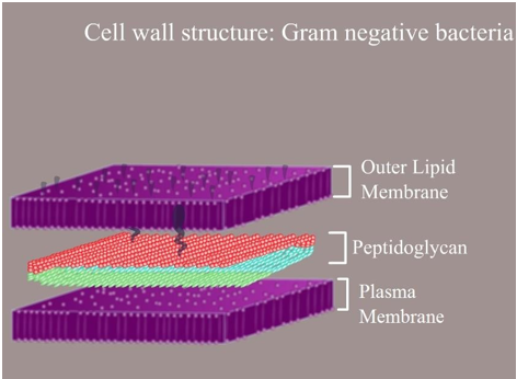
Gram-negative bacteria
have a thinner layer of peptidoglycan (10% of the cell wall) and lose the crystal violet-iodine complex during decolorization with the alcohol rinse, but retain the counter stain Safranin, thus appearing reddish or pink
Gram-negative bacteria
They also have an additional outer membrane which contains lipids, which is separated from the cell wall by means of periplasmic space
The cell wall of Gram-negative bacteria is often a virulence factor that enables pathogenic bacteria to cause disease.
The virulence of Gram-negative bacteria is often associated with certain components of the cell wall, in particular, the lipopolysaccharide ( otherwise known as LPS or endotoxin).
In humans, LPS elicits an innate immune response characterized by cytokine production and activation of immune system. Inflammation occurs as a result of cytokine production, which can also produce host toxicity.
Medical Relevance of Gram Negative Cell Wall:
Summary