Med Imaging- Lungs Pt. 2
1/51
There's no tags or description
Looks like no tags are added yet.
Name | Mastery | Learn | Test | Matching | Spaced |
|---|
No study sessions yet.
52 Terms
Walls of the bronchi thickened by inflammation or infection, may be diffuse or focal
Bronchiectasis
CXR in affected individuals is often normal or shows non-specific findings
Bronchiectasis
Bronchiectasis
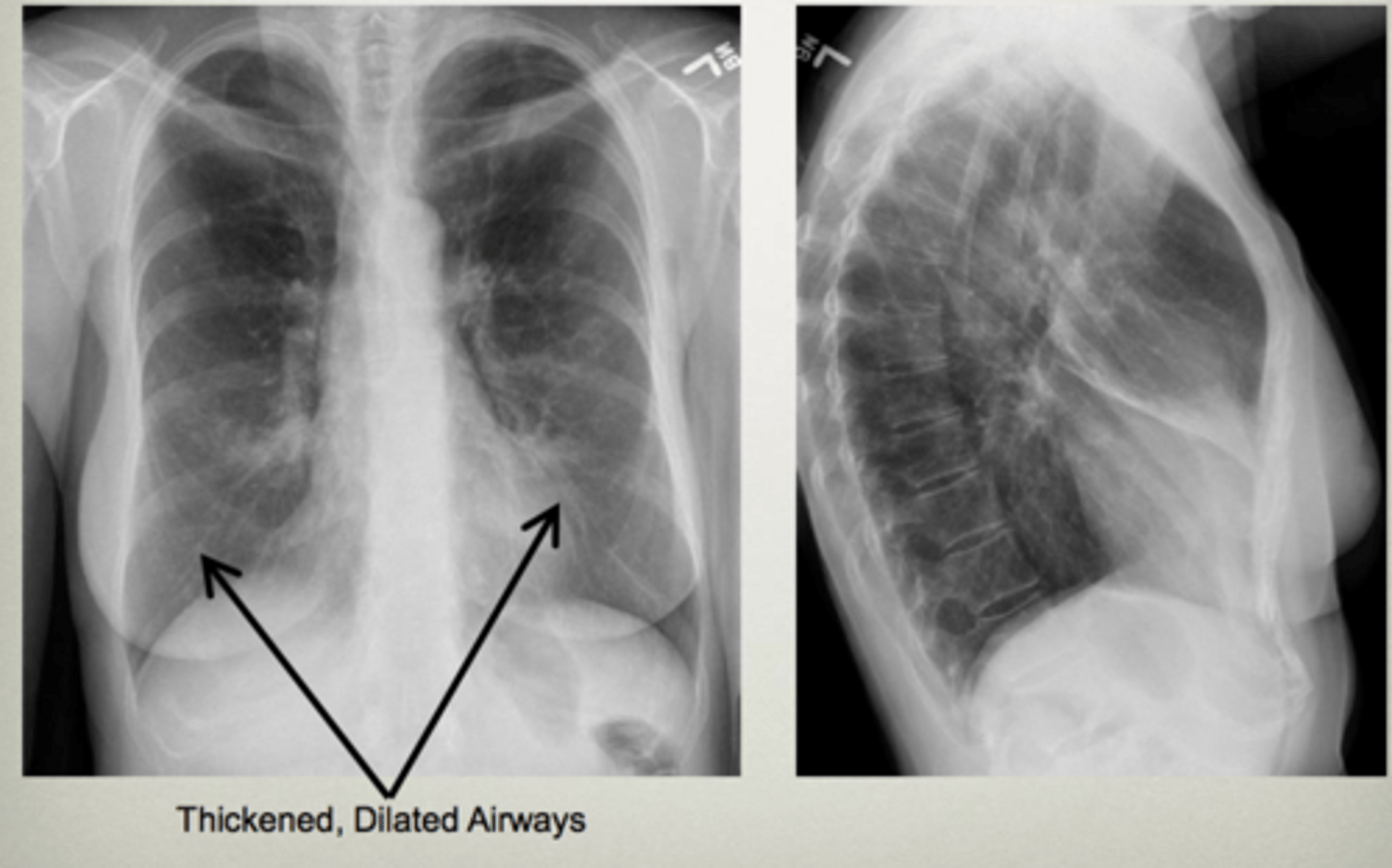
Tram track lines
Bronchiectasis
Condition that can cause bronchiectasis
Cystic fibrosis
Lung tissues with air space but no alveoli thats less than 1 cm in diameter
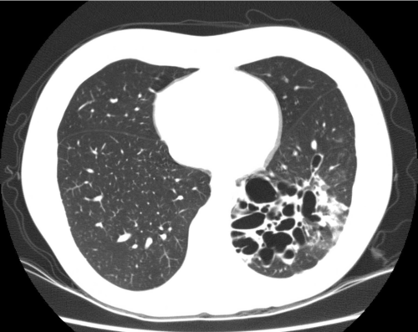
Bleb
Lung tissues with air space but no alveoli thats greater than 1 cm in diameter
Bullae
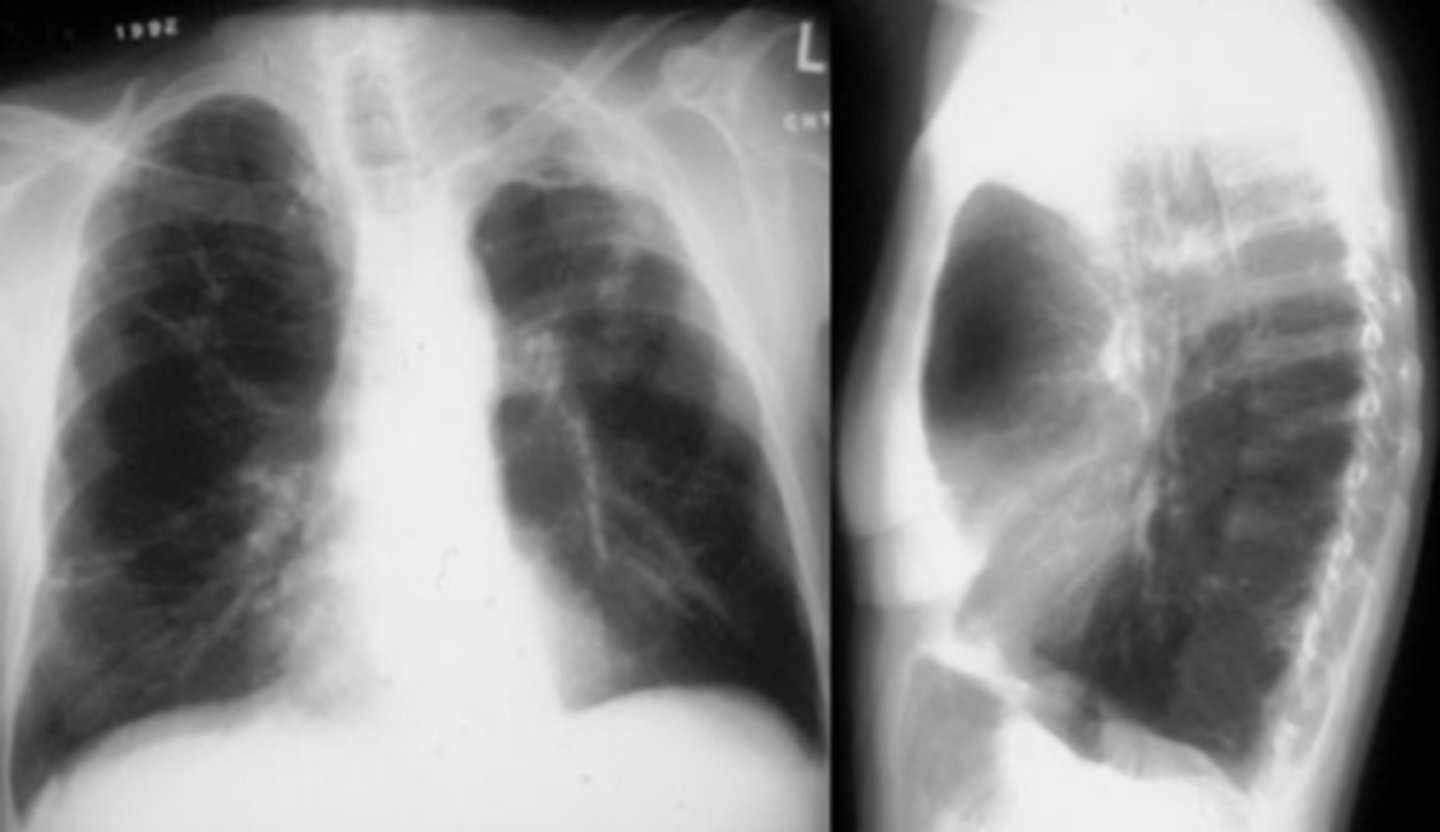
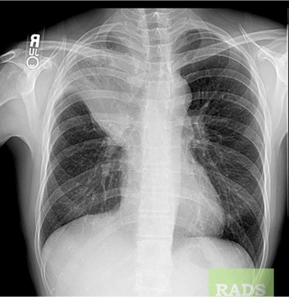
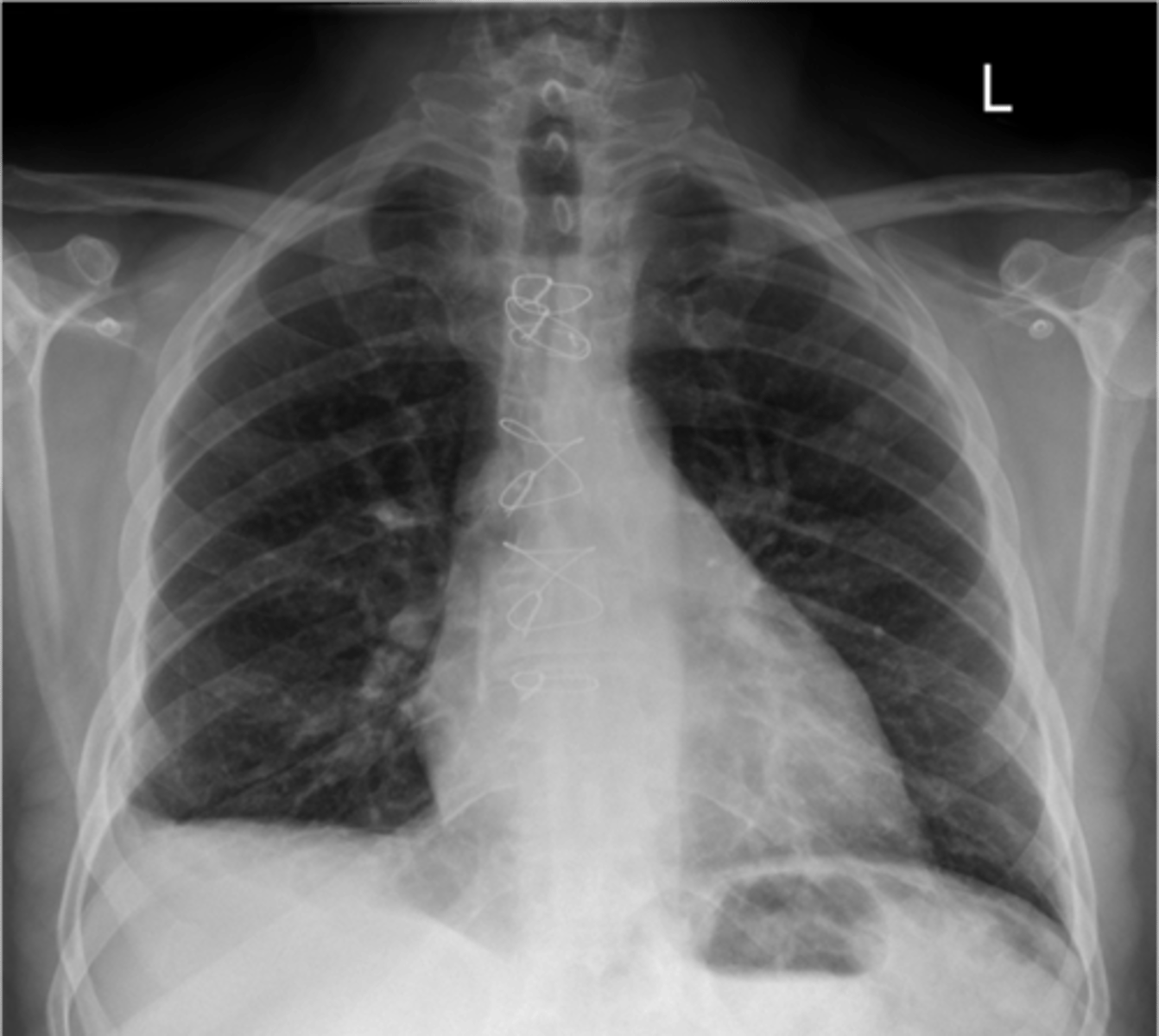
RUL bullae
Flattening of hemidiaphragms
& blunting of costophrenic angles, Increased AP diameter (barrel chest), bullae or large air cavities indicates
hyperinflation
COPD/Emphysema
COPD/Emphysema
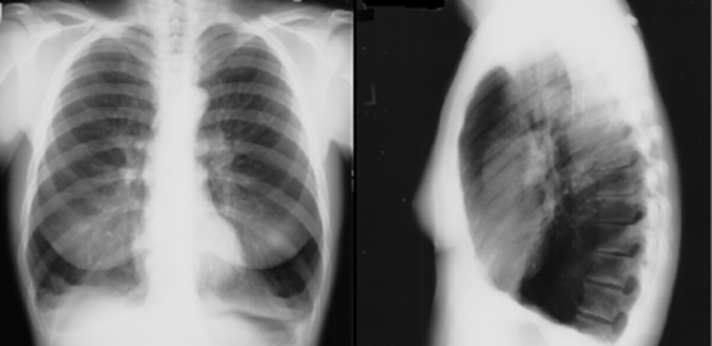
Occurs when alveoli become deflated (complete or partial collapse) or possibly filled with alveolar fluid
Atelectasis
Atelectasis
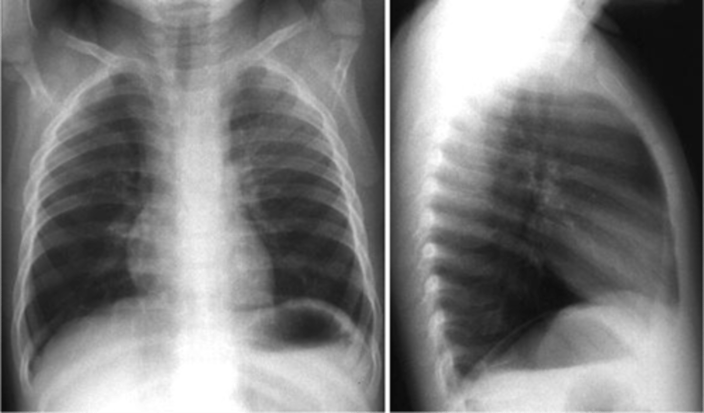
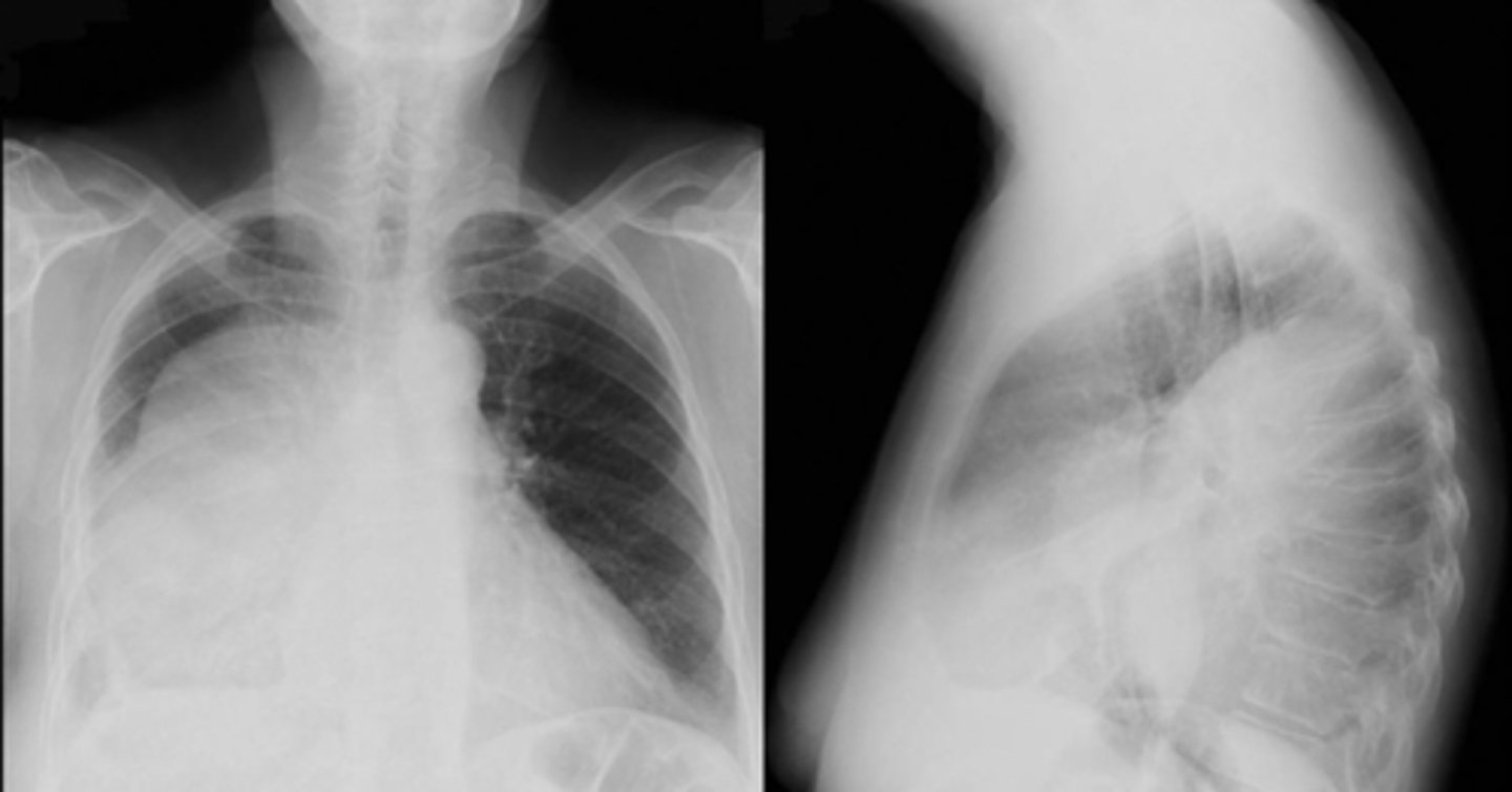
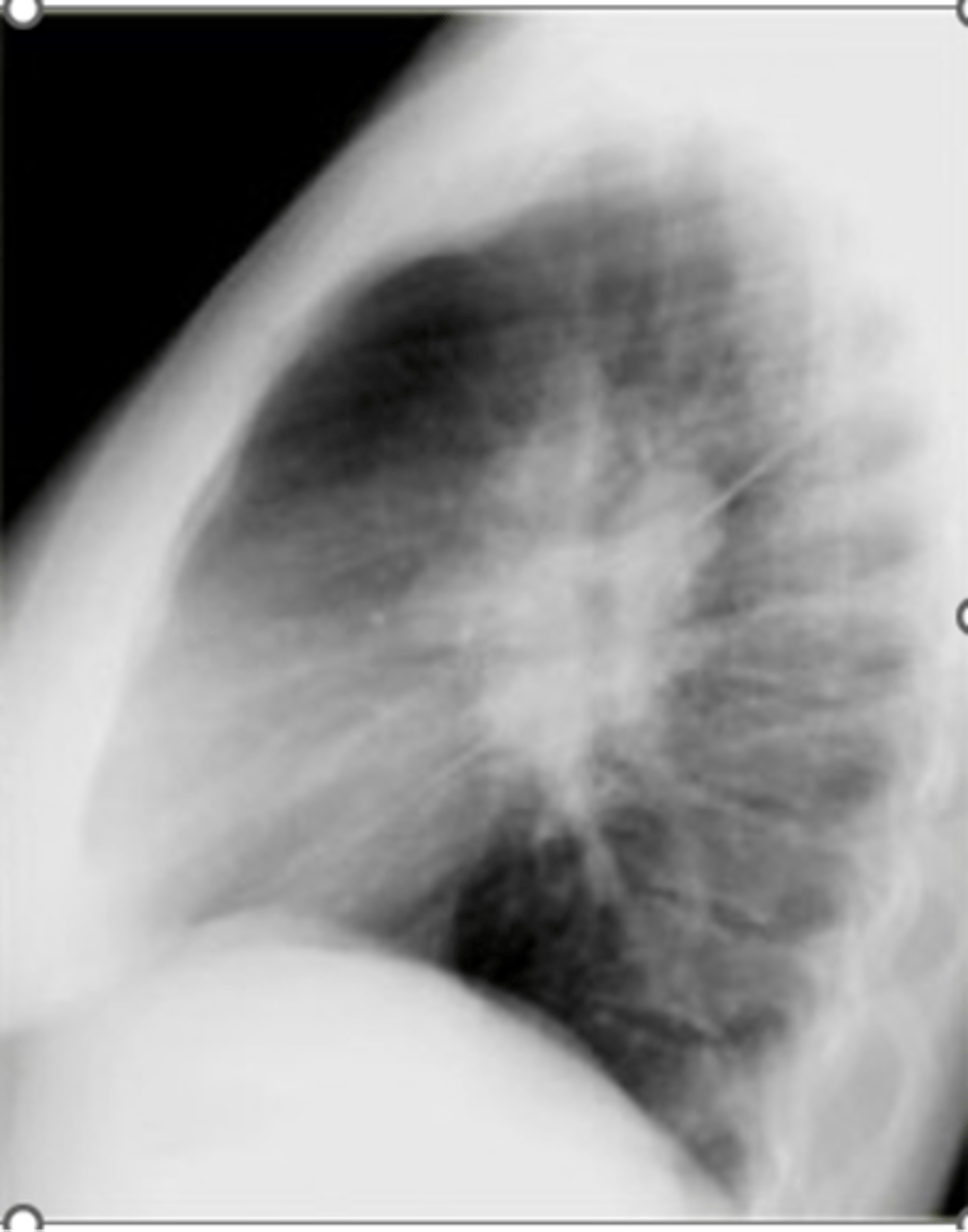
Atelectasis (RUL)
Linear atelectasis (often seen post-op)
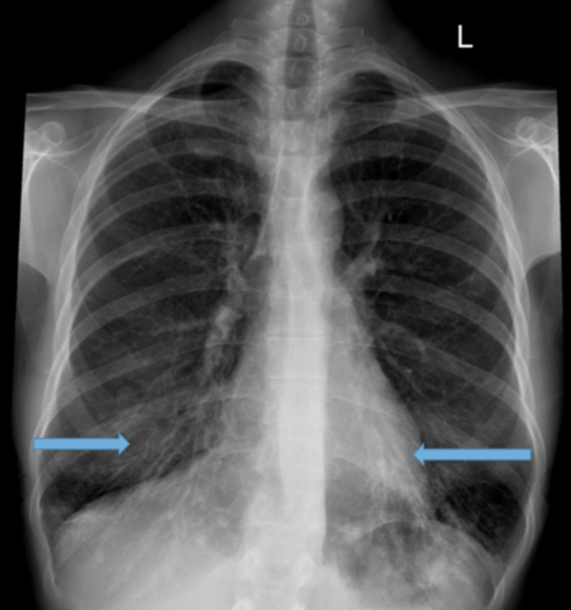
Asthma (hyperinflation with tram lines)
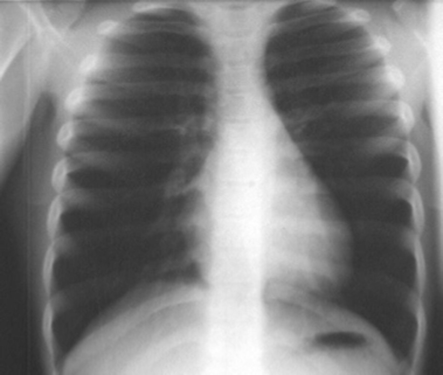
asthma
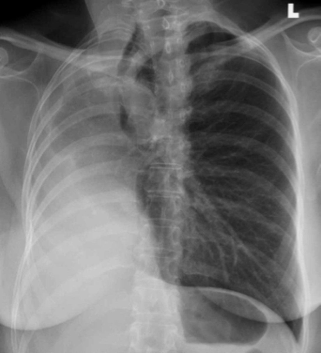
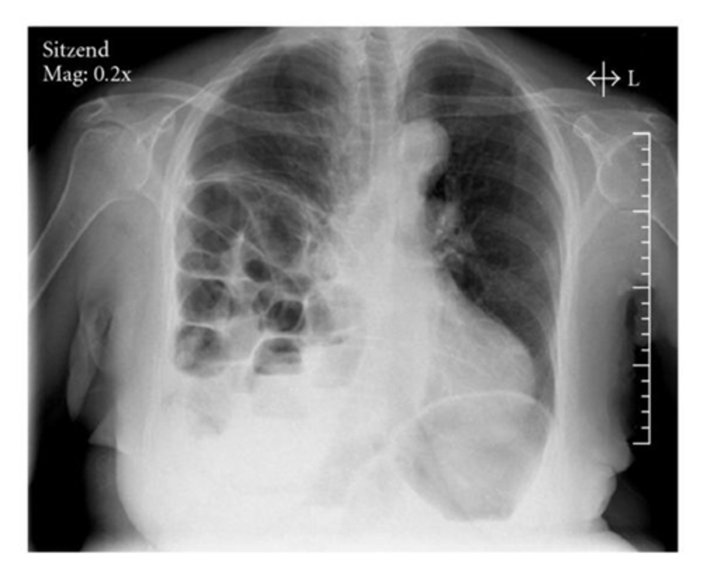
Air in the pleural space that causes collapse of the lung usually seen in the apices if pt is upright
Pneumothorax
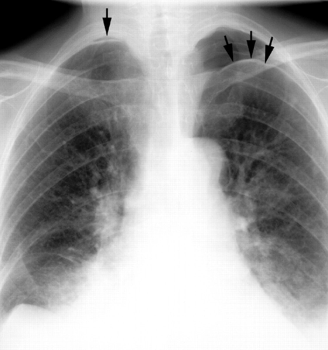
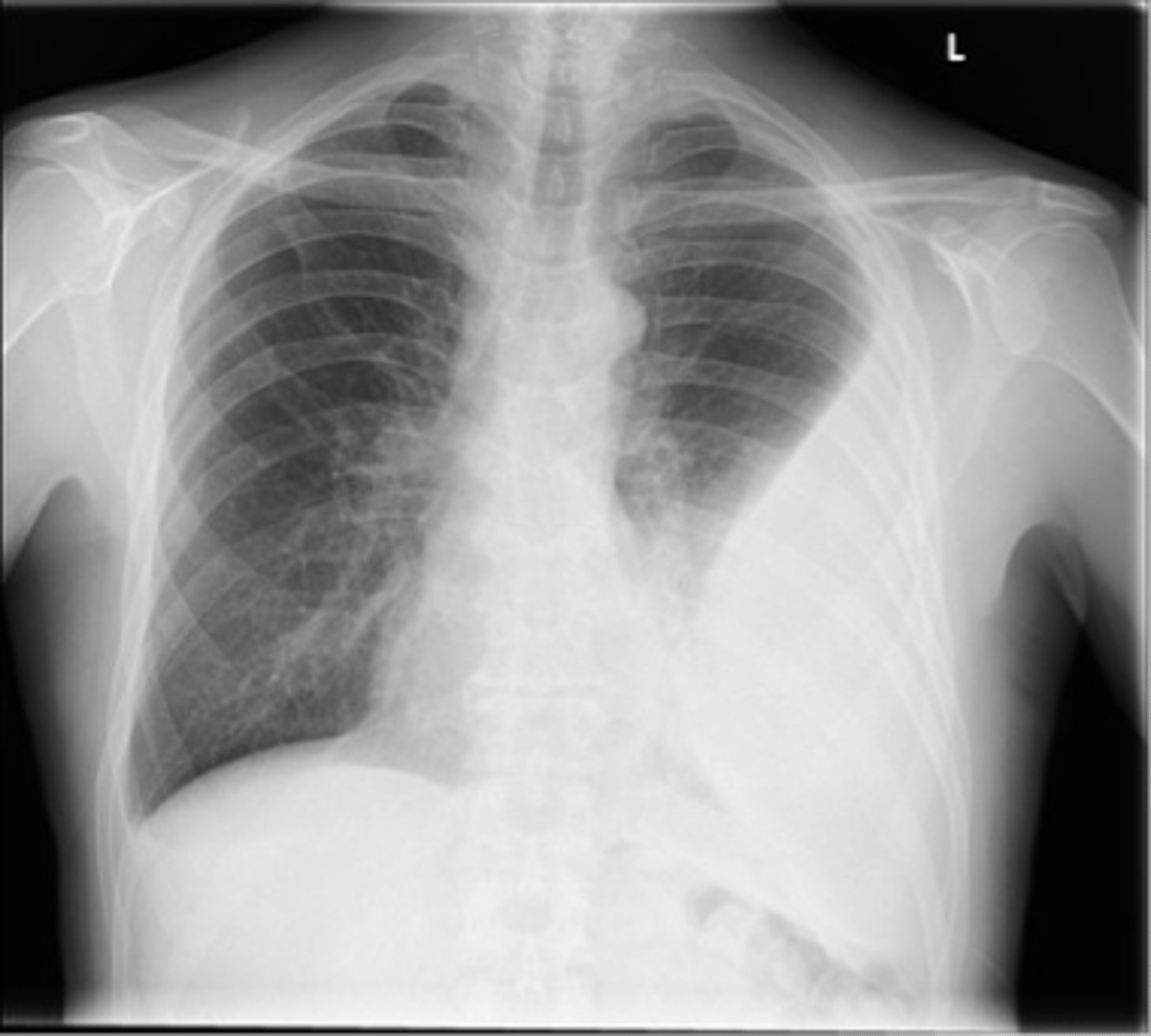
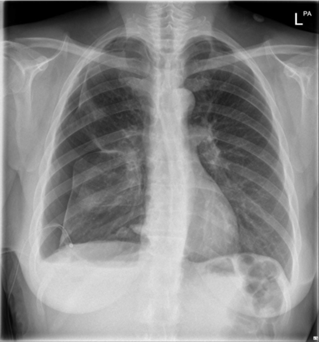
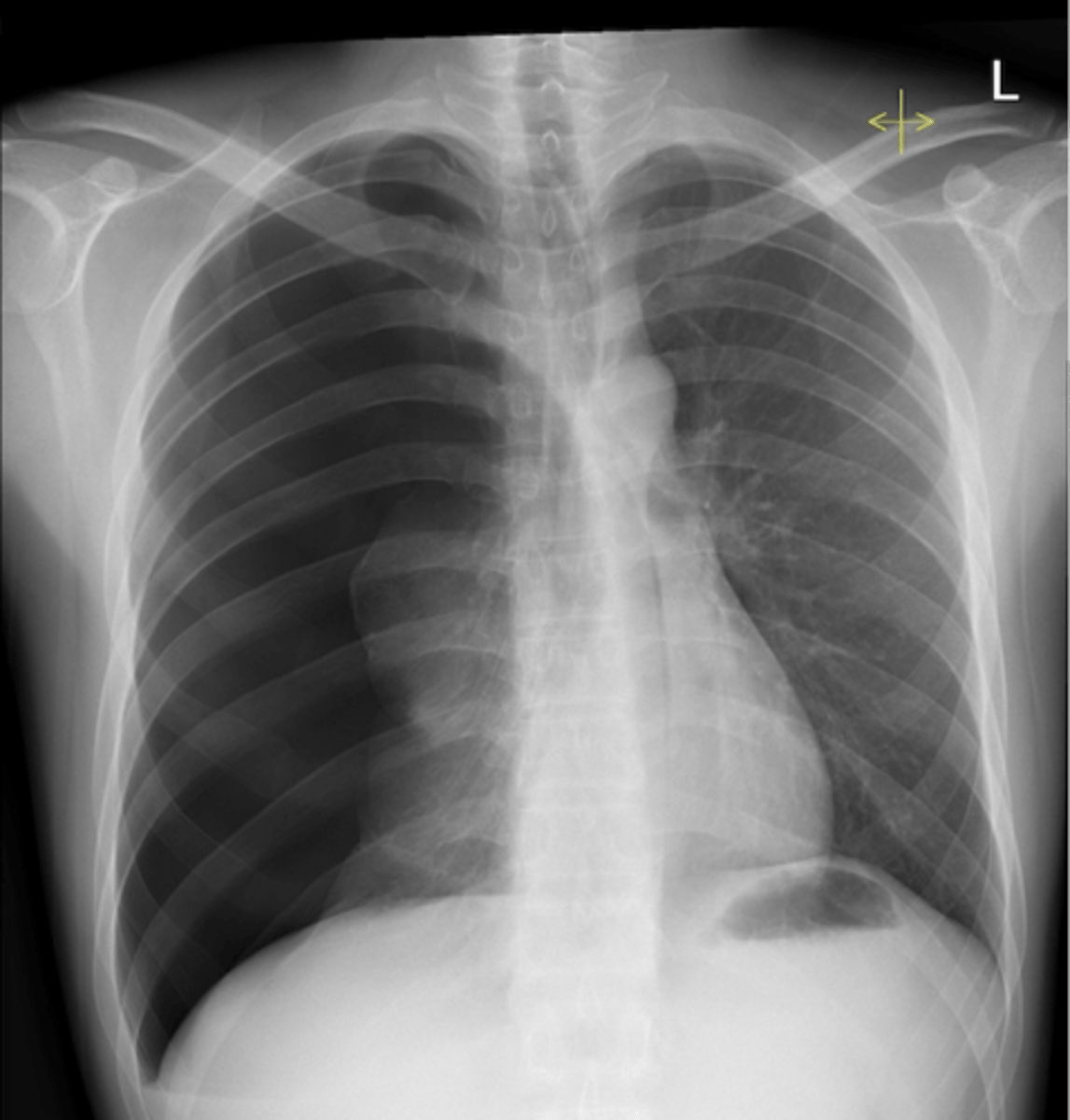
Pneumothorax (apices)
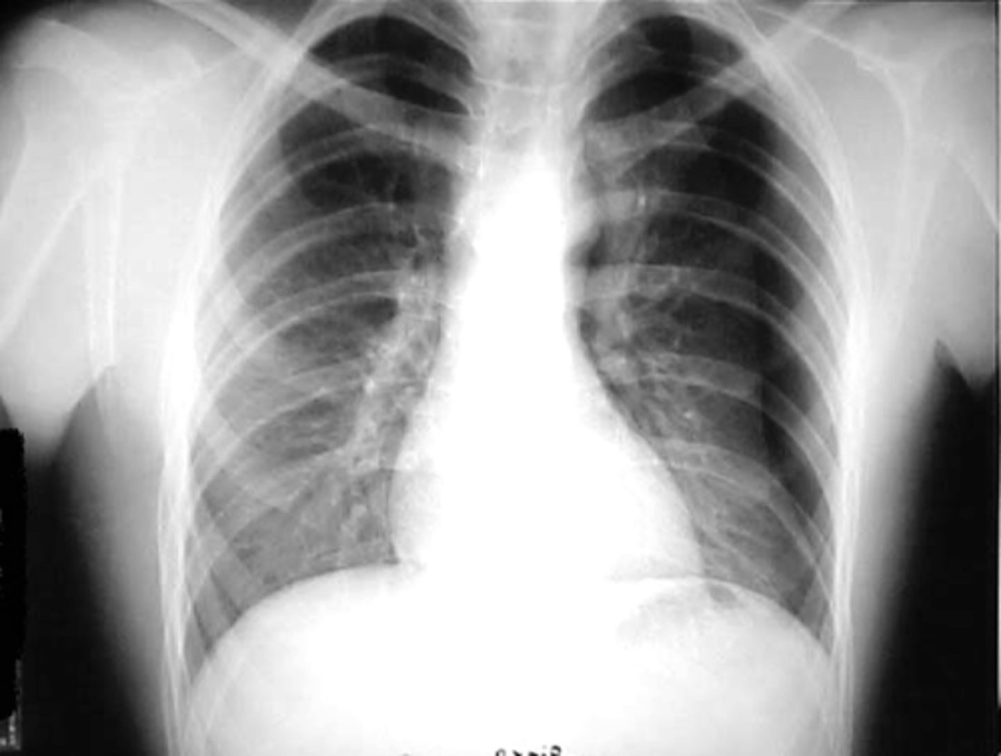
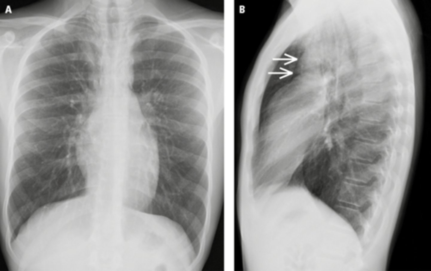
Pneumothorax (moderate, non tension)
Tension pneumothorax
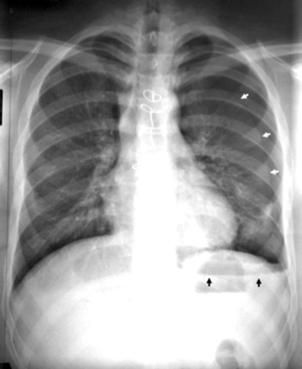
Hemopnuemothorax
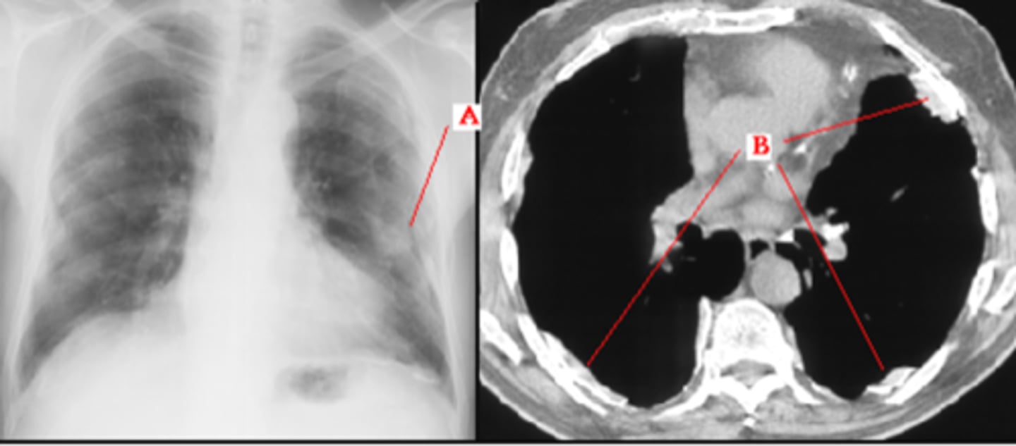
Pneumomediastinum (mediastinal emphysema)
Pneumomediastinum
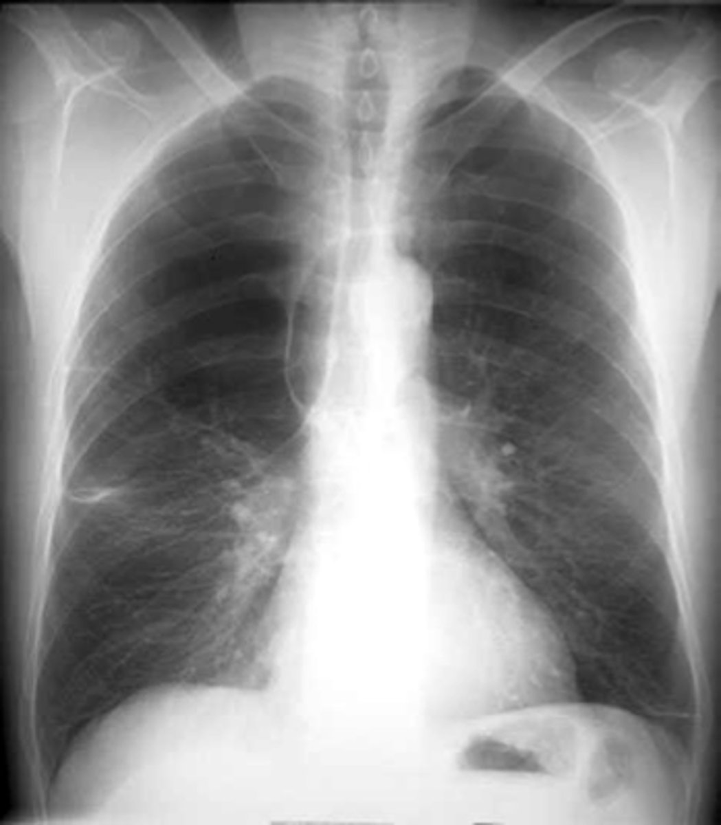
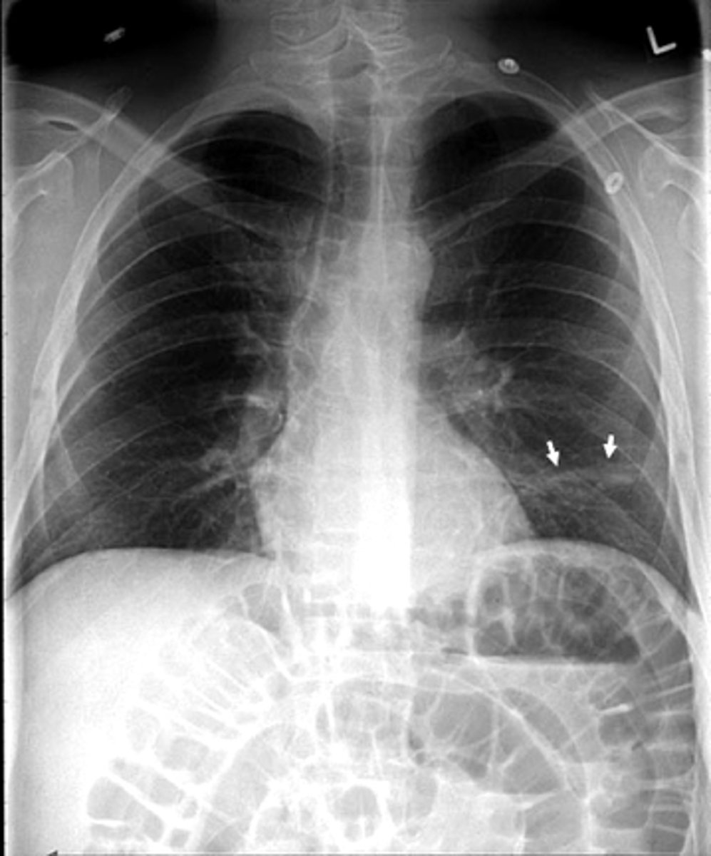
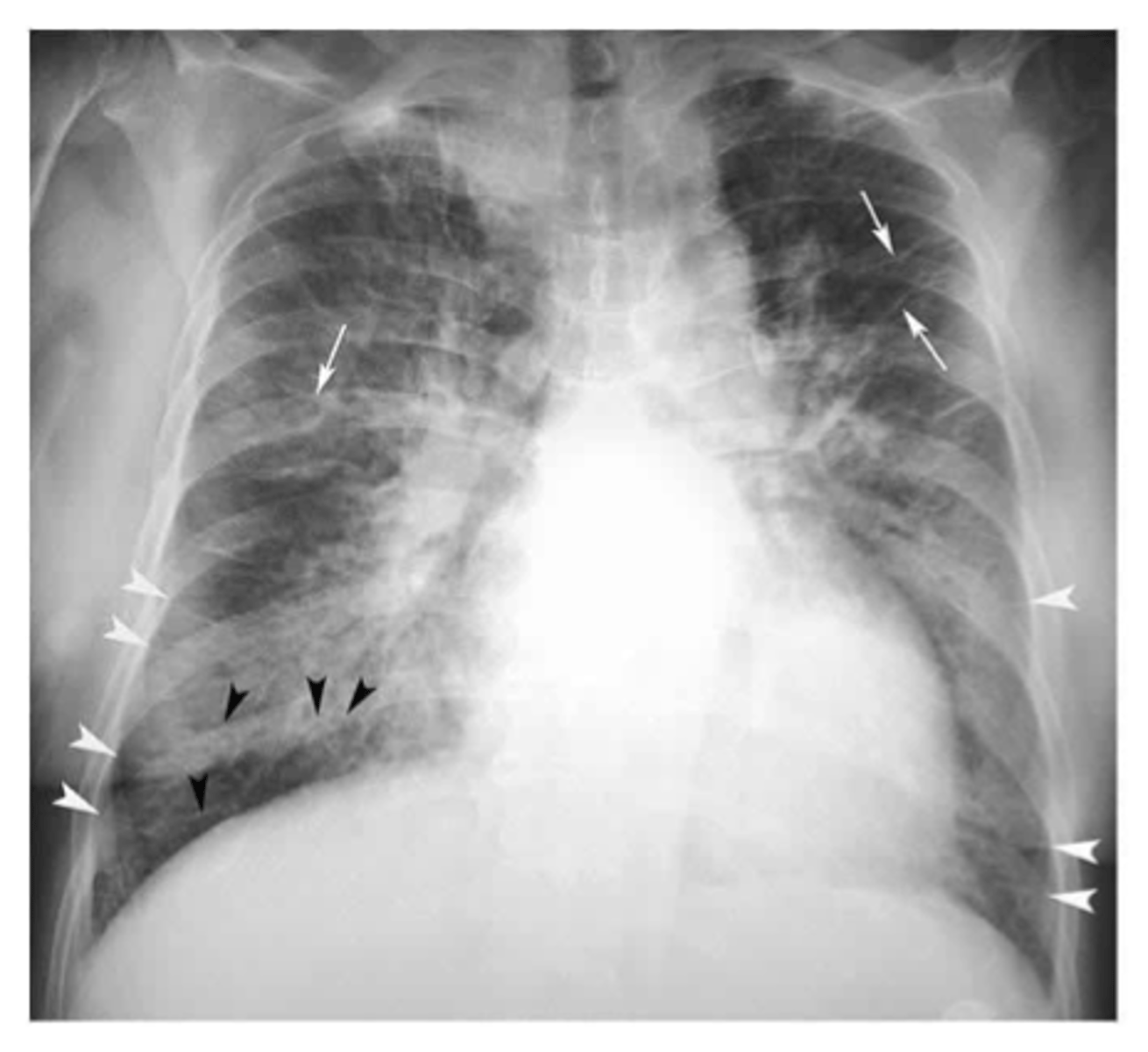
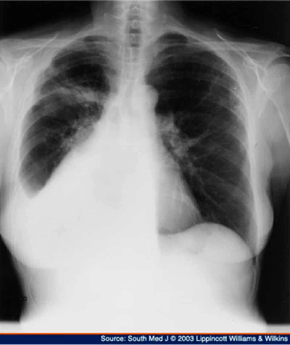
Pleural effusion
Pleural effusion
Pleural effusion (right sided)
Empyema
Empyema
Pleural classifications and masses
Diaphragmatic rupture
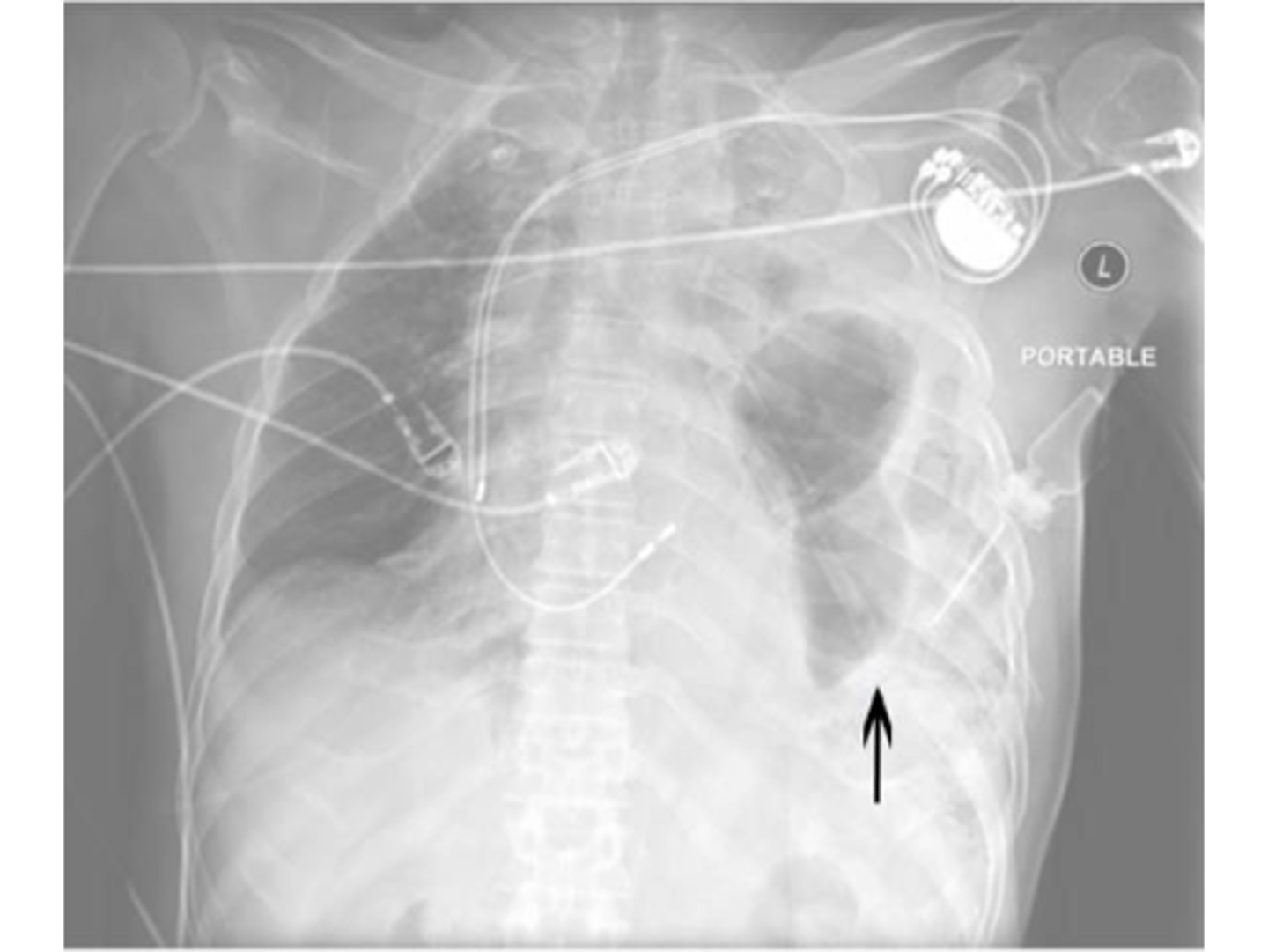
Diaphragmatic rupture
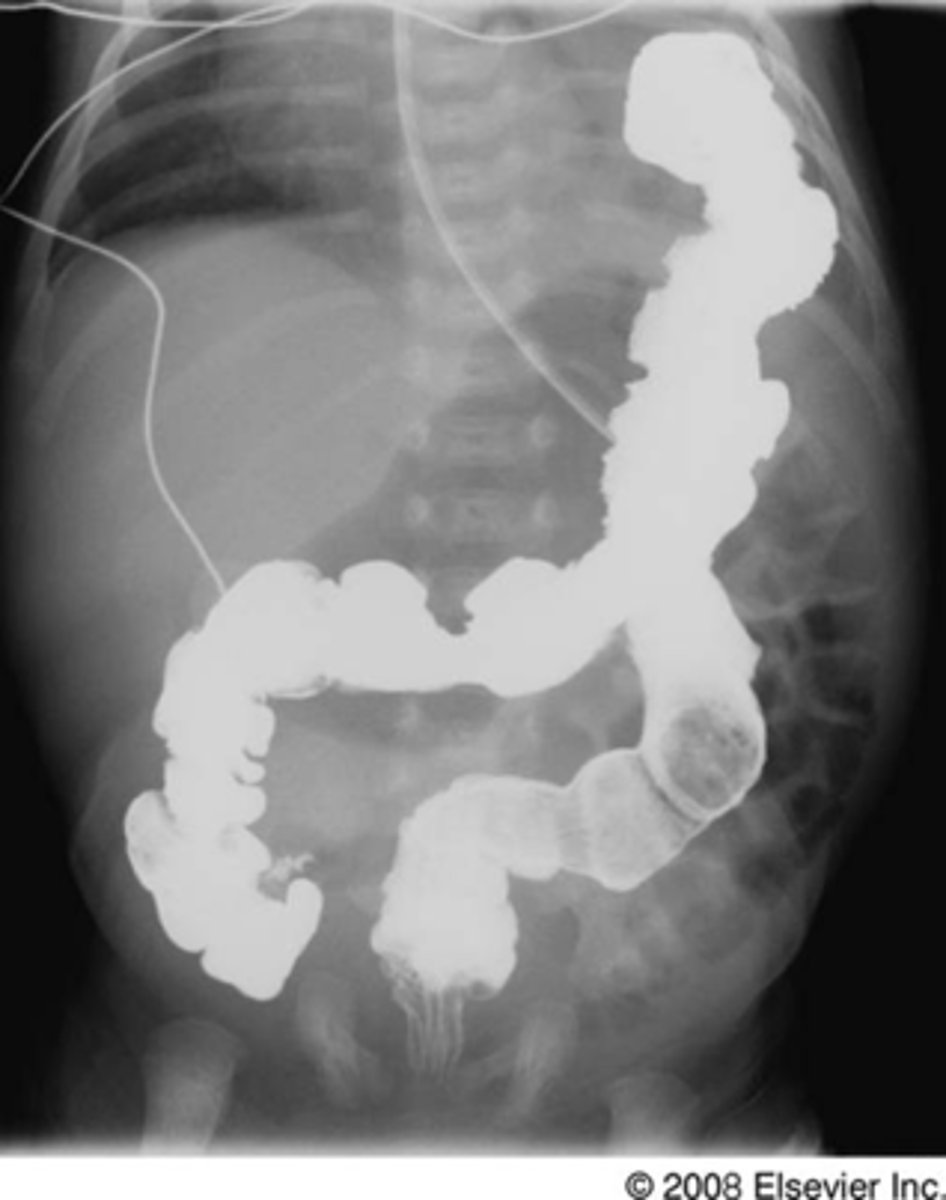
Diaphragmatic hernia
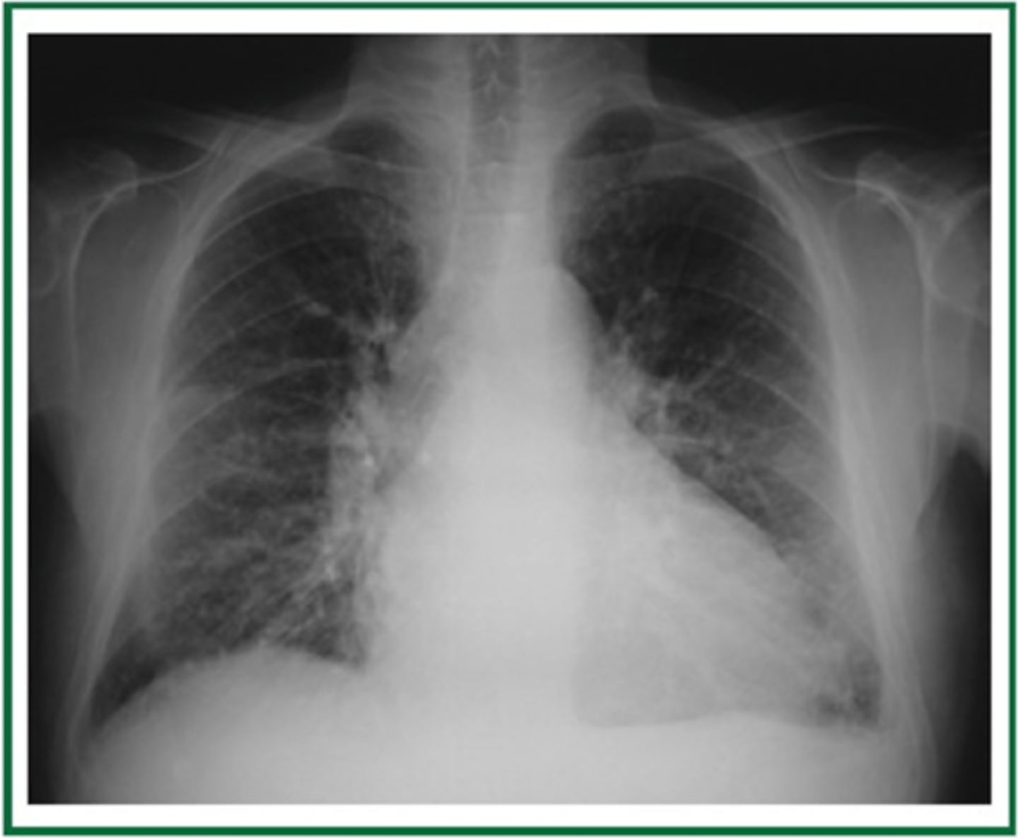
congestive heart failure
congestive heart failure
Most common anterior mediastinal mass
Thymoma
Most frequent cause of a middle mediastinal mass
Enlarged lymph nodes
Middle mediastinal masses
Posterior mediastinal masses
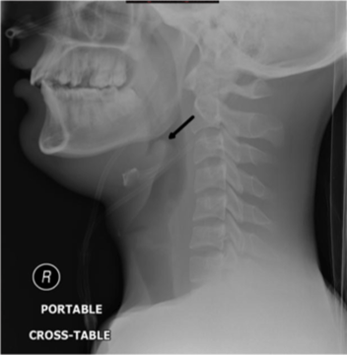
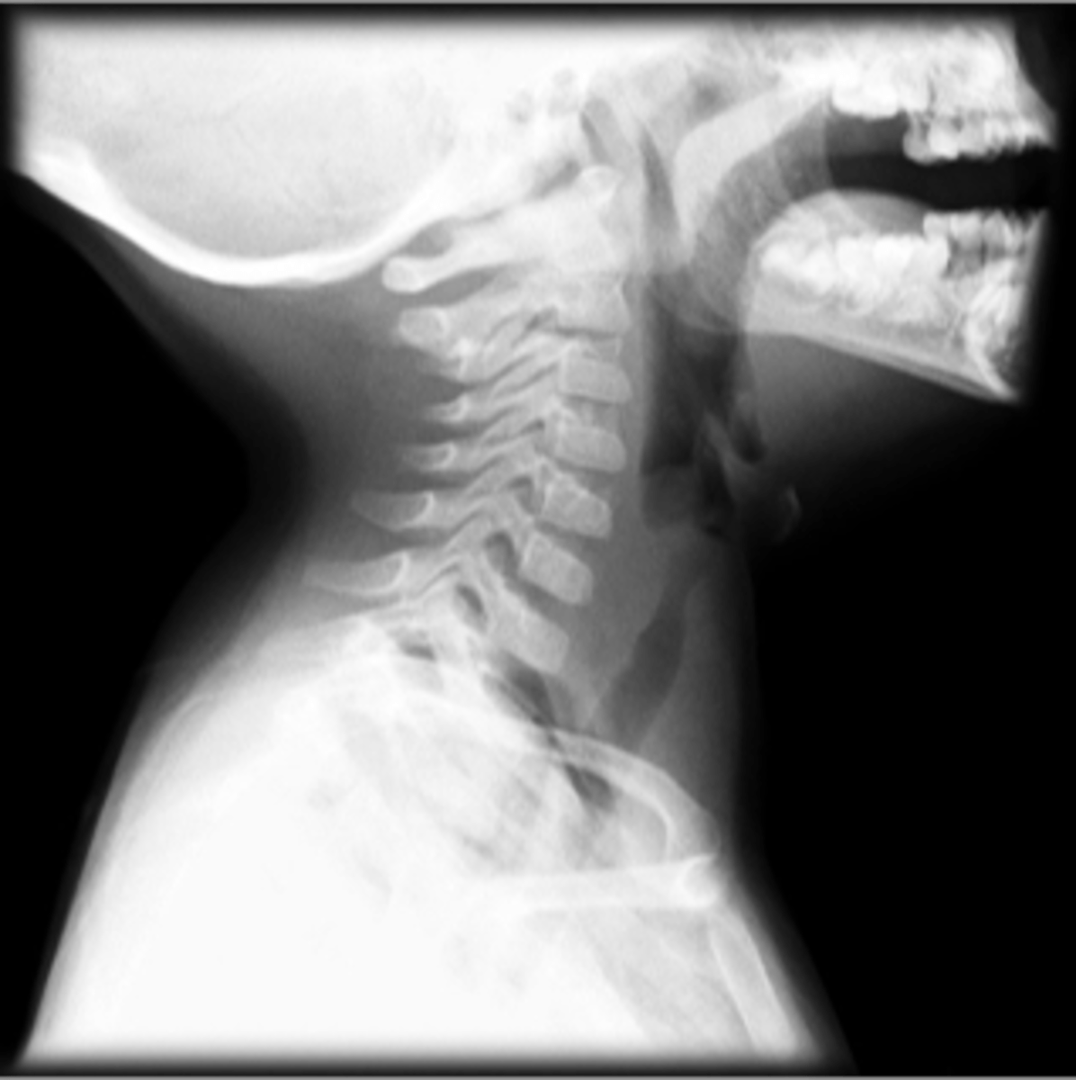
Epiglottitis (thumb sign)
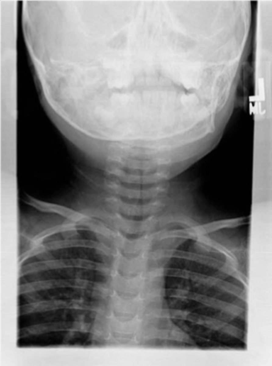
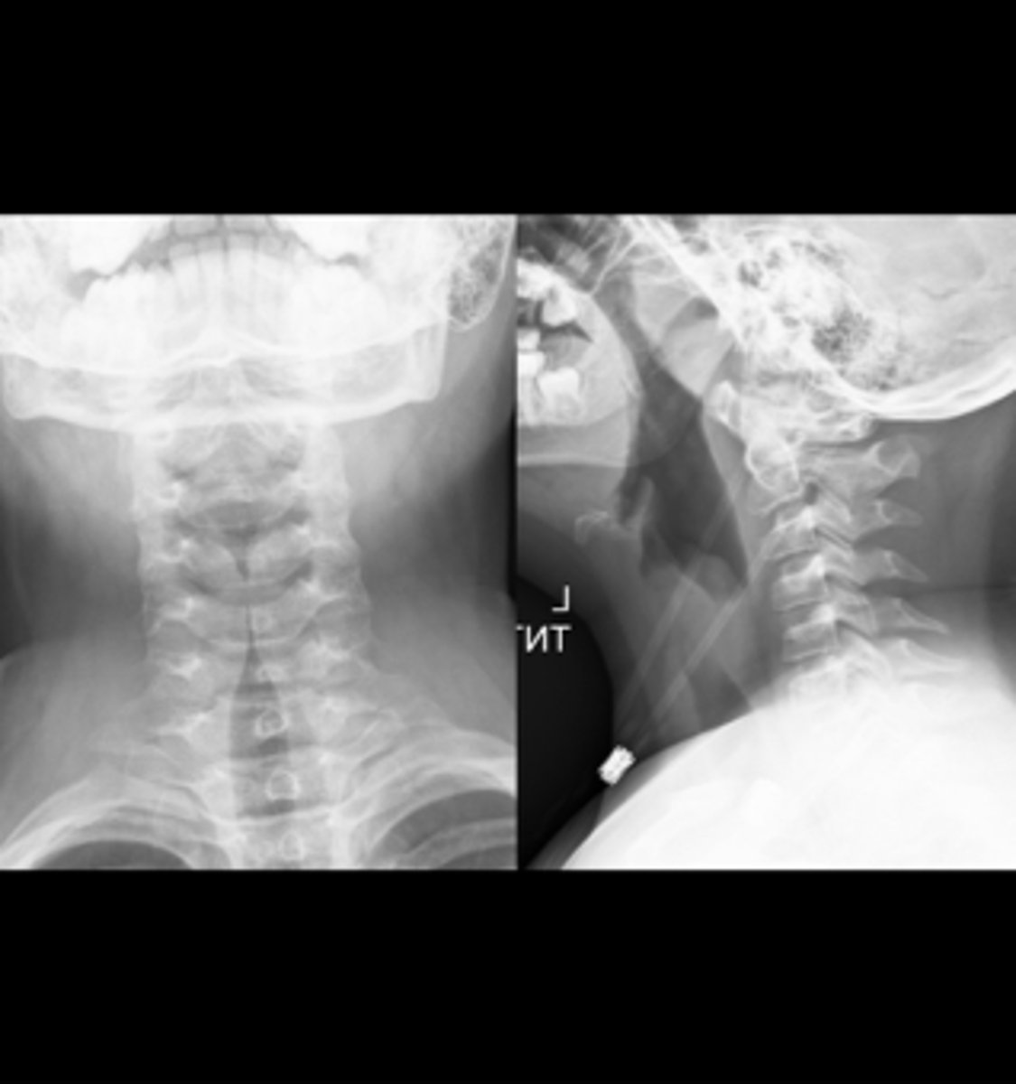
steeple sign (croup)
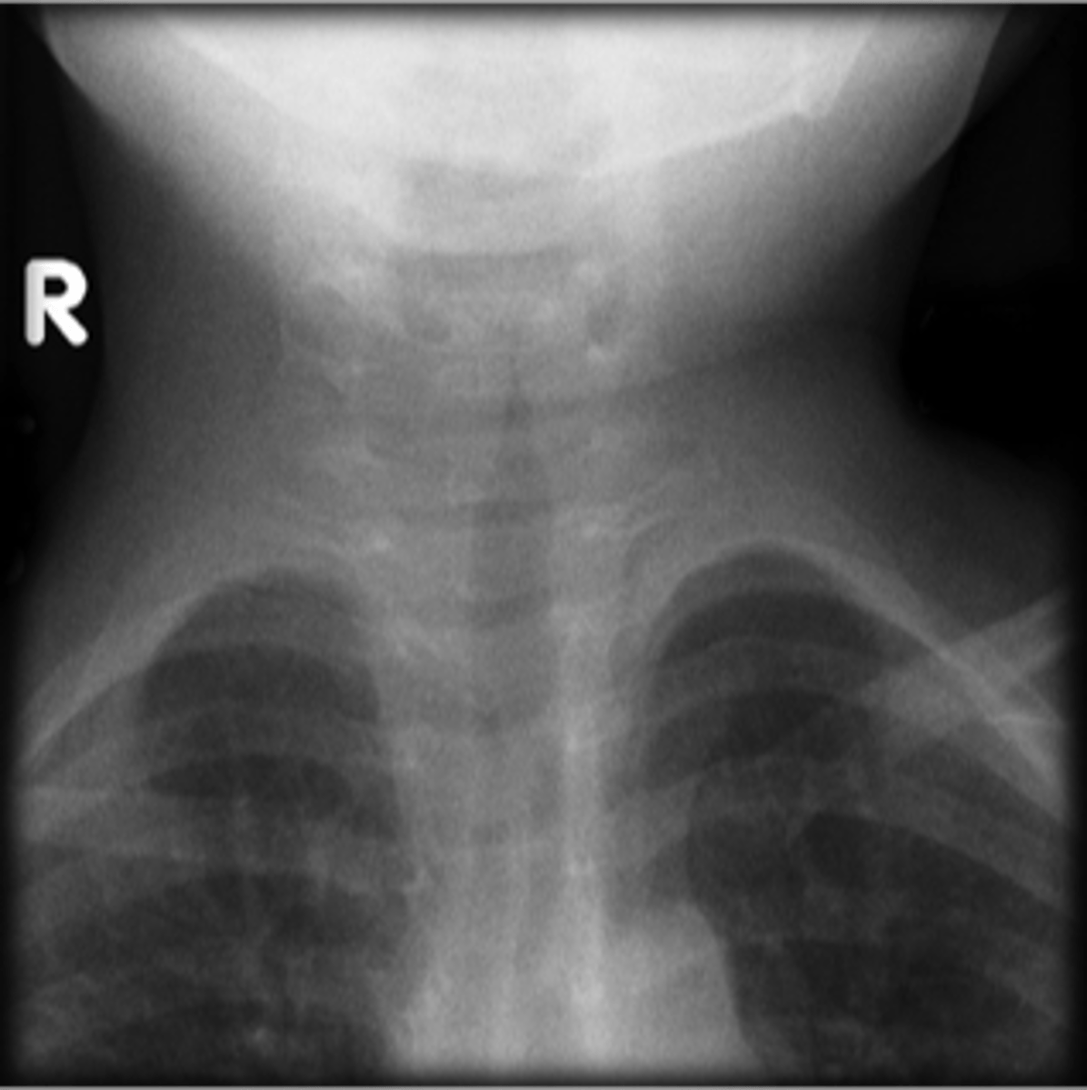
steeple sign (croup)
Croup
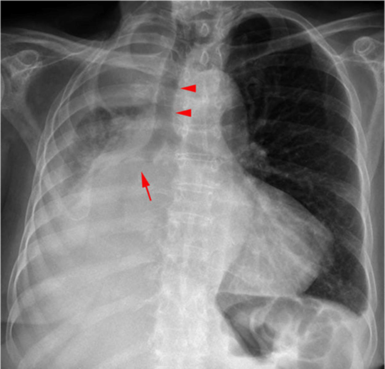
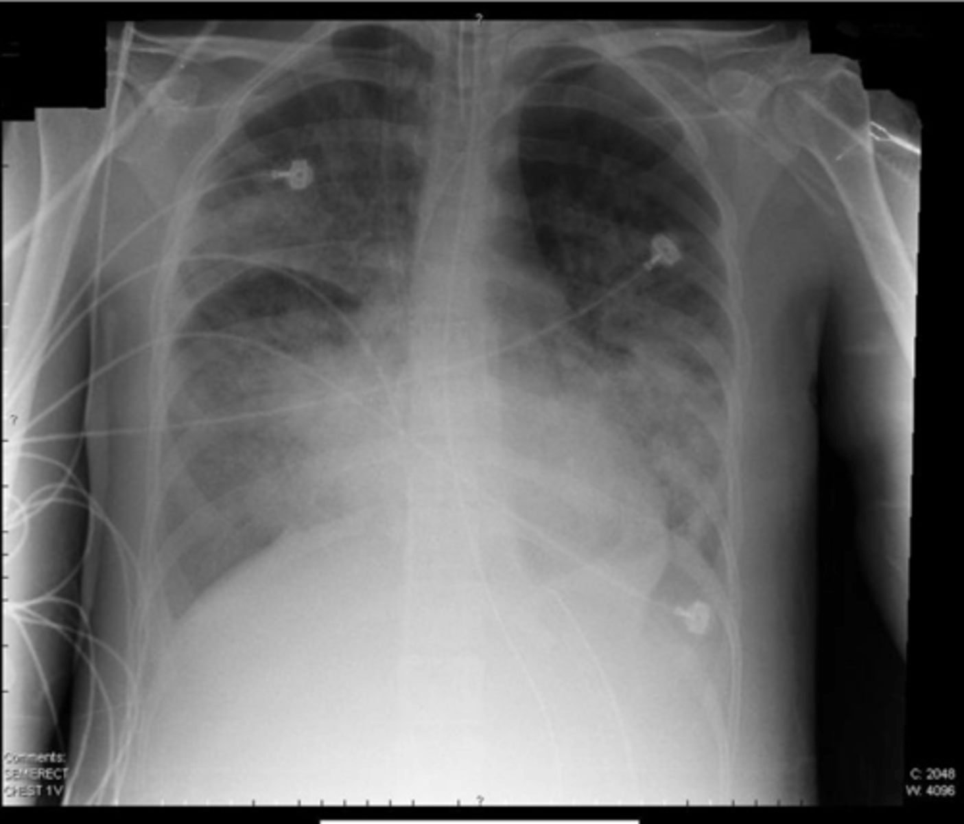
Right sided atelectasis
Right sided non tension pneumothorax
Atelectasis (right)
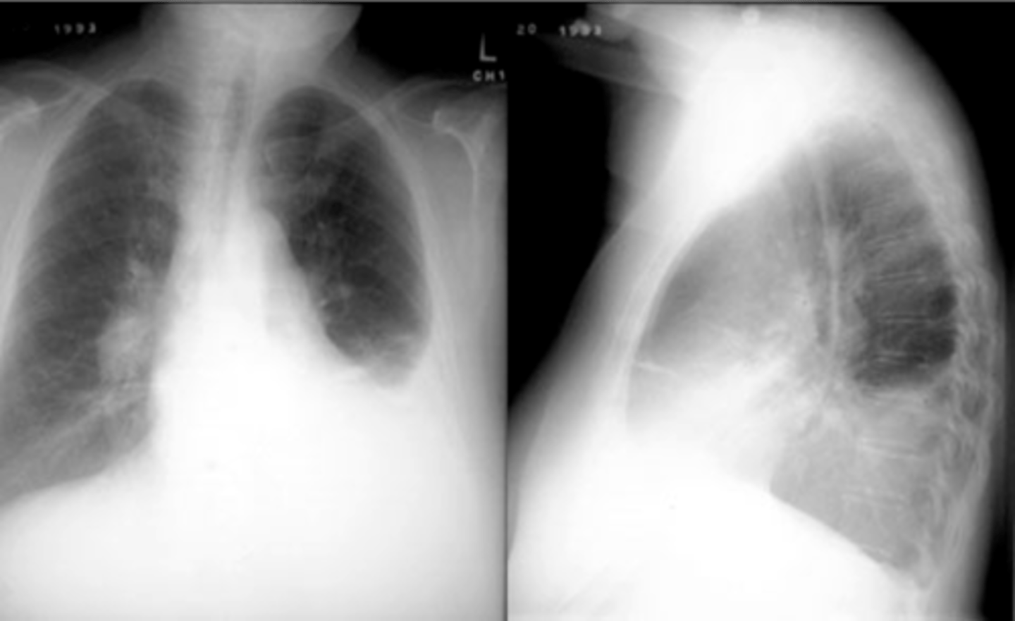
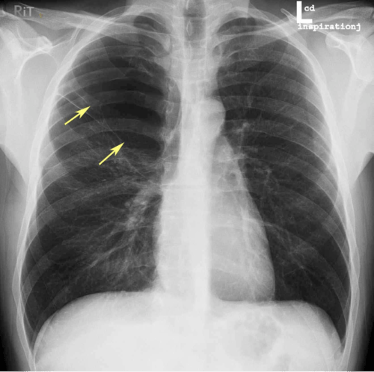
Bullae RUL
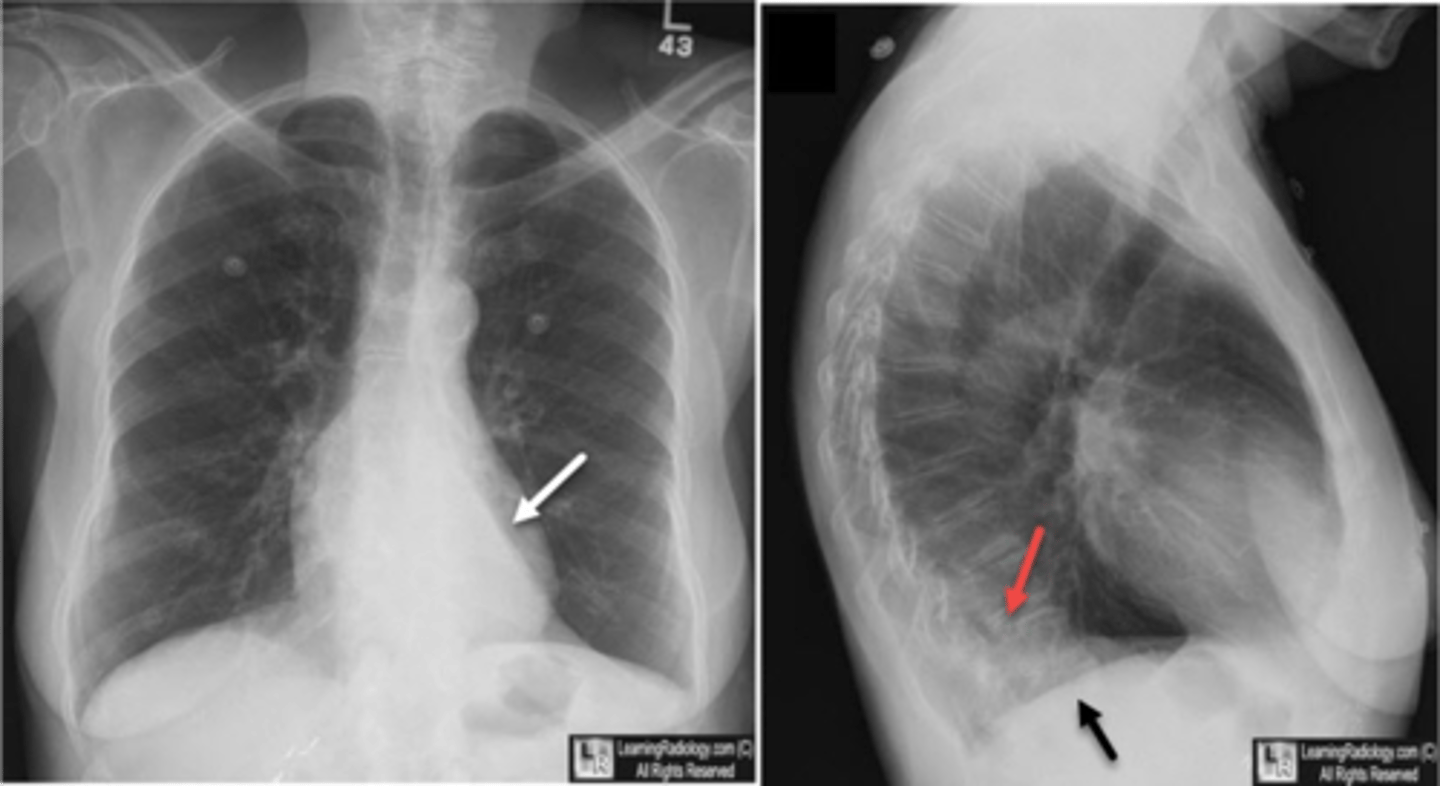
Atelectasis LLL
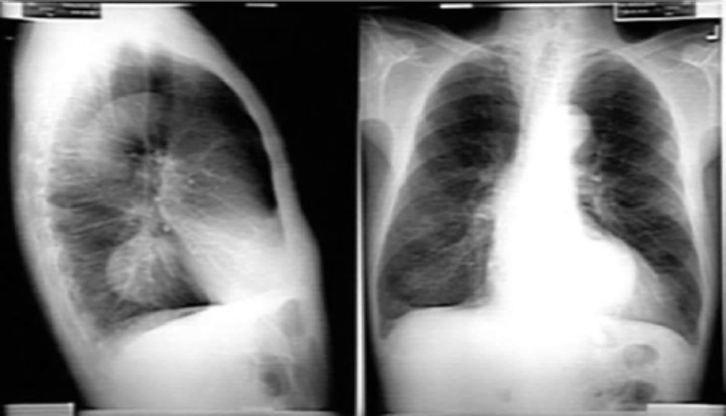
CHF - severe alveolar infiltrate
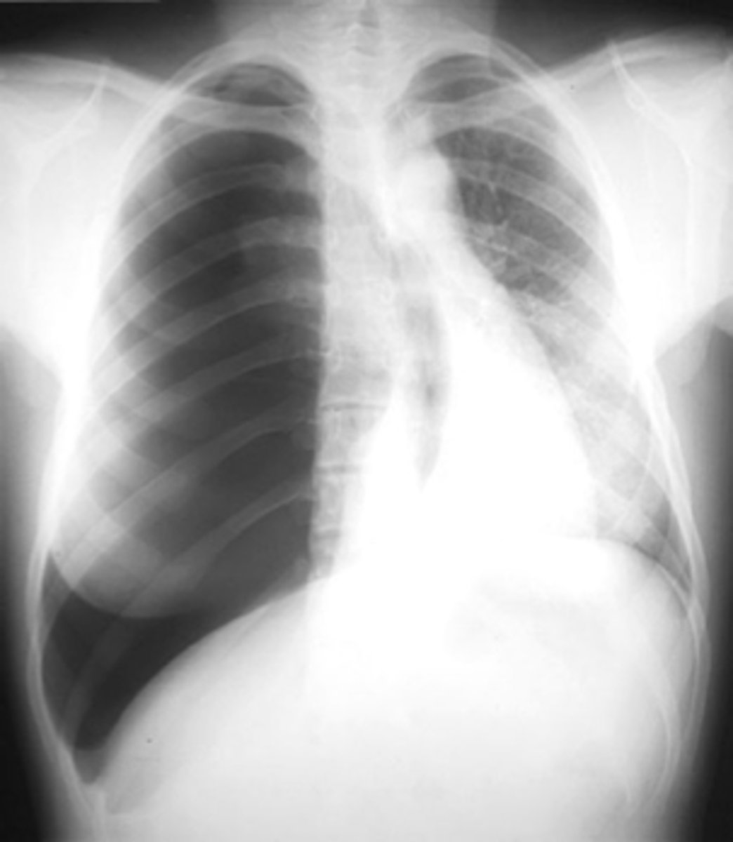
Tension pneumothorax (right)
Steeple sign- group