Med Imaging- Lungs Pt. 1
1/61
There's no tags or description
Looks like no tags are added yet.
Name | Mastery | Learn | Test | Matching | Spaced | Call with Kai |
|---|
No analytics yet
Send a link to your students to track their progress
62 Terms
Alveolar spaces are filled with some material such as blood, pus, fluid, or cells
Alveolar (localized)
tissue outside of the alveoli are affected
Interstitial (diffuse)
Alveolar infiltrates are either
fluffy or complete consolidations
Air bronchogram
Air bronchogram sign
air filled bronchi become visible against a background of dense lung tissue
Kerley A lines
periphery (interstitial infiltrate)
Kerly B lines
base (interstitial infiltrate)
vertebral bodies are lighter (white) than normal on lateral x-ray view
spine sign
Aspiration pneumonia shows as an
alveolar infiltrate
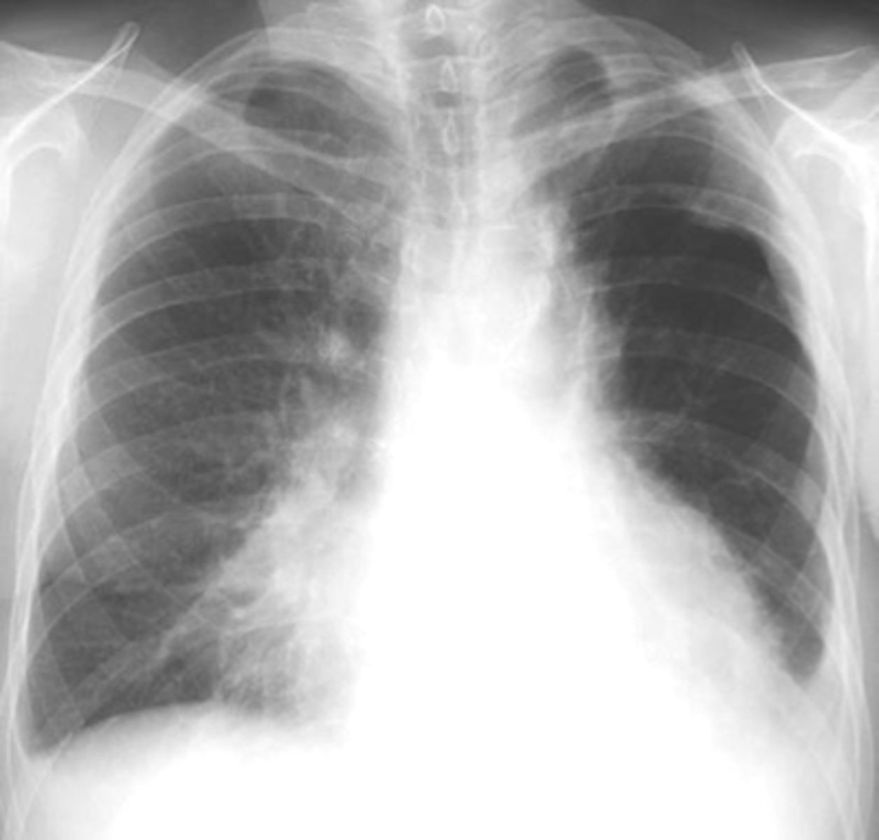
Enlargement of hilar or mediastinal lymph nodes is present in
primary active TB
calcified nodes are associated with
TB
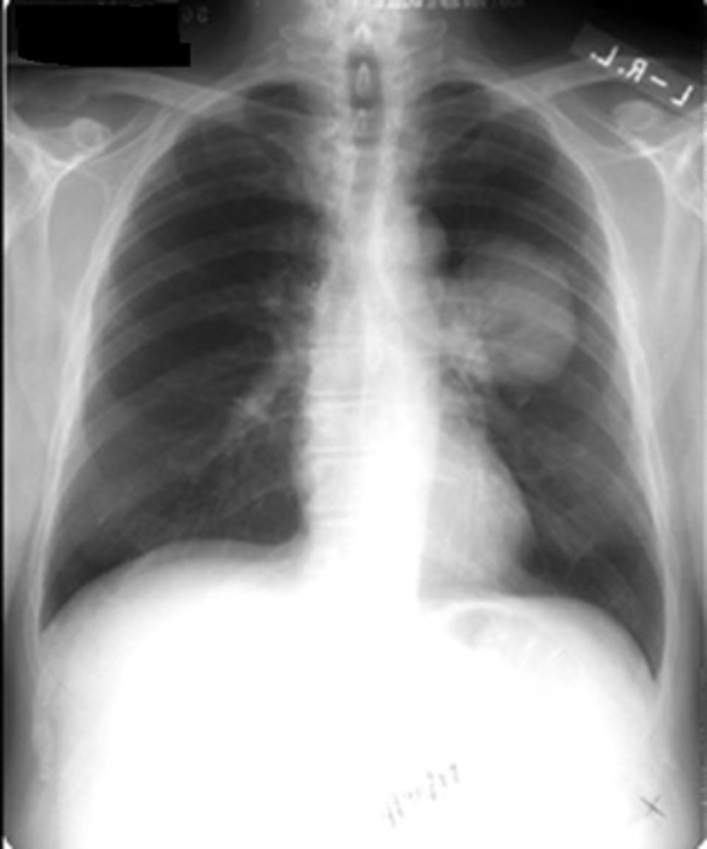
less than 3cm, calcified, round well defined borders, solid-no cavity
Benign solitary pulmonary nodule
great than 3 cm, not calcified, irregular shape, cavitated, growth in past 2 years
malignant solitary pulmonary nodule
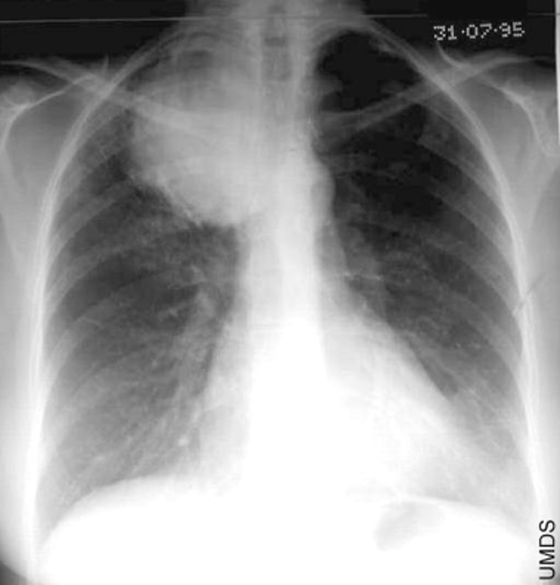
most common type of lung cancer and usually occur peripherally
Adenocarcinomas
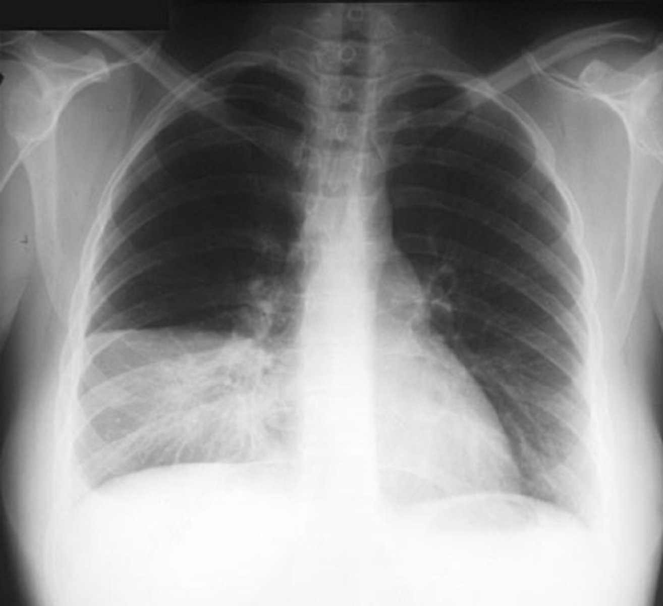
what lung cancer usually occurs centrally and tend to cavitate
squamous cell carcinomas
what lung cancer often presents as an indistinct hilar or perihilar mass
small cell carcinomas
what lung cancer occurs peripherally or centrally and grow rapidly with early metastases- poor prognosis
non-small cell carcinomas
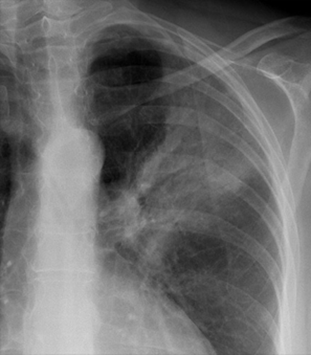
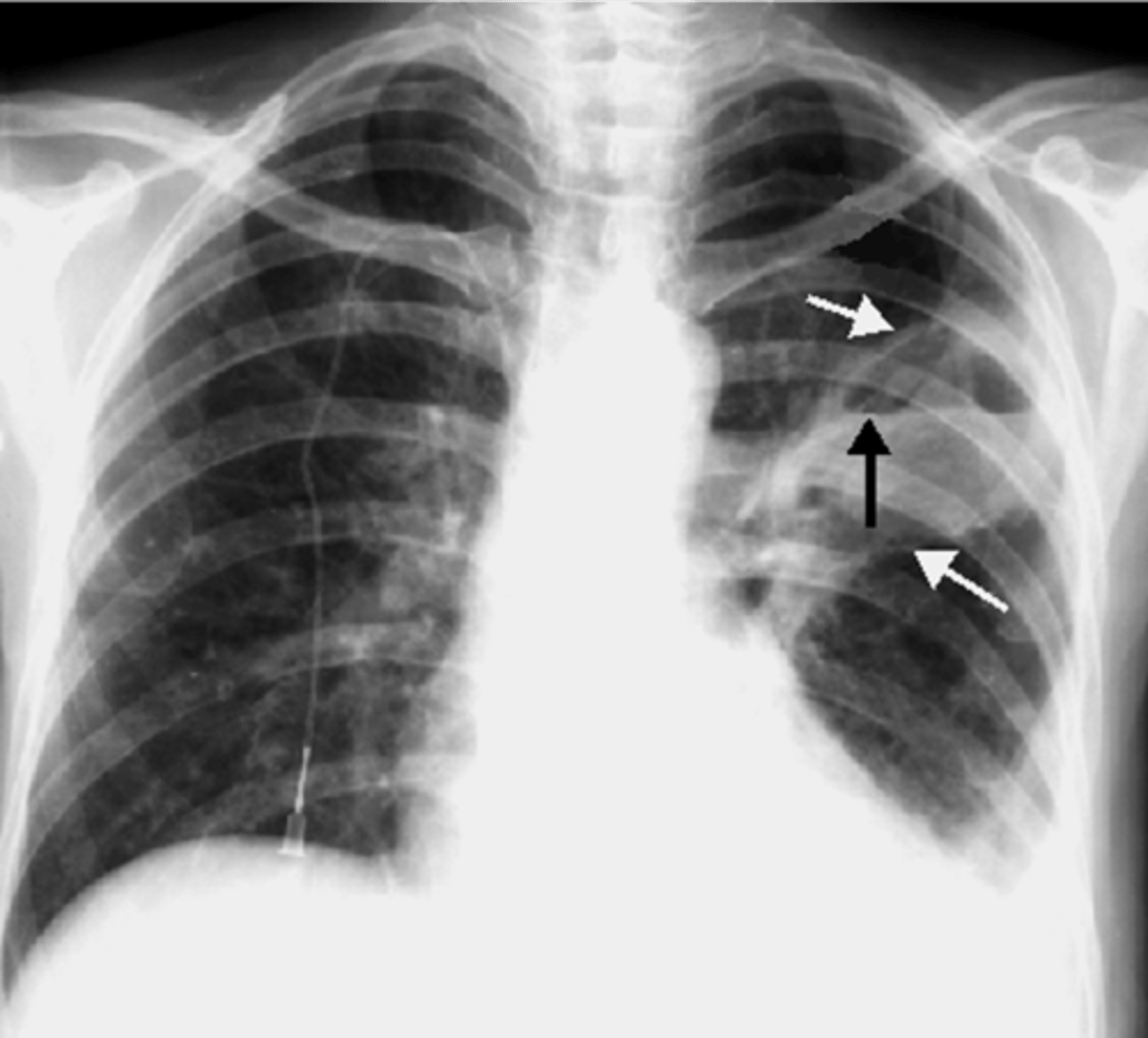
Arise in the superior sulcus of the lung as an apical soft tissue mass
lung cancer- pancoast tumor
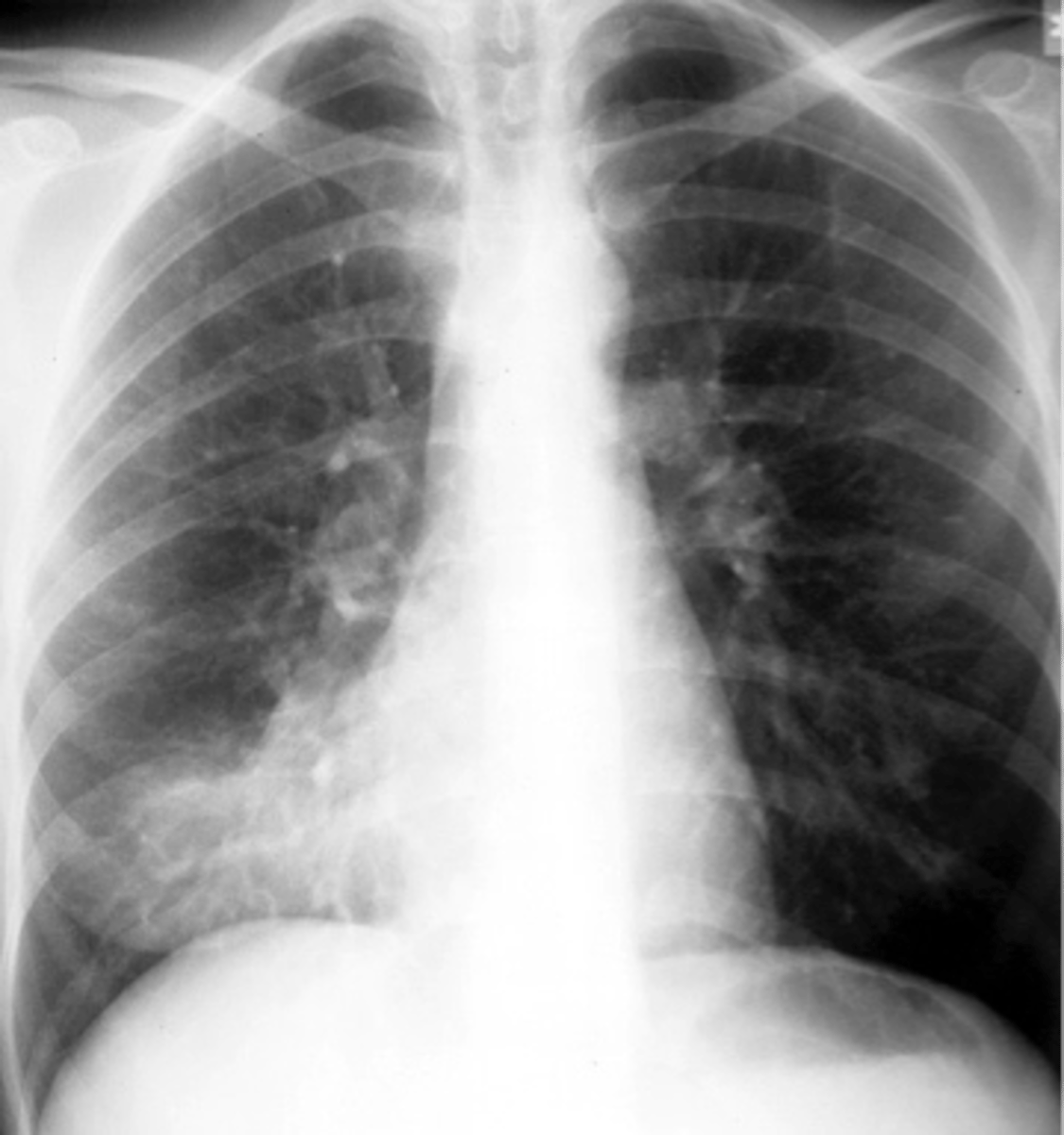
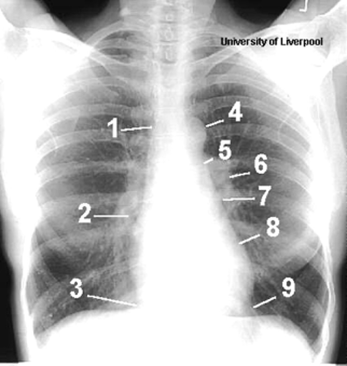
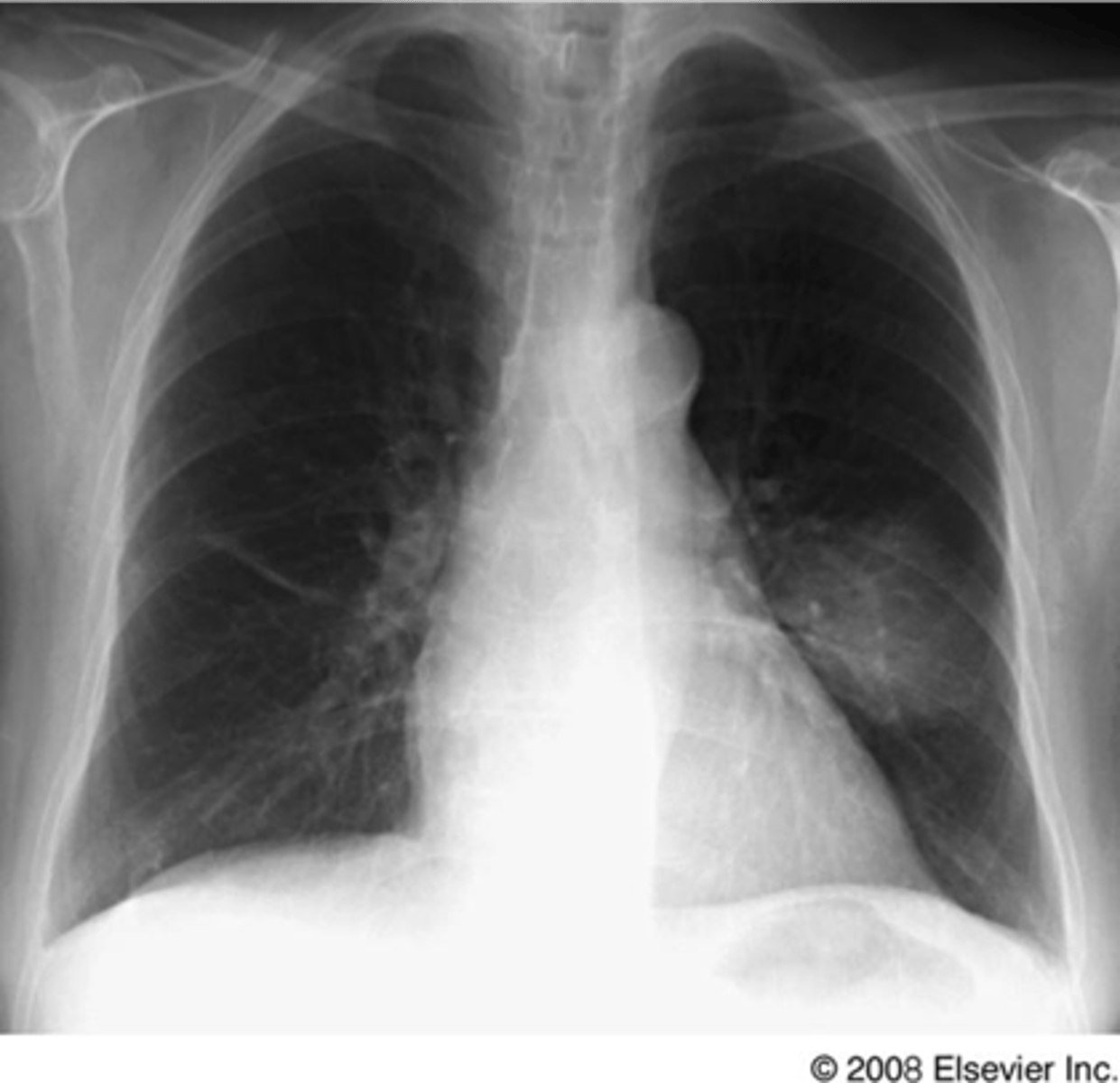
1
superior vena cava

2
right atrium

3
inferior vena cava

4
aortic arch or knob

5
left pulmonary trunk

6
left pulmonary artery

7
left atrium

8
left ventricle

9
left cardiophrenic angle
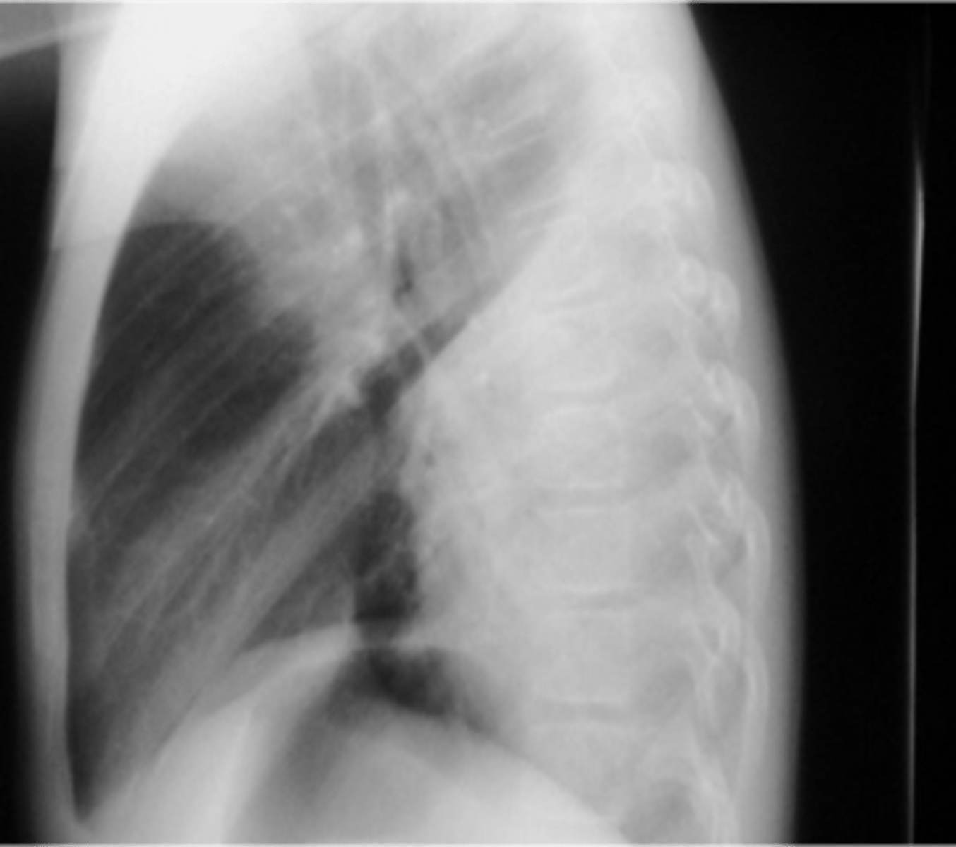
A
trachea
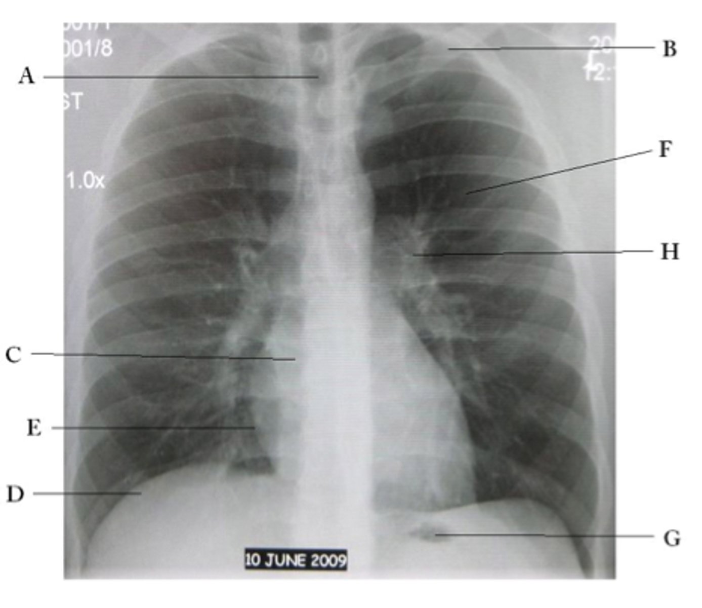
B
Clavicle

C
Right atrium

D
Diaphragm

E
Cardiophrenic angle

F
left upper lobe

G
gastric bubble

H
Left hilum

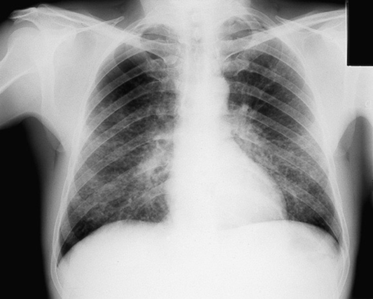
Reticular interstitial infiltrate (fine)
Reticular interstitial infiltrate (coarse)
Nodular Interstital infiltrate
Reticulonodular interstitial infiltrate
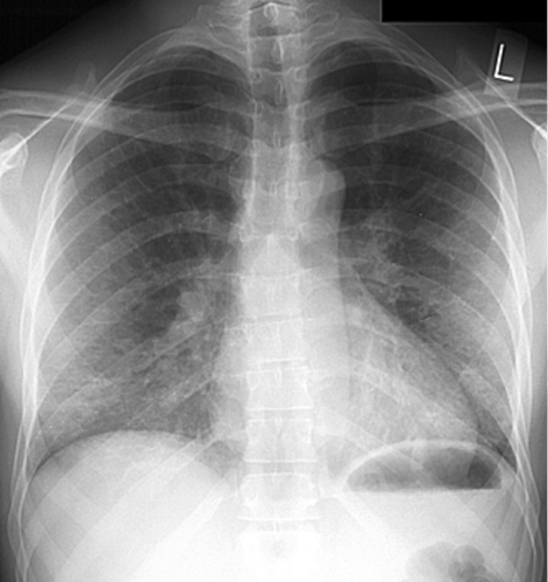
community acquired pneumonia
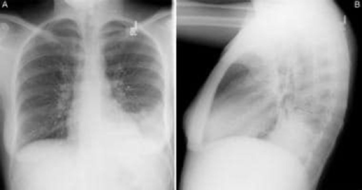
Silhouette sign

spine sign
CAP- interstitial
CAP- round
Aspiration pneumonia

lung abscess
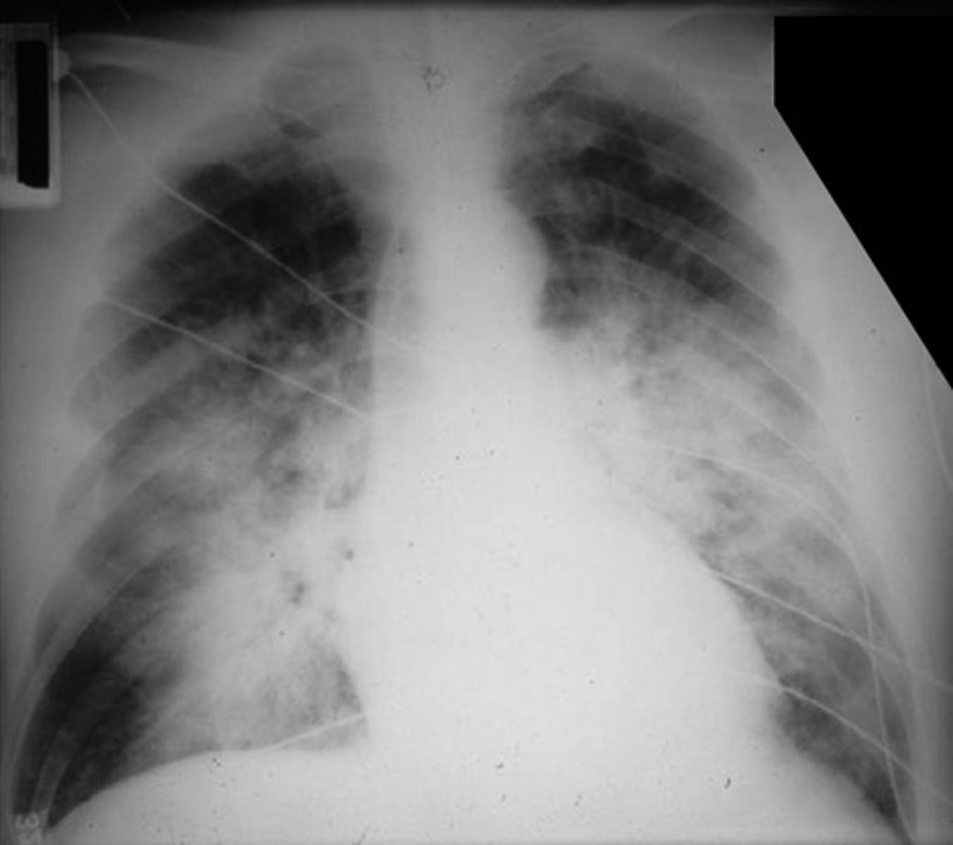
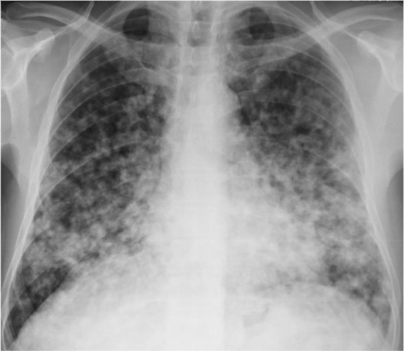
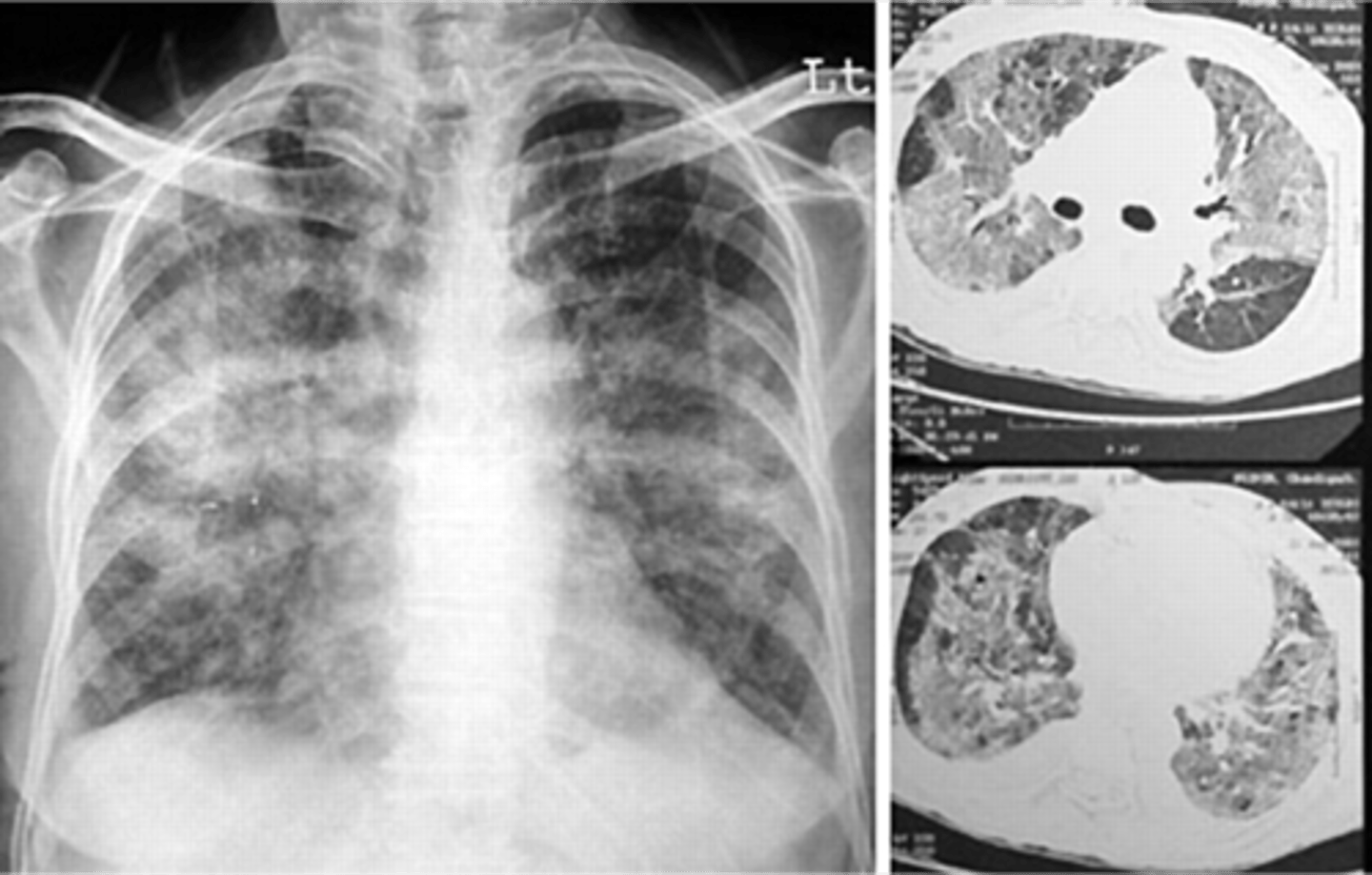
ARDS
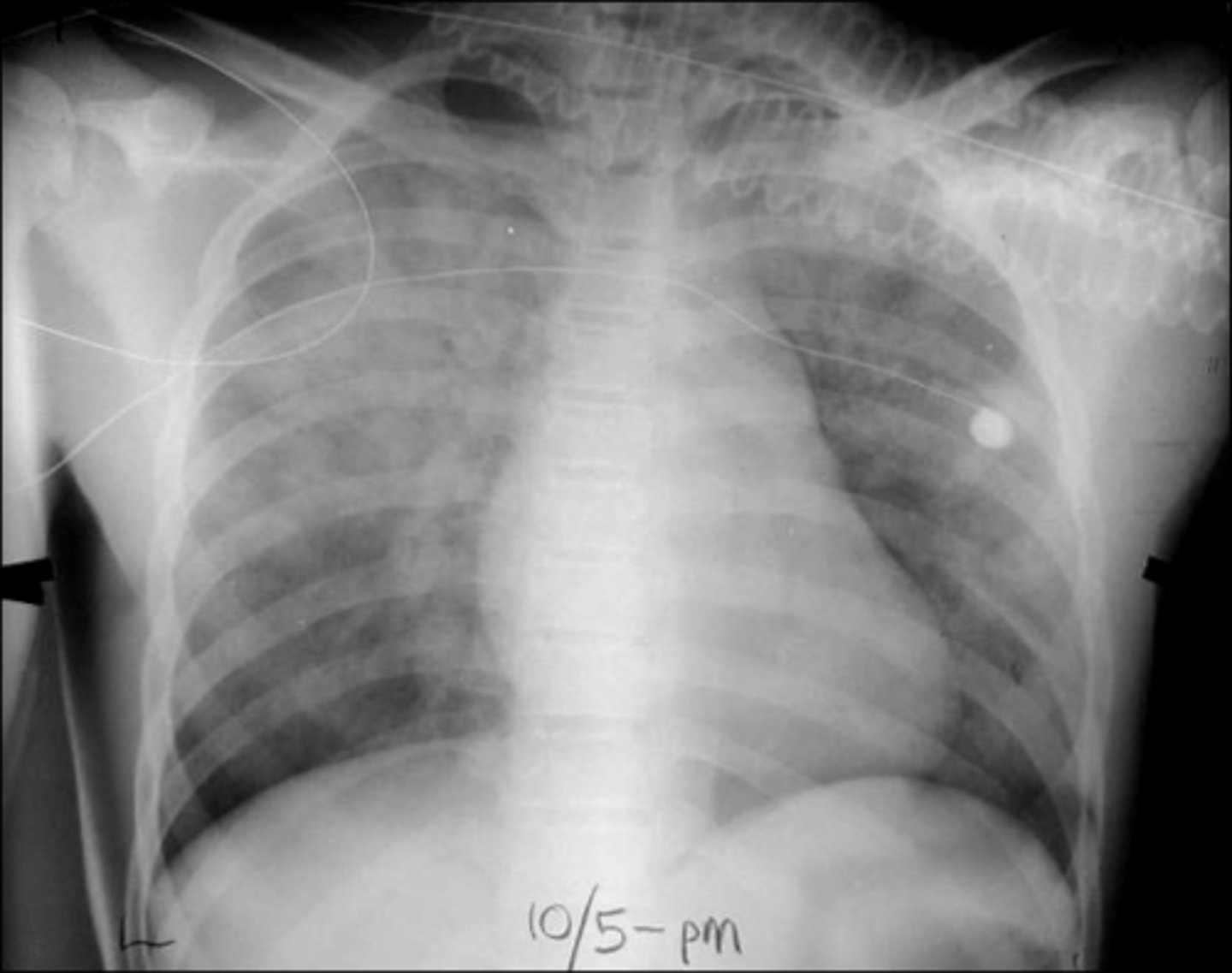
ARDS
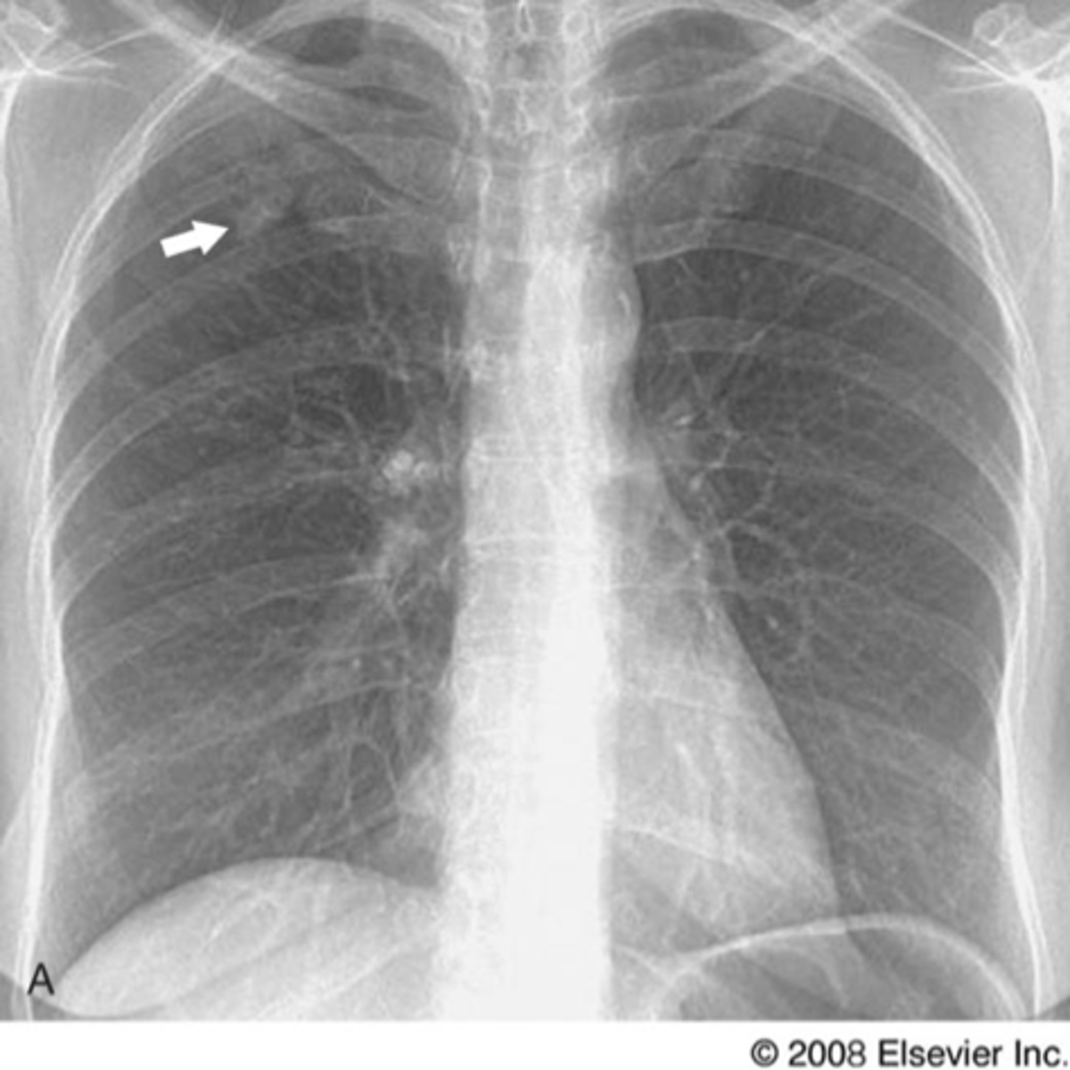
Solitary pulmonary nodule
Lung cancer- adenocarcinoma
Lung cancer- squamous cell carcinoma
Lung cancer- non small cell carcinoma
Pancoast tumor
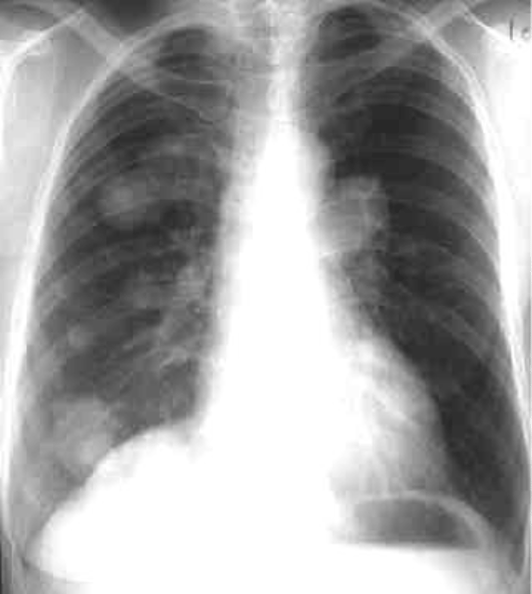
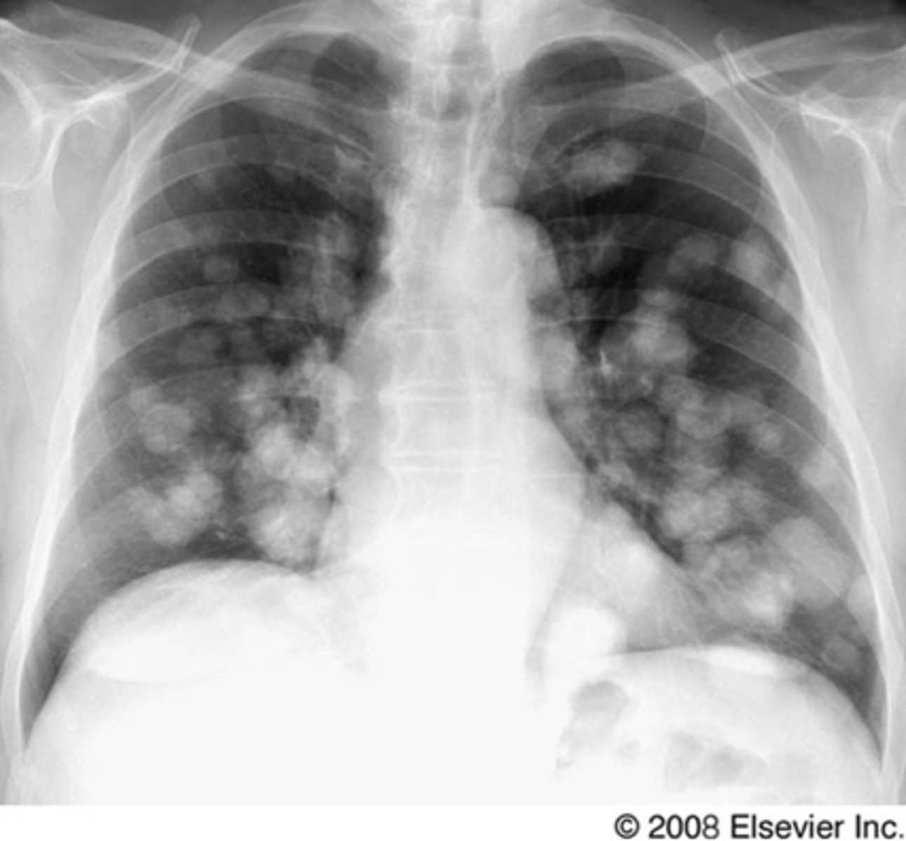
Lung cancer- metastatic disease
Silhouette sign right cardiac border- RML infiltrate
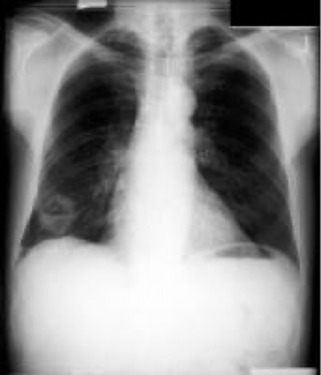
Bilateral interstitial infiltrates
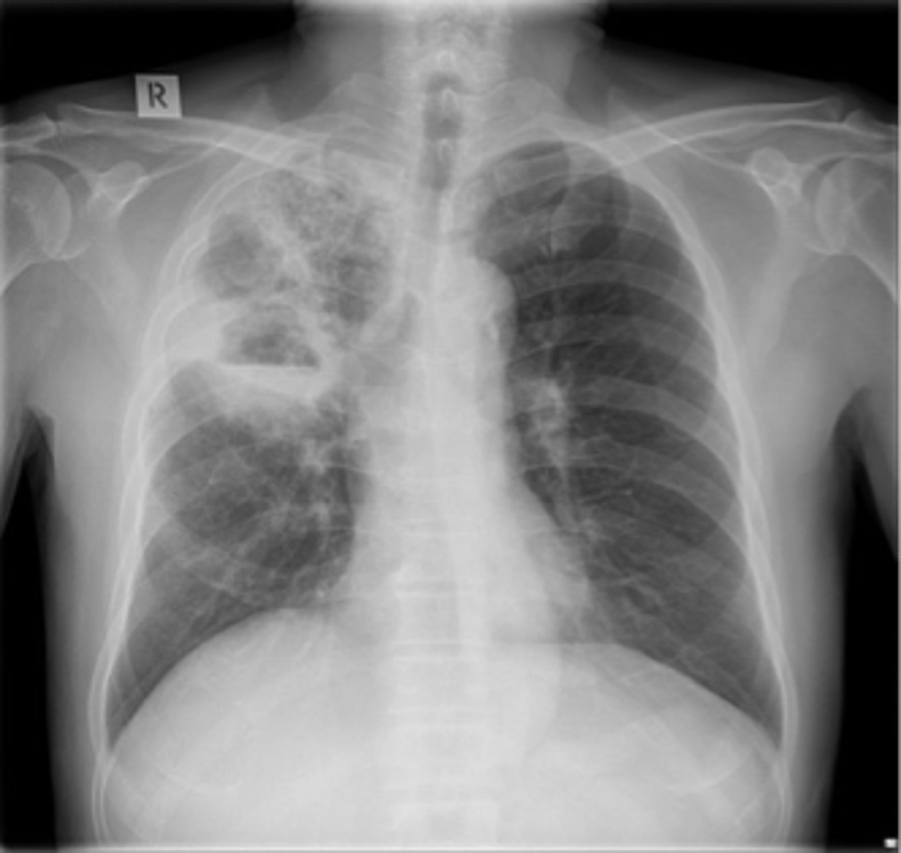
squamous cell carcinoma
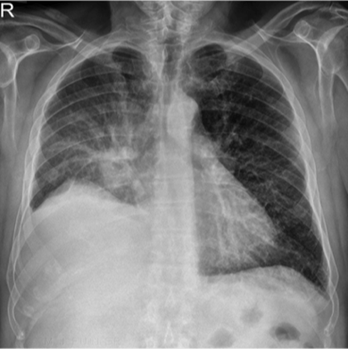
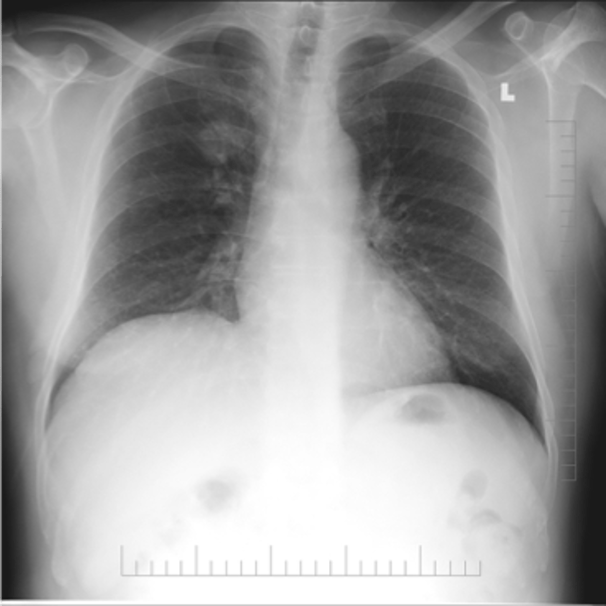
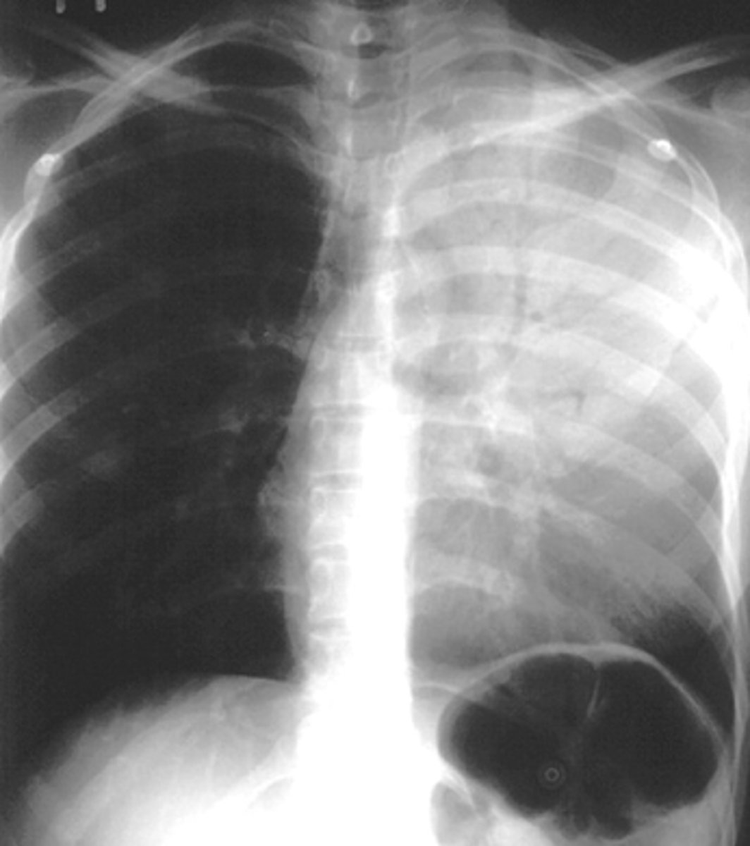
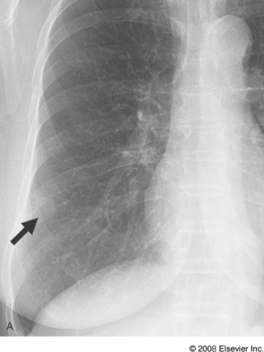
Bilateral pleural effusion
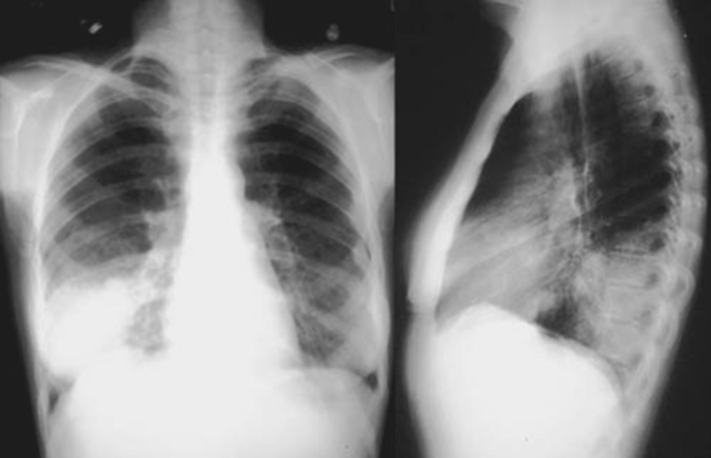
air bronchogram- alveolar consolidation LUL
Multiple nodules- metastatic
Nodule
Silhouette sign right cardiac border, RML