Anatomy: Cardiovascular System
0.0(0)
Studied by 3 peopleCard Sorting
1/30
Earn XP
Description and Tags
Last updated 7:43 AM on 11/15/22
Name | Mastery | Learn | Test | Matching | Spaced | Call with Kai |
|---|
No analytics yet
Send a link to your students to track their progress
31 Terms
1
New cards
Cardiovascular system function
Transport (via the blood)
nutrients and metabolic waste
CO2 and O2
Hormones
Heat
Etc
nutrients and metabolic waste
CO2 and O2
Hormones
Heat
Etc
2
New cards
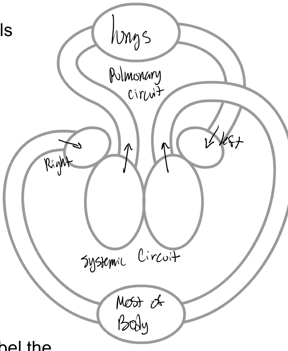
Circulatory routes
Pulmonary circuit= delivers blood to and from the lungs= for external respiration
Systemic circuit= delivers blood to and from the rest of the body= For internal respiration
Systemic circuit= delivers blood to and from the rest of the body= For internal respiration
3
New cards
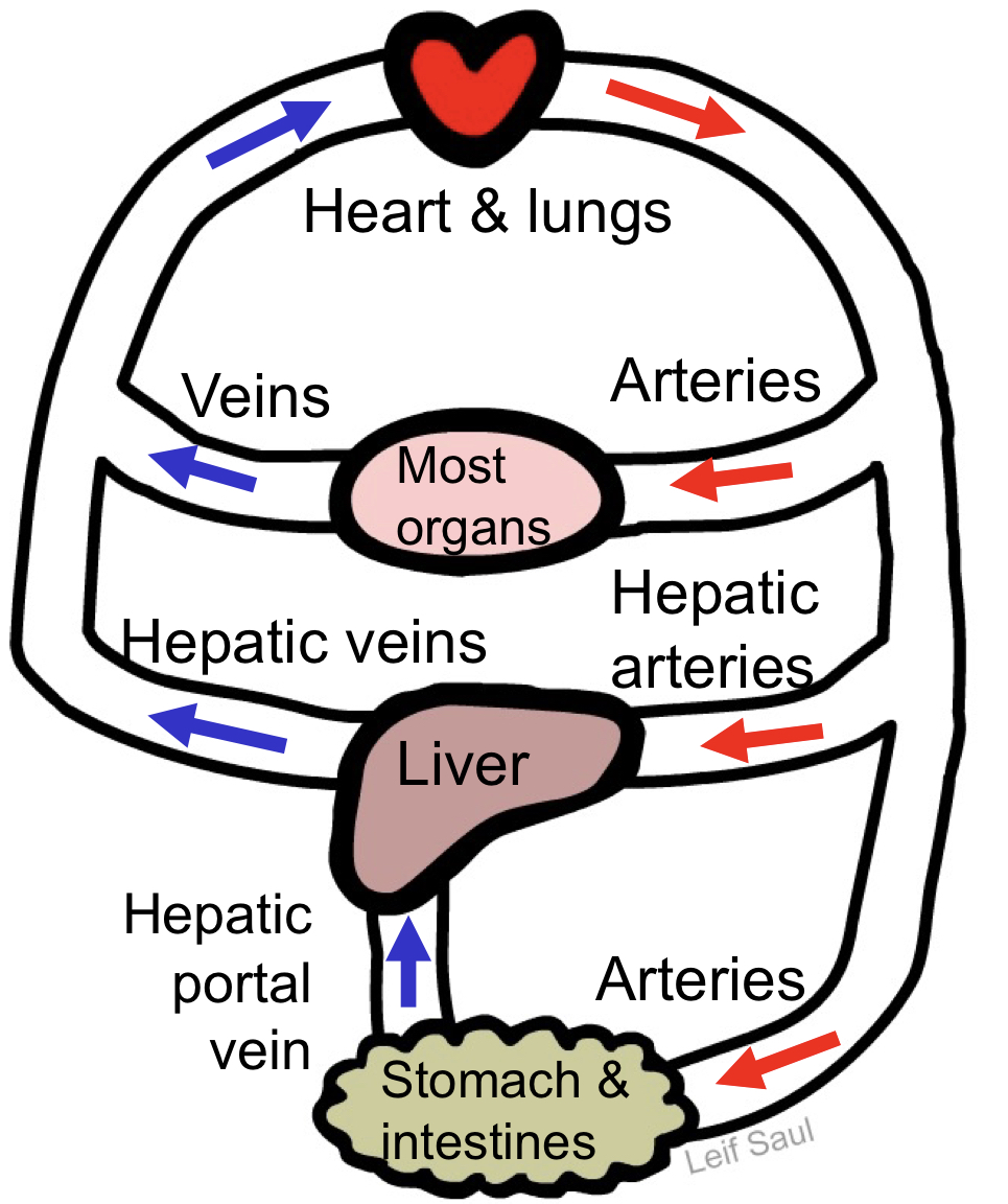
General circulatory principles
Capillary beds- where exchange takes place
Artery- delivers blood from the heart to capillary beds (oxygenated blood- most arteries not all)
Vein- delivers blood from capillary beds
Back to the heart (most veins)
Or to another capillary bed (these veins are portal veins)
Deoxygenated blood- most veins (not all)
Artery- delivers blood from the heart to capillary beds (oxygenated blood- most arteries not all)
Vein- delivers blood from capillary beds
Back to the heart (most veins)
Or to another capillary bed (these veins are portal veins)
Deoxygenated blood- most veins (not all)
4
New cards
Hepatic portal system
5
New cards
The heart
A muscular pump that circulates the blood
Surrounded by pericardial cavity in mediastinum
Surrounded by pericardial cavity in mediastinum
6
New cards
Coverings of heart (superficial to deep)
The heart is enclosed in pericardium
Fibrous pericardium (outer)- not serosa
Parietal layer of serous pericardium
Pericardial cavity- contains serous fluid
visceral layer of serous pericardium
Fibrous pericardium (outer)- not serosa
Parietal layer of serous pericardium
Pericardial cavity- contains serous fluid
visceral layer of serous pericardium
7
New cards
Cardiac tamponade
Compression of the heart due to excess fluid in pericardial cavity
8
New cards
The wall of the heart (from outer to inner)
Epicardium= visceral layer of serous pericardium
Myocardium- cardiac muscle layer
Endocardium- has simple squamous epithelium (endothelium) lines inner heart (including valves)
Myocardium- cardiac muscle layer
Endocardium- has simple squamous epithelium (endothelium) lines inner heart (including valves)
9
New cards
Chambers and vessels overview
10
New cards
Chambers and vessels
Atria (receive blood from veins)
Right atrium: receives deoxygenated blood from: inferior and superior vena cava; coronary sinus- returns blood from heart tissue
Left atrium: receives oxygenated blood from: pulmonary veins
Ventricles (eject blood from heart)
Right ventricle: pumps deoxygenated blood to: pulmonary trunk -> pulmonary arteries
Left ventricle: pumps oxygenated blood to:
aorta- coronary arteries (supplying blood to heart tissues) are branches of aorta
11
New cards
Heart valves
Prevent backflow of blood
Atrioventricular valves
Semilunar valves
Mnemonic: tri before you bi
Atrioventricular valves
Semilunar valves
Mnemonic: tri before you bi
12
New cards
Atrioventricular valves (AV valves)
Between atrium and ventricle
Tricuspid (R AV) valve- between the right atrium and ventricle
Bicuspid (mitral, L AV) valve- between left atrium and ventricle
These valves held in place by chordae tendinae, which are anchored to papillary muscles- prevents eversion (prolapse)
Tricuspid (R AV) valve- between the right atrium and ventricle
Bicuspid (mitral, L AV) valve- between left atrium and ventricle
These valves held in place by chordae tendinae, which are anchored to papillary muscles- prevents eversion (prolapse)
13
New cards
Semilunar valves (SL valves)
Between great arteries and ventricles
Aortic SL valve- between left ventricle and aorta
Pulmonary SL valve- between right ventricle and pulmonary trunk
Aortic SL valve- between left ventricle and aorta
Pulmonary SL valve- between right ventricle and pulmonary trunk
14
New cards
Heart sounds in each heartbeat
First heart sound (“lub”)= closing of both AV valves when L and R ventricles begin contracting
Second heart sound (“dup”)= closing of both SL valves when L and R ventricles begin relaxing
Second heart sound (“dup”)= closing of both SL valves when L and R ventricles begin relaxing
15
New cards
Conducting system
Heart muscle has intrinsic rhythm
Conducting system= specialized cardiac muscle cells
Initiates electrical signal (“firing”)
Signals heart chambers to contract in proper sequence
gap junctions spread signal from one cardiac muscle cell to another
Conducting system= specialized cardiac muscle cells
Initiates electrical signal (“firing”)
Signals heart chambers to contract in proper sequence
gap junctions spread signal from one cardiac muscle cell to another
16
New cards
Sequence of conduction
Sinoarterial node (SA node)= pacemaker
Note: all cardiac muscle cells can spontaneously fire, but SA node cells have fastest rate
Atrioventricular node (AV node)
Bundle of His (AV bundle)
Bundle branches
Purkinje fibers
Note: all cardiac muscle cells can spontaneously fire, but SA node cells have fastest rate
Atrioventricular node (AV node)
Bundle of His (AV bundle)
Bundle branches
Purkinje fibers
17
New cards
Disorders of conducting system
Heart block= damage to AV node or bundle of His (the only path from atria to ventricles)
Signal doesn’t reach ventricles
Ventricles still beat but at a slower pace
Artificial pacemaker restores normal function
Signal doesn’t reach ventricles
Ventricles still beat but at a slower pace
Artificial pacemaker restores normal function
18
New cards
Blood
A type of connective tissue
Components of blood:
Plasma= fluid with dissolved nutrients, etc.
Erythrocytes (red blood cells)- carry oxygen
Leukocytes (white blood cells)= immune cells
Platelets= cell fragments for clot formation
Components of blood:
Plasma= fluid with dissolved nutrients, etc.
Erythrocytes (red blood cells)- carry oxygen
Leukocytes (white blood cells)= immune cells
Platelets= cell fragments for clot formation
19
New cards
Function of blood vessels
Capillary- allows diffusion between blood and other tissues
Artery= carries blood away from heart
Vein= carries blood away from capillary beds
-eventually back to heart
Artery= carries blood away from heart
Vein= carries blood away from capillary beds
-eventually back to heart
20
New cards
General structure of blood vessel wall
1. Tunica intima (has endothelium= a simple squamous epithelium)
2. Tunica media (smooth muscle, collagen, elastin- all circularly arranged)
3. Tunica externa (collagen, elastin- all longitudinally arranged)
2. Tunica media (smooth muscle, collagen, elastin- all circularly arranged)
3. Tunica externa (collagen, elastin- all longitudinally arranged)
21
New cards
Arteries structure
(Compared to veins and capillaries)
Subject to highest pressure
Thicker walls- mainly due to thicker tunica media
More elastic
Subject to highest pressure
Thicker walls- mainly due to thicker tunica media
More elastic
22
New cards
Types of arteries
Elastic arteries= conducting arteries
largest artery is 1 cm to 1 in wide
Thick wall, highest elastin content
Very elastic- smooths out pressure fluctuations
Muscular arteries- most of the named arteries
0.3 mm to 1 cm wide
thickest tunica media relative to vessel diameter
Regulate blood pressure and distribution
Arterisles
Smallest arteries, 0.01 to 0.3mm
Regulate blood pressure and distribution
23
New cards
Capillaries structure
Facilitates diffusion
Wall only has tunica intima (mostly endothelium)- very thin wall
Tiny: capillary diameter
Wall only has tunica intima (mostly endothelium)- very thin wall
Tiny: capillary diameter
24
New cards
Capillary beds
Precapillary sphincters open when tissue is active- lets blood in the capillaries
Precapillary sphincters close with tissue in active- shuts off exchange
Blood still travels though metarteriole and thoroughfare channel
Precapillary sphincters close with tissue in active- shuts off exchange
Blood still travels though metarteriole and thoroughfare channel
25
New cards
Types of capillaries
Continuous capillaries
Fenestrated capillaries
Sinusoid capillaries
Fenestrated capillaries
Sinusoid capillaries
26
New cards
Continuous capillaries
Many tight junction between endothelial cells
In brain:
Completely sealed by tight junctions
all molecules must go across membrane of endothelial cell
least leaky of all
Blood-brain barrier
In most organs (muscles, lungs, skin, etc.):
Not completely sealed by tight junctions
Small molecules can pass through intracellular clefts (where tight junctions are absent)
27
New cards
Fenestrated capillaries
Have fenestrations= holes through endothelial cells (in other aspects, similar to continuous capillaries)
Allows more rapid exchange of small molecules
Kidney, endocrine glands, intestines, synovial membranes
Allows more rapid exchange of small molecules
Kidney, endocrine glands, intestines, synovial membranes
28
New cards
Sinusoid capillaries
have fenestrations
Intercellular clefts are large (very few tight junctions)
Allows exchange of proteins and cells, lots of fluid
Liver, lymphoid organs (spleen, red bone marrow)
Intercellular clefts are large (very few tight junctions)
Allows exchange of proteins and cells, lots of fluid
Liver, lymphoid organs (spleen, red bone marrow)
29
New cards
Vein structure
Very low pressure system
thinner walls than arteries- less smooth muscle and elastin- collapsible
Larger lumen then arteries- blood reservoir, ≈65% of total body blood
Have valves to prevent backflow of blood
thinner walls than arteries- less smooth muscle and elastin- collapsible
Larger lumen then arteries- blood reservoir, ≈65% of total body blood
Have valves to prevent backflow of blood
30
New cards
Types of veins
Venules (small veins)- receive blood from capillary beds
veins (other than venules) receive blood from venules
Portal veins- deliver blood from capillary bed to capillary bed (ex: hepatic portal vein)
veins (other than venules) receive blood from venules
Portal veins- deliver blood from capillary bed to capillary bed (ex: hepatic portal vein)
31
New cards
Mechanisms of enhancing venous return
Return of blood to heart is slow because of low pressure
Need ways to enhance blood return to heart
E.g., skeletal muscular pump: pressure changes (and valves) drive blood back to heart
Need ways to enhance blood return to heart
E.g., skeletal muscular pump: pressure changes (and valves) drive blood back to heart