mechanics of breathing
1/28
There's no tags or description
Looks like no tags are added yet.
Name | Mastery | Learn | Test | Matching | Spaced | Call with Kai |
|---|
No analytics yet
Send a link to your students to track their progress
29 Terms
what is the main muscle used in respiration?
diaphragm
what are the other muscles used in quiet respiration?
- external intercostal muscles
- internal intercostal muscles
what does the diaphragm consist of?
muscles fibres and central tendinous portion
what happens to diaphragm during inspiration? (4)
- diaphragm contracts and moves downwards
- the attached parietal pleura descends
- the visceral pleura also descends so the airways and alveoli expand
- air is sucked into the lungs
what happens to the diaphragm during expiration?
diaphragm relaxes and moves upwards
how is air expelled from the lungs in terms of muscles?
recoil of the elastic tissue in lungs
what happens to the ribcage and intercostal muscles during inspiration?
- external intercostal muscles contract and move the ribcage upwards and outwards
- the joints between posterior ends of the ribs and the transverse processes of the vertebrae enable the lower ribs to swivel upwards and outwards
what does the upwards and outwards movement of the ribcage cause?
- increases the lateral and anteroposterior diameter of thorax
- increases thoracic volume and making the negative pressure of the lungs more negative so air can be sucked in
what extra is used in forced respiration?
the accessory muscles of respiration
what are the accessory muscles of respiration? (4)
- abdominal (oblique, transversus and rectus abdominis)
- sternocleidomastoids
- scalene muscles
what innervates the diaphragm?
phrenic nerve (C3-C5)
what innervates the intercostal muscles?
intercostal nerves (T1-T12)
what are the microscopic parts of skeletal muscle biggest to smallest? (5)
- muscle
- fascicle (portion of muscle)
- muscle fibre (cell)
- myofibril (bundles)
- sarcomere (short units of myofibril)
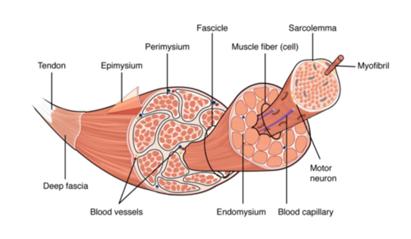
what is the structural unit of a muscle?
muscle fibre (cell)
what is the functional unit of a muscle?
sarcomere (z line to z line)
what are the two components of the sliding-filament theory?
actin and myosin
which is the thicker filament?
myosin
which is the thinner filament?
actin
what are the bands/zones/lines involved in the sliding-filament theory?
- A-bands
- I-bands
- H-zone
- Z - line
- M-line
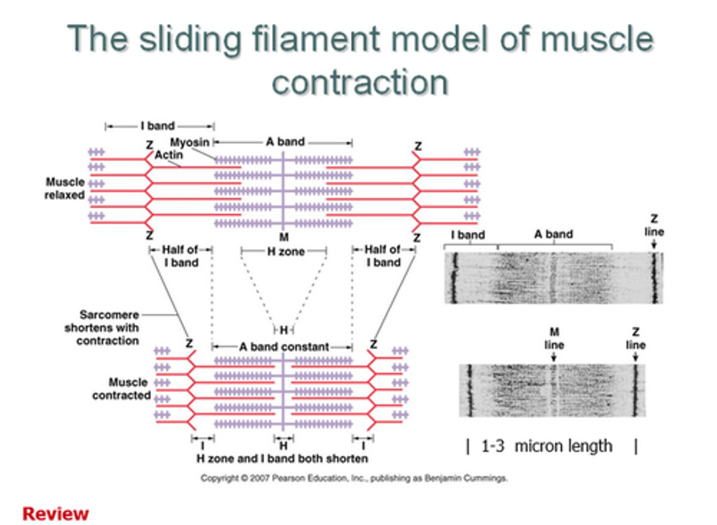
where do the A-bands cover?
entire myosin filament
where do the I-bands cover?
non-overlapping sections of actin filament
what is the H-zone?
non-overlapping sections of myosin filament
what is the Z-line?
end of sacromere
what is the M-line?
middle of sacromere
what happens to the A-band during contraction?
stays the same length
what happens to the I-band during contraction?
gets shorter
what happens to the H-zone during contraction?
gets shorter
how is are skeletal muscles stimulated to contract? (7)
- an action potential arrives at a neuromuscular junction
- causes an opening of voltage-gated calcium channels
- Ca2+ enter the cell
- causes vesicles containing acetylcholine to release contents into synaptic cleft
- ACh causes an influx of Na+ into muscle fibre causing depolarisation
- the depolarisation activates voltage-sensitive sodium channels
- causes an action potential in skeletal muscle fibre
how does the skeletal muscle contract? (7)
excitation-contraction coupling:
- depolarisation at neuromuscular junction
- conducted down t-tubules
- influx of calcium ions into sarcoplasm from sarcoplasmic reticulum
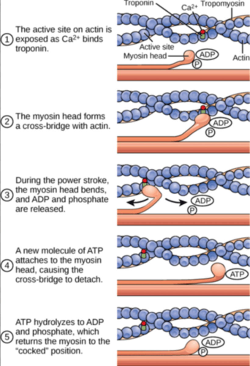
- calcium binds to troponin C causing a change in conformation that moves tropomyosin away from myosin head binding sites on the actin filaments
- this allows the myosin head to bind to the actin, forming a cross-link
- a power stroke occurs as myosin heads pivot in a 'rowing motion' moving the actin past the myosin towards the M line
- ATP then binds to the myosin head causing it to release the actin so the process can repeat