3. Digenea, cestoda, acanthocephala
1/23
Earn XP
Description and Tags
Helminths, TREMATODES & CESTODES! Digenea is subclass acc. to book, but a class acc. to lecture! Morphology, localization, imp. species, geo, epidemiology, LC, patho, CS, immune response of organism, pathology, dg, therapy and prevention. NB! Classification is according to the blue book!! (not notes/pres)... (different in qs.1)
Name | Mastery | Learn | Test | Matching | Spaced | Call with Kai |
|---|
No analytics yet
Send a link to your students to track their progress
24 Terms
Platyhelminthes, classification, characteristics, life cycle
Phylum: Plathyhelminthes (flatworms)
Subphylum: Trematoda, cercomeromorpha (from lecture)
Class: Trematoda, Monogenea, Cestoda (from lecture → digenea is class)
Subclass: Digenea
DIGENEA - Trematodes (flukes)
Morphology general:
Dorsoventr. flattened, bilateral symmetry, triploblastic (3 germ layer)
Acelomate (no body cavity)
No circulatory/resp. system → diffusion instead
incomplete dg. system (No anus)
Excretory system = protonephridia with flame cells
NS: ladder-like + cephalization
Mostly hermaprhrodites (except schistomatidae - separate)
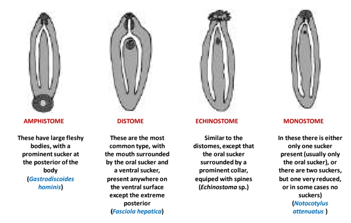
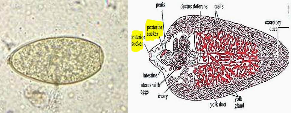
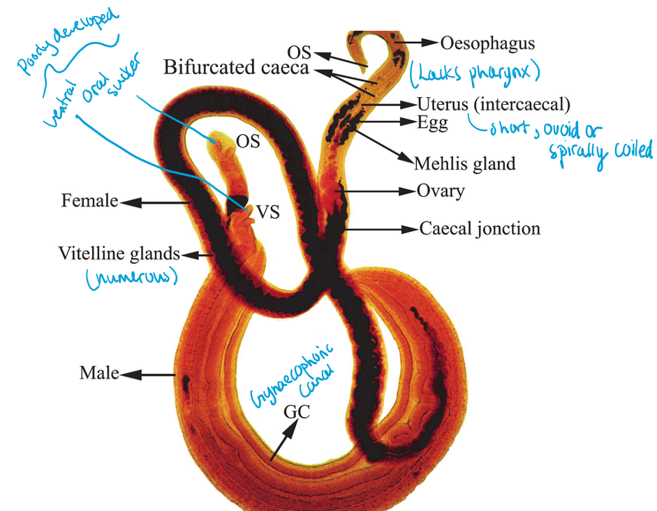
Digenea specific: Two suckers - Oral sucker & ventral sucker (acetabulum) → for attachment. Tegument (absorbs nutrients). Male 2 testes, female one ovary + uterus, yolk from vitelline glands. Different shapes/position of the reprod. organs acc. to species.
Schistosoma: elongated trematodes, separate sexes (male larger, holding female within a groove formed by a folding of the male body) - canalis gynecophorus. Found within circulatory system (ex. S.mansoni).
Location: Endoparasites. Found in most vertebrate groups (fish, reptile, birds, mammals) - FH, causing highly pathogenic infections.
Can be in most internal organs (lungs, bladder, blood) but majority are found in GIT, or closely linked organs like bile duct and liver.
Worldwide.
Digenea Life cycle: 2-4 different hosts for Digenea, several larval stages.
IIH: 1st: Snail (always), 2nd: aquatic animal, Asexual reproduction
FH: vertebrate, eats the 2nd IH. Sexual reprod.
Eggs: Flukes release (non-embryo or embryonated) eggs into the environment. Most have an operculum (not Schistosoma). Develops outside the host.
Digenea larval stages:
Miracidium → Sporocyst → rediae → cercariae (Furcocercaria in Schistosoma) → metacercaria (encysted stage, infective)
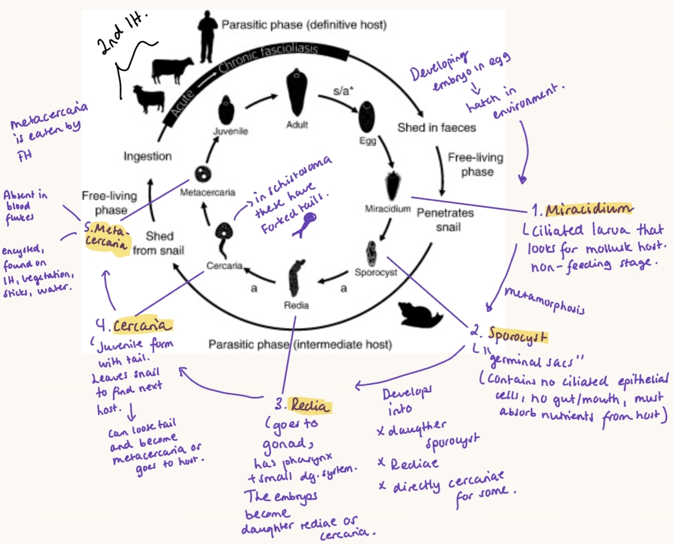
Digenea development:
Embryogony: egg → miracium, some hatch free in water, others only when eaten by IM.
Parthenogony (Asexual): inside IM, from miracidium → sporocyst, rediae and cercariae - 3 options, which is to either go to FH (schistosomes), go to 2nd IH and encyst as metacercariae or attach to aquatic vegetation and encyst.
Cystogony: development of metacercariae. In some species, this is missing. MC can either go to FH, remain in external environment, or go into 2nd IM. Transform into mecocercaria + MC are made in 3rd IM.
Maritogony: final maturation in FH, becoming adult.
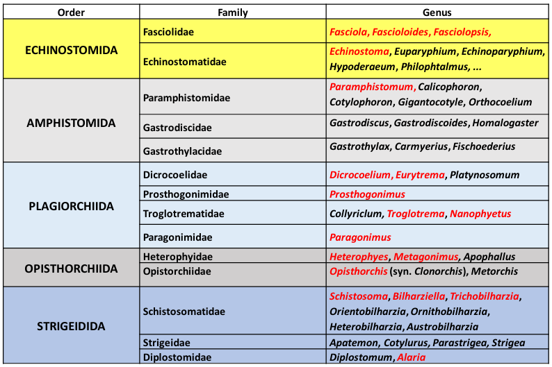
Classification of Digenean Trematodes:
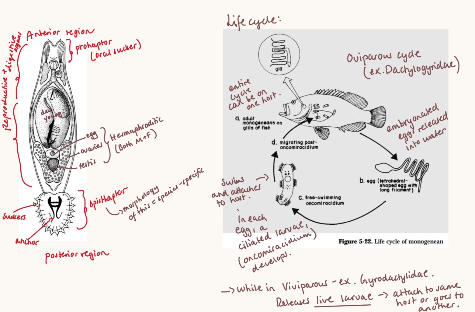
Class: MONOGENEA
Genra: Gyrodactylus (skin&fins), Dactylogyrus (Gills), Diplozoon
Species: Dactylogyrus extensus - in hatchery fish, economic important, large anchors on opisthaptor, lives on gill filaments → heavy infection cause blood loss, erosion, sec. infections, mucus in gills → suffocation.
Location: Infect gills, skin and fins of fish (ecto), some can be endoparasitc - bladder, mouth of turtles/amphibians. Worldwide.
Most are host-specific. Effects on host - insignificant, but some can cause death.
Morphology: spindle/circular, length from few mm to 3cm, Attaches to host by opisthaptor (Hooks/suckers), oral sucker (prohaptor).
hermaphroditic
Life cycle: Direct, with a single host. May be ether Ovi- or vivi-parous. (Egg or live young).
2.Fasciolosis of mammals (Fasciola, Fascioloides, Fasciolopsis).
Subclass: Digenea (Under class Trematoda)
Order: Echinostomida, suborder: Fasciolata
Family: Fasicolidae (Zoonotic)
Genus: Fasciola, Fascioloides, Fasciolopsis (Trematodes - high vet imp.!)
General:
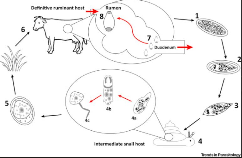
Fascolidae family is in liver, gall bladder, intestine. LC with 1 IH (fresh water snail). Cause fasciolosis in mostly ru, but sometimes humans.
Worldwide distribution, F.gigantica & Buski more in Asia, F.magna more in America.
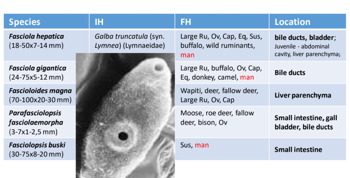
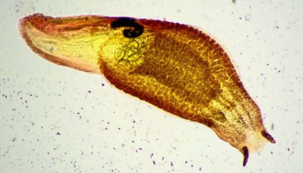
FASCIOLA SPP.
Species (both - ru, pigs, man):
F. hepatica (common liver fluke)
Location: Bile ducts, bladder. Juvenile - abd.cavity, liver parenchyma
F.Gigantica
Location: Bile ducts
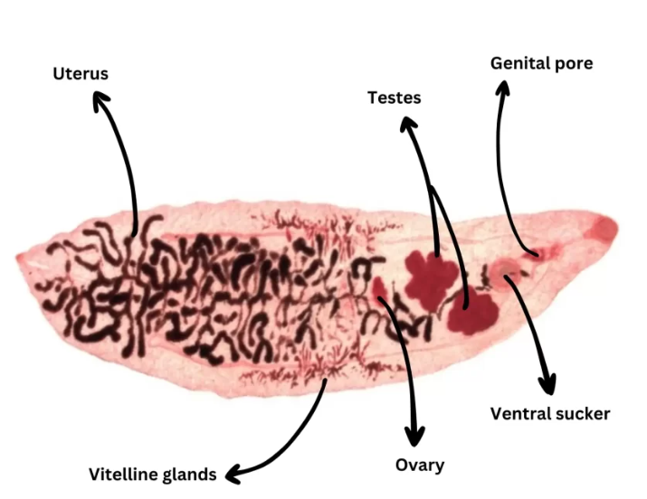
Morphology:
Leaf-shaped, large, apical cone on one end, branched testes + branched ovary. 2 suckers (oral + ventral)
Acetabulum → uterus → ovary → testes
gigantica is longer, slender, more rounded post. end
Eggs: Large, oval, symmetrical, 2 thin shells with operculum at one pole, unembryonated, yellow/brown color.
IH: Lymnaea truncatula (Freshwater snail) - F.Hepatica
F.Gigantica: Lymnaea auricularia
Transmission: Orally, contaminated water/grass with MC.
needs wet environment. MC lives best on grass/hay at temp. under 20deg. Dies at higher. Low zoonosis. Humans get MC from typ. water etc.
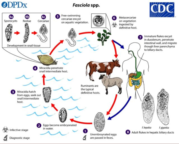
Life cycle: Indirect. 1 IH.
Eggs passed in feces → embryonates in water (Embryogony) → miracidium hatches → goes to snail - sporocyts → redia → cercaria (Parthenogony - asexual reprod.) → cercaria leaves snail and encysts on vegetation as metacercaria (Cystogony) → FH eats MC on grass/water plants.
Maritogony (inside FH) - Juvenile from MC excyst in intestine, wall → liver → enters bile ducts and matures → eggs appear in feces - 3months after infection.
Pathogenesis & CS (Pathogenesis depends on ingested number):
Juvenile migration in liver → traumatic hepatitis + hemorrhage
Adults in bile ducts → cholangitis + fibrosis
Cathepsins released by adult → liver damage, inflammation
Cattle: calcification of bile ducts & gall bladder enlargement. Heavy - subm. edema, red. milk yield & quality.
Clinical forms:
Acute (>2000): heavy infection, esp. sheep → severe hemorrhage/abd.bleeding, sudden death
Subacute → hemorrhage + fibrosis, non-specific signs
Chronic (most typical, -500): anemia, weight loss, emaciation, bottle jaw (submand. edema).
F.Gigantica - similar, fibrosis, cholangitis.
Diagnosis: sedimentation, Serology (ELISA, IHA - Ab in serum from 3rd week post-infection), CS, PM.
Sheep - do not gain resistance to rei-infection, cattle do to a certain level.
Treatment: Praziquantel, Albendazole, mebendazole.
Acute: Stop grazing, flukicide (diamfenetid), subacute/chronic: flukicide (notroxynil)
Control: treatment of hosts, reduce snail habitats, heat treatment for plants
Other:
Fascioloides magna (large american liver fluke, not zoonotic - low): Pseudocysts in liver.
Deer/cattle: less pathogenic, encapsulated in liver
sheep/goat: continuous migration - truamatic hepatitis, often fatal.
vascular lesions (thrombosis), dies after 6m.
More rounded post. end. up to 10cm.
Triclabendazole
Fasciolopsis Buski (Asian giant intestinal fluke).
FH: man/pig, in SI. Zoonotic
generally little pathology, can lead to irritation/obstruction, malabs. Intoxication. Severe → circulatory failure.
praziquantel
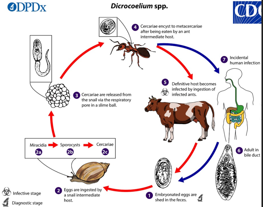
3. Dicrocoeliosis of ruminants (Dicrocoelium, Eurytrema, Platynosomum)
sub-Class: Digenea (Under class Trematoda)
Order: Plagiorchida
Family: Dicrocoelidae
Genus: Dicrocoelium, Eurytrema, Platynosomum
General:
Location: liver, gall bladder, bile ducts and sometimes pancreatic ducts
FH: reptiles, birds, mammals. (Incl. humans, zoonotic potential but RARE).
Usually indirect LC iwth 2 IH: 1st: Land snail, 2nd: Insect (ex.ant)
Eggs: small, dark brown, operculated + embryonated
Compared to Fasciola - smaller, less acute liver migration damage (no liver migration through parenchyma)
worldwide.
Does not require wet environment, eggs survive long on dry pasture. Massive infection of snail/ant occur during grazing season.
Morphology:
Adults: Small, slender, transparent, well-developed suckers, body mostly filled with uterus containing eggs.
Acetabulum → testes → ovary → uterus (different from fasciola)
no spines on cuticle (diff. from fasciola)
Eggs: Small, oval/symmetrical, operculum, 2 thick shells, embryonated (with miracidium), dark-brown.
Patho&CS:
Flukes live in bile ducts → inflammation, fibrosis, enlargement
Usually mild/subclinical, unless heavy infection.
if signs do occur - anemia, emaciation, red. milk/wool prod., signs similar to fasciolosis.
Dg: sedimentation
Treatment: Triclabendazole, Reinfection common (No strong imunity)
1) D.Dendriticum (Worldwide, not America/Australia)
FH: Ru, camels, rabbit, man, horse (Mostly bile ducts of sheep), IH2:Ant
spear/Lance shaped.
LC: Eggs from host → eaten by land snails → hatch in intestine, miracidia goes to gut wall → sporocyst produce daughter sporocyst → cercariae → leaves and accumulate in the mantle chamber → “slime balls” with a lot of cercariae → released on plants → eaten by ants. Snail release cercariae in slime balls → eaten by ants → brain → metacercariae encyst there → changes the behavior, so when temp. drops in the evening, ants climbs to tips of grass → FH ingest them while grazing.
in FH: NO liver tissue migration. Juveniles migrate directly through ductus choledochus (bile duct opening)
NO REDIA!
2) Eurytrema Pancreaticum
FH: RU (mainly in pancreatic ducts!)
IH2: Grasshopper
Oval/fusiform, large suckers (oral/ventral)
Patho/LC: similar but mainly pancreatic involvement.
3) Platynosomum Fastosum
FH: cats (esp. outdoor cats) (Found in bile & Pancreatic ducts)
IH: 1st snail, 2nd: Lizards/geckos/toads
Looks like D.dendriticum, but has testes horizontal rather than tandem (one behind the other).
CS: non-specific like Vomit/diarrhea, icterus. Possible death. Hepatic dysfunction → not eating.
Praziquantel - for treatment
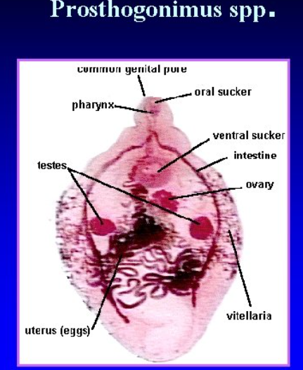
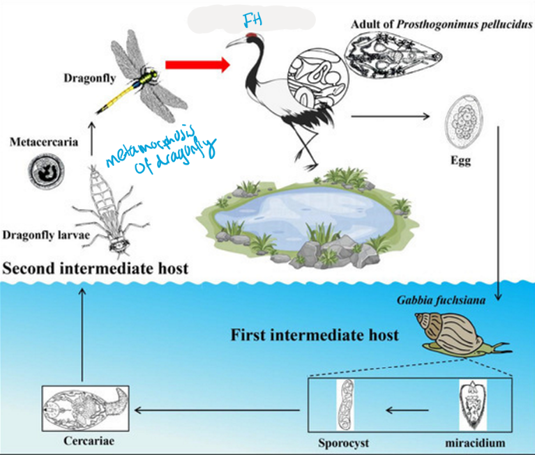
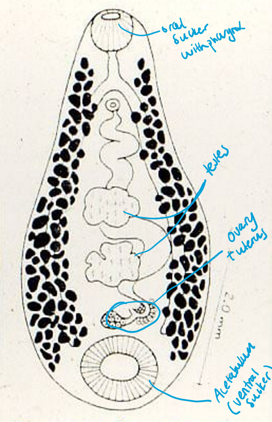
4. Prosthogoniosis and other trematodonoses in birds
SubClass: Digenea (Under class Trematoda)
Order: Plagiorchida
Family: Prosthogonimidae
Genus: Prosthogonimus
Worldwide. Considered to be most pathogenic trematodes of poultry in Europe/America.
Important species:
Prosthogonimus Ovatus (Fowl, geese, wild birds)
P. Macrorchis (Poultry, ducks)
Organ damage, prevents egg laying in some cases
P.pellucidus (fowl, duck, wild birds)
P.cuneatus (duck, goose)
Mainly affect: Bursa fabricii & Oviduct, sometimes post.intestine (Peullucidus)
Morphology - adult:
Flat, transparent, oval/pear shaped. spines on cuticle.
Uterus occupies ½ of body on posterior part
Ovary contains multiple lobes, bw. ventral sucker and testes. 2 Suckers. Testes is misaligned in many.
Eggs: Small, oval, 2 thin shells, small button on the other side, miracidium inside (embryonated). yellow/brown.
FH: ducks, chickens, geese, wild birds
IH: 1st: Water snail, 2nd: Dragonfly nymph
Life cycle: unembryonated Eggs released into water → water snail → hatch into miracidium → sporocyst → cercaria → released and goes into anal openings of dragonfly nymph (breathing movements of these insects) → bird eats dragonfly → MCencyst in SI → migrates to oviduct/bursa → matures → atrophy of bursa.
Pathogenesis:
Irritation in oviduct → acute inflammation + Retroperistalsis
Leads to yolk, albumen, bacteria + parasites to enter abdominal cavity → egg yolk peritonitis. Can be fatal.
Produce abnormal eggs, discharges of albumen from cloaca
Clinical signs:
Early: Soft-shelled or shell-less eggs, decr. egg production
Later: cloacal discharge, dirty feathers around claoca, pendulous abdomen, weak, legs far apart when walking
Severe: Peritonitis, cyanotic comb/wattles, death
PM: Inflamed oviduct, cheesy/yolk material in abdomen (lumen), adhesions/cheesy mass of organs - in case of peritonitis. Can also be congestion&hemmorrage.
Diagnosis: Fecal examination (eggs), PM
Treatment: albendazole, fenbendazole, praziquantel.
Control - avoiding humid places, where dragonly nymphs can develop.
Other Bird Trematodes:
Blood flukes:
Bilharziella Polonica → “Swimmer`s itch” water poultry, dermatitis.
Intestinal flukes:
Echinostoma revoltum → intestine/cecum
Plagiorchis arcuatus → bursa/oviduct
Triglotrema acutum → can penetrate bone/brain
Opistorchis simulans → gall bladder of duck/geese
5. Paramphistomatidosis of ruminants.
SubClass: Digenea (Under class Trematoda)
Order: Amphistomida
Family: Paramphistomidae
Genus: Paramphistomum, Calicophoron
Worldwide, Europe
Main species: Paramphistomum cervi (most common), Calicophoron daubneyi, P. ichikawai, P. microbothrium - are most pathogenic
Morphology:
Amphistomida: Amphi (on both sides), stoma (mouth) - 2 suckers (anterior, larger posterior).
Pear-shape, small, light pink/red (Hb), testes usually at middle
Same organs as rest of Digenea class.
Eggs: Oval, operculum, large, 2 shells, unembryonated, colorless. similar to F.hepatica, but are a bit longer.
Hermaphrodites, complete reproductive system.
Location: rumen, juvenile in small intestine (duodenum/abomasum)
Infection per: orally (MC)
Epidem: Common in ru, may survive for years in humid places, MC - resistant shell. Continuous source of snail infection. Snails - can shed a high amount of cercariae. Outbreaks typ. occur during drier months, when snails concentrate around water sources. Older animals may develop immunity over time. Small immunity amount after infection (like F.hepatica).
FH: Ruminants (Fish, amphibians, reptiles, birds, mammals)
IM: Water snail (Planorbidae)
Life cycle: Similar to Fasciola Hepatica.
embryonated egg in feces → Miracidium → snail → cercaria → MC on plants → Ru eats MC
excyst in duodenum (juvenile) → goes to rumen to become adult.
Pathogenesis:
Juveniles in SI: attach to mucosa → erosion/necrosis, hemorrhage, enteritis/duodenitis
Adults in rumen: bleeding, nodules, damages papillae - papillary atrophy, decr. digestion/appetite.
CS:
Acute/intestinal (usually due to juveniles): Severe diarrhea, anorexia, Thirst, anemia, edema (hypoalbumin), weak, death possible
Chronic/rumen (adults): often mild/no signs.
Diagnosis: sedimentation methods, PM - pink flukes in duodenum. Pathology such as fluid diarrhea, finding flukes in feces.
Treatment: closantel, albendazole
control of snails - difficult, Molluscicide, pasture control.
Gigantocotyle explanatum– found in biliary duct of large ruminants (wild and domestic). Recently come to Europe (Italy), originally found in Asia, Africa and south America. Life cycle is the same.
6.Opistorchiida (Opistorchis, Clonorchis, Metagonimus, Heterophyes)
SubClass: Digenea (Under class: trematoda)
Order: Opistorchiida
General of the order:
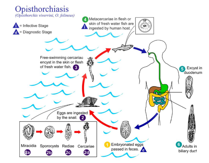
All are food-borne liver or intestinal flukes
Infection = Raw/undercooked fish (SUSHI)
FH: Human, fish-eating mammals
IH: 1st: snail, 2nd: Fish, amphibians, for M.yokogawai = sweet fish/cyprinid fish, For Heterophyes = also frog.
Epidemiology - fishermen at high risk, defecation in water → contamination
Dg: Eggs in feces
Treatment: Praziquantel
1) Family: Opistorchiidae (Liver flukes)
Genus: opistorchis/Clonorchis
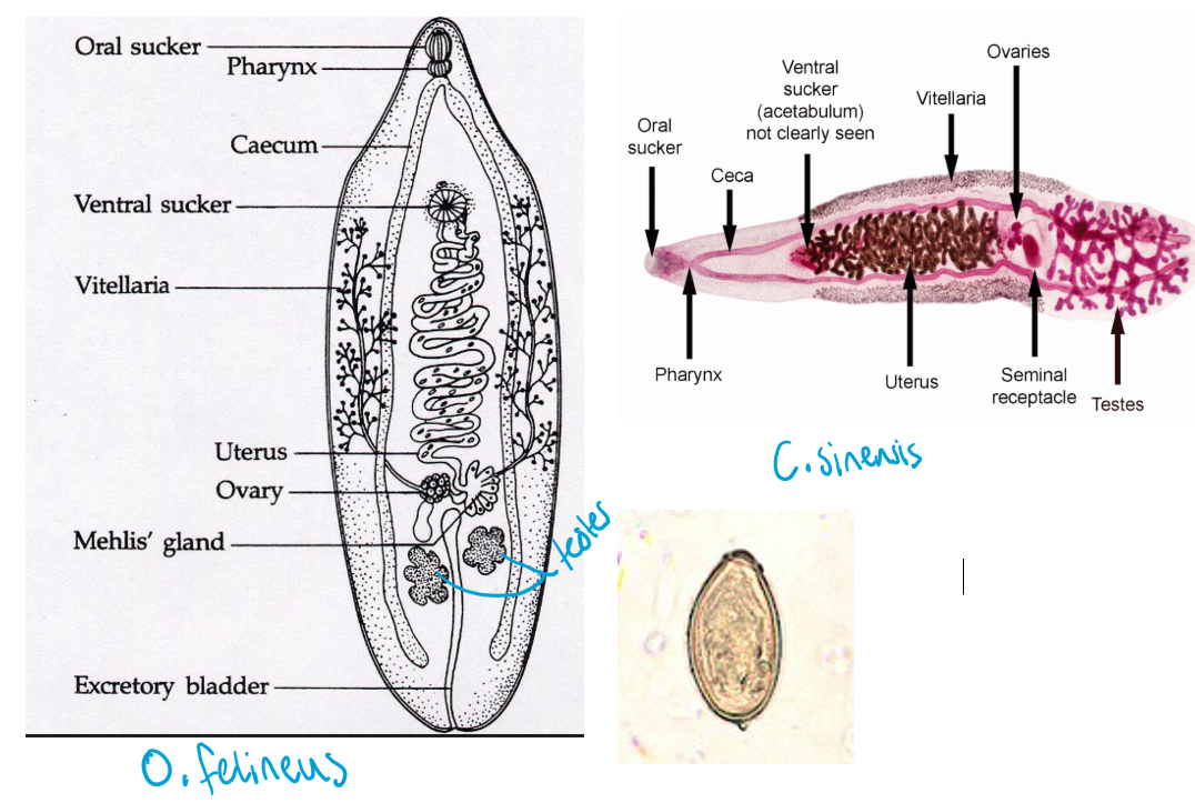
Important species: Opistorchis felineus (europe - cat liver fluke), O.viverrine, Clonorchis sinensis (All - Asia)
Location: Bile ducts, liver, gall bladder (Zoonotic)
Morphology:
Weakly developed suckers, 2stk - oral + smaller ventral
O.felineus - lobed testes
Clonorchis sinensis - like F.hepatica, also bigger than opistorchis. Trea-like branched testes + vitelline follicles different distributed.
Egg: S, Oval, asym., operculum, 2 shells, embryo (miracidium), yellow
Life cycle:
Embryonated eggs in feces → snail → sporocyst → rediae → cercariae (1000+) → infect fish and encyst → metacercariae under skin/muscle → FH eats raw fish → MC excyst in intestine → matures in bile duct.
Pathogenesis:
Juvenile stages: Hepatitis, due to liver parenchyma migration.
Adult: cholangitis, pericholangitis (bile duct inflammation + tissues around, enlargement of bile ducts), pancreatic damage
Clinical signs:
Enlarged & painful liver (pressure sensitive)
icterus, ascites (advanced case)
2) Family: Heterophyidae (Intestinal Fluke)
Genus: Heterophyes, Metagonimus
Species: Heterophyes Heterophyes, Metagonimus Yokogawai
Location: SI
A) H.Heterophyes (Asia)
Morphology:
Very small intestinal fluke, teardrop/pear shape, oral sucker, strong ventral sucker with spines
simple intestinal system, does not have cirrus pouch.
Genital sucker with incomplete circle of small rods
Life cycle (Same pattern as liver flukes): Egg → snail intestine → cercariae → fish → MC → human eat raw fish → MC excyst in SI, mature.
Pathogenesis & CS: Mild intestinal inflammation mostly.
Heavy infection: Diarrhea, abdominal pain (colicky)
Rare severe: eggs can migrate → Heart or CNS damage (fatal cases)
B) Metagonimus yokogawai
Same general LC/signs as H.heterophyes, more toxic inflammatory effect in intestine, invading intervillous space, severe local inflammation by toxin release of MC.
Morphology: Spined tegument, genital pore, scaly surface. Abdominal sucker is more ventral from middle.
FH: Dogs/cats, humans, rats/pigs. Zoonotic!
7.Schistosomosis in Mammals.
Subclass: Digenea (Under class trematoda)
Order: Strigeidia
Family: Schistosomatidae
Genus: Schistosoma (Blood flukes)
also: S.bovis
Typical:
Separate sexes (Gonochorists)
Female is in longitudinal groove of male (Gynecophoral canal)
Adults live in bv. (intravascular parasites)
Morphology:
Female is thin and long, male is shorter and broader.
Eggs: embryonated, has spine/thorn
Furcocercariae: Forked tail
Epidem: Tropical/subtropical areas, Common in Sub-saharan Africa. Infection linked to contaminated freshwater, poor sanitation, freshwater snails - contact with water infested by cercariae.
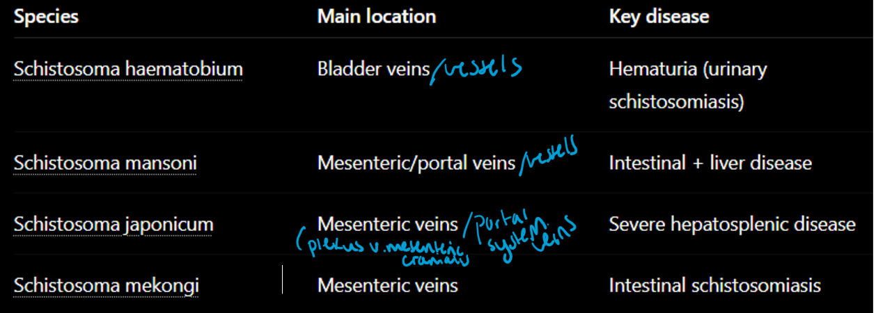
Location: BV - mesenteric, portal veins, bladder venous plexus
FH: Human, mammals, birds (Zoonotic)
IH: freshwater snails
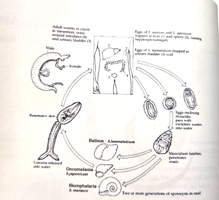
LC: Eggs released in feces/urine → Miracidium hatches in water → snail → sporocyst → furcocercariae → into human skin (by penetration glands - branders organ) → loses tail → schistosomula → migration (skin - blood - lungs - liver - final veins) → adults mate, eggs.
ONLY sporocyst & Furcocercariae/schistosomulae present.
Species differences:
S.haematobium: eggs are released with feces/urine. Cause hematuria & Urinary schistosomiasis.
S.Mansoni: Eggs moved from lumen of intestine, shed with feces. Causes hepatosplenomegaly, portal hypertension, ascites.
“Spider veins” on abdomen surface in humans
S.japonicum: eggs in feces, more severe liver disease -gradual enlargement in acute stage. Cause liver fibrosis - liver shrinks + hardens, edge granulated in chronic. enlarged spleen, GI bleeding, thrombocytopenia.
Pathogenesis: Disease is mainly IMMUNOLOGIC - signs are rarely seen except in heavily infected. damage caused mostly by eggs trapped in tissues.
Clinical phases:
Skin penetration phase - cercarial dermatitis, itching, rash, petechia
Migration phase - non-specific signs, fever, cough, sweat, diarrhea + pulmonary congestion. (can be no symtpoms).
Acute phase - Intestinal species (Mansoni, japonicum) - bloody diarrhea/feces, urinary (haematobium) - hematuria
Chronic - Hepatic dysfunction, fatigue, chronic bloody diarrhea with mild abd. pain and lethargy.
Sometimes, affecting CNS - lesions, like flaccid paraplegia, myelitis (Mansoni&Haematobium).
Diagnosis: Eggs in feces/urine, serology, rectal biopsy, PM
Treatment/Control: Praziquantel. Avoid contaminated water, snail control, sanitation.
Canine schistosomiasis (Heterobilharzia americana)
In dog - north america, mesenteric veins
same diagnosis, same treatment - similar to schistosomatidae.
Swimmer`s Itch (Schistosome dermatitis)
Caused by bird schostosomes - trichobilharzia, gigantobilharzia, ornithobilharzia (Duck, geese)
Humans are accidental host
itchy allergic skin rash after swimming (cercarial dermatitis)
8.Other trematodoses of veterinary importance (Paragonimus, Alaria, Troglotrema, Nanophyteus)
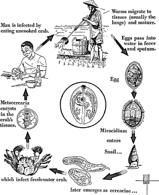
1) Paragonimus (Lung fluke)
family: Paragonimidae (order: Plagiorchiida) under Digenea.
Lung trematode, chronic lung disease (Paragonimosis)
Species: P.westermani, Pulmonalis, Kellicotti (cats)
Location: Mainly lungs, may migrate to brain, spinal cord, brain (rare)
Morphology: Thick cuticle with small spines (imp. for dg.)
Geo: Worldwide
FH: human, car, pig, ru (zoonotic potential)
IH: 1st: Snail, 2nd: Crustaceans (crab/crayfish)
LC: Eggs released in feces → miracidium → snail (sporocyst → redia → cercariae) → infect crayfish/crabs → metacercaria develops → FH eats 2nd IH → juveniles migrate to lungs → adults (eggs can be coughed up and swallowed).
Pathogenesis & CS:
Adult flukes - inflammation, granulomas & lung cysts
acute: non-specific signs, fever, weak, abd.pain, cough, sweating, bleeding/inflamamtion in tissues (or can be asymptomatic). Lungs - encapsulated cysts.
Chronic: Cysts with adult parasites, eggs + purulent fluid, patients with taste of fish in their mouth. Granulomas form around the cysts, walls - calcify.
P.pulmonalis: parasitic cysts (only one with this, others have no cysts, but migrate in pleural cavity), severe
P.westermani: Inflammatory response, granulomas formation, migrate into heart + brain → death. CS: chronic cough, breathing issues, blindness, imbalance + epilepsy.
Kellicotti - chronic intermittent cough, rusty sputum.
Severe migration - brain/heart → blindness, epilepsy, imbalance, death
Diagnosis: eggs in sputum, fecal examination, history (eating crayfish), radiographs may also help (adult fluke in lungs)
Therapy: Praziquantel, albenda/fenbendazole (health checks of crayfish can help)
2) Genus Alaria
Family: diplostomidae, order: Strigeidida, under Digenea.
Species: A. alata, A.canis
Location: SI
Morphology:
flattened body, characteristic anterior flaps (pointed processes), oral + ventral suckers - for nutrients which leaves through a single pore. Bifurcate intestine.
Eggs: operculated, unembryo, large, yellow/green, 2 shells
FH: Car, IH: 1st: water snail, 2nd: tadpole (baby frog)
Paratenic host: Water snake
Life cycle: Eggs → miracidium → snail → furcocercaria → tadpole → mesocercaria. Then - directly eaten by car or transferred to paratenic host → mesocercaria migrate (lungs - trachea - swallowed - intestine)
CS: usually non-specific or mild
3) Genus: Troglotrema
Family: troglotrematidae, Order: Plagiorchida
species: T.acutum (Central Europe)
Location: Nasal sinuses of cats/car, under skin of birds
Morphology: Tear-drop shape, strong suckers
IH: Snails&Frogs
Patho/CS: bone perforation, penetrate into brain
cause osteitis, severe pain
Yellow-white nasal discharge, painful sinus disease
Treatment: Poor prognosis, can try using triclabendazole, often euthanasia.
4) Genus: nanophyetus
Family: Nanophyetidae (or troglotrematidae acc. to lecture), Order: Plagiorchiida
species: Nanophyetus Salmincola
Location: crypts of SI.
Morphology: tiny, large testes opposite each other
FH: fish eating mammals (dog/cat, human)
IH: 1st: freshwater snail, 2nd: salmonid fish
LC: snail → cercaria infect salmon fish → MC in fish muscle/kidney → mammal eat raw salmon → adult develop in intestine.
The parasite is NOT linked with clinical disease, but it serves as the vector of Neorickettsia helminthoeca - causative agent of salmon poisoning (fatal in dogs)
9.Morphology & Classification of CYCLOPHYLLIDA
Class: Cestoda (tapeworms)
Subclass: Eucestoda
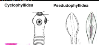
Orders: Pseudophyllida & Cyclophyllida
General: Endoparasites/internal parasites affecting liver or digestive tract, body: Dorsoventrally flattened, segmented, elongated
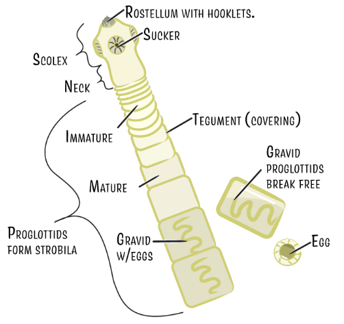
Body structure:
1) Scolex (head) - Attachment organ, to intestinal wall
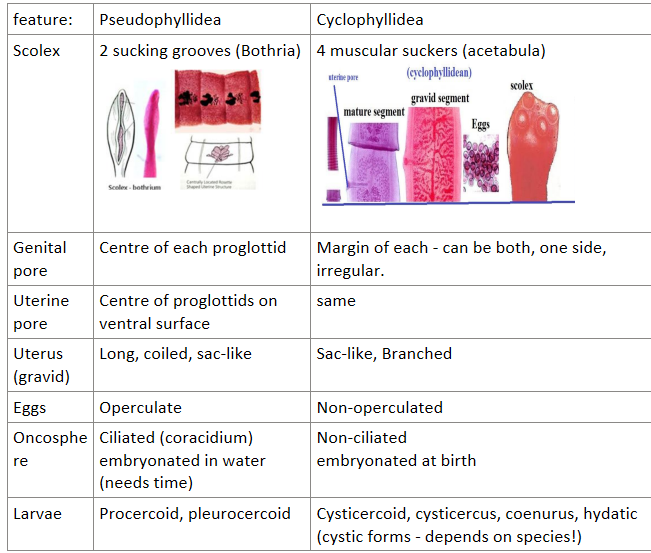
cyclophyllidea have: 4 muscular suckers (acetabula), sometimes rostellum with hooks for attachment
Pseudophyllidea: weakly muscled grooves (bothria, 2per scolex)
2) Neck - Makes new proglottids (strobilation - asexual formation)
3) Strobila - chain of segments = proglottids. That may extend for a few or thousands. Usually very long - occupying entire SI.
types: immature, mature, gravid
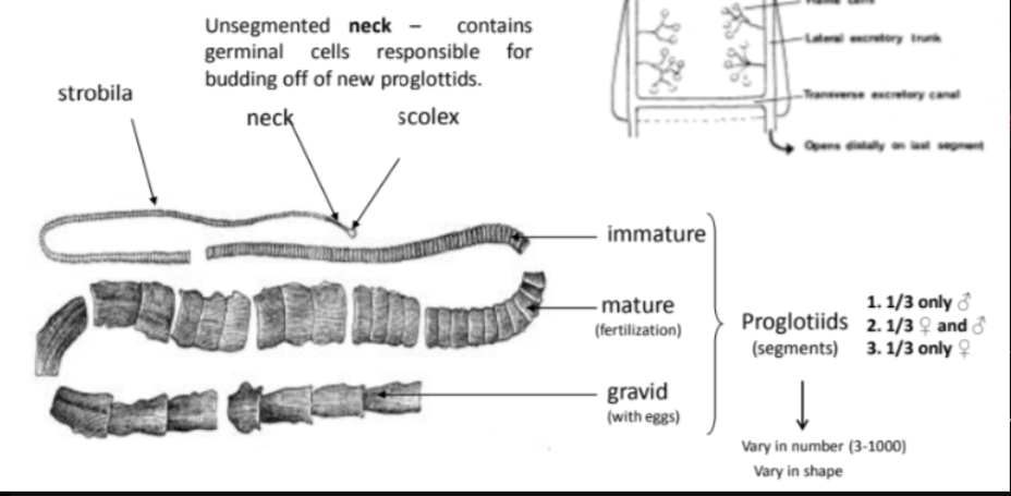
Digestive system: Completely absent. Nutrients absorbed through tegument (outer body wall - similar as in trematodes, role in nutrient absorption, surface has microvilli/microtriches).
Nervous & Excretory system: Similar to trematodes - has cerebral ganglion in scolex (main nerve center). Flame cell (protonephridial) excretory system.
Reproductive system:
Proglottids - are budded off from the head and neck region. These are hermaphroditic, egg producing units. In cross-sections the body is seen to be filled with parenchyma. Muscle layers separate the body into outher cortex and inner medulla. The reproductive organs are usually in the medulla. Each proglottid = male + female organs - internal fertilization.
In some species, eggs are shed coninuously and leave hosts body in feces. in others, the eggs are stored until proglottid is filled and entire proglottid is shed. Taenia species detach entire proglottid, while anoplocephala or hymenolepis have eggs released (by proglottid disintegrating/rupturing).
Eggs develop into embryos with hard shell - that do not hatch until eated by suitable IM host.
Male system: many testes, vas deferens to sperm duct, in some the vas enlarges to form seminal vesicle for storing sperms before going to cirrus sac, cirrus (copulatory organ). Some species - cirrus armed with spines. Similar in cyclo + pseudo. The male genital pore joins female to open to the outside by genital atrium.
Female system (differs in the orders):
Uterus = blind duct, NO uterine pore, vitelline glands make a cluster in post. part. Eggs are retained in the uterus → expands + degeneration of other sexual organs → gravid segment.
Eggs: non-operculated, embryonated when released - at birth for cyclophyllidean, Contains hexacanth embryo = Oncosphere (6-hooked larva).
Proglottid development: Immature → mature → gravid (filled with eggs, may detach and pass in feces).
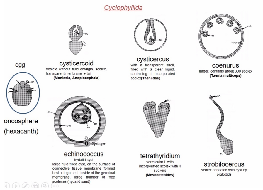
10: Life cycle and Description of Larval stages of Cyclophyllida
Cyclophyllidean eggs are: embryonated when released
contain: oncosphere (= hexacanth embryo) - bilaterally symmetrical, Has 3 pairs of hooks (6 total)
General LC: Embryonated eggs released by FH → IH eats egg → hatches inside IH → oncosphere goes into intestinal wall → tissue migration → into larval stage (Metacestode) → FH infected by eating tissues of IH (containing larval cysts)

Usually only 1 IH
Larvae are often the pathogenic stage, while adults are harmless
Most cestode larvae are some form of liquid filled bladder with invag. scolex.
Larva | Key feature | Example |
|---|---|---|
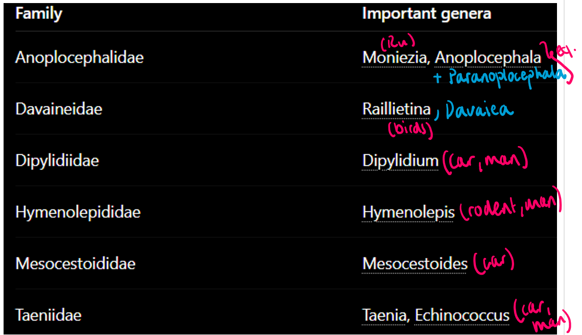
Cysticercoid | Small, non-invag. scolex, very small/no cavity. | Dipylidium caninum, Moniezia, anoplocephala |
Cysticercus | One scolex invag. into itself in fluid bladder | Taenia |
Coenurus | large fluid-bladder with many invag. scolices to wall | T. multiceps |
Tetrathyridium | Elongated larva, deeply invag. scolex. | Mesocestoides |
Strobilocercus | Scolex (not invag. when mature) + segmented tail (strobila) | T. taeniaeformis |
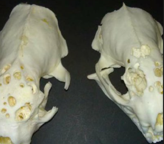
Hydatid cyst/echinococcus | Many protoscolices - produced from germinal layer of cyst or by forming brood sacs (endo/exogenous budding), large-fluid filled. FH infected by IM with larvocysts in tissue. | Echinococcus |
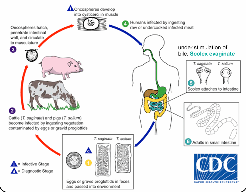
11.Taenia saginata and bovine cysticercosis
Family: Taeniidae (under order cyclophyllidea)
Species: Taenia saginata
General of the family:
Large intestinal tapeworms
Larval stages - most important in vet med.
Most taenia have indirect LC with dog, cat, man as FH. Other domestic/wild animals as IH. In FH - eggs are shed with feces, often still inside gravid segments. The eggs are directly infective for IH - can be that for months in moist/cool environment.
Eggs:
Spherical, small, gelatinous outer coat, thick striated embryophore, oncosphere inside with hooklets visible (hexacanth), 3 hard shells
Eggs of - T.saginata & solium - alike
T.SAGINATA:
Morphology:
Adult: Largest taenia species, length of 4-10m.
Scolex - 4 suckers, no hooks, no rostelleum.
Unarmed scolex
Proglottids: 1000-2000 segments. Gravid uterus - up to 30 lateral branches with many eggs - twice as many as T.solium. Segments are longer than wide, actively leave host.
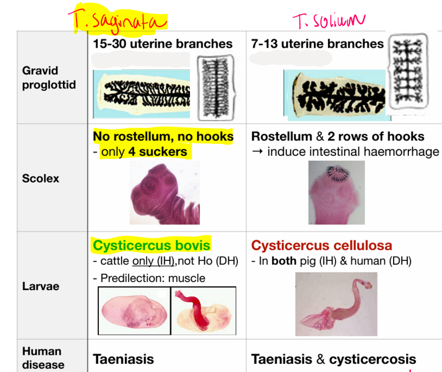
Location: Adults are in SI of man, Larva are in striated muscle of ru (cattle) - tongue, heart, masticatory muscles, intercostal muscles.
Larval stage: Cysticercus Bovis
FH: human
IH: Cattle
Geo: worldwide, more common in areas of poor sanitation. Raw/undercooked beef consumption. Still present in Europe - economic losses, despite EU directives for meat inspection. Main reason - low sensitivity of current meat inspection protocols + survivial of eggs in the environment. Assumed that water streams/surface are potentially contaminated with eggs. Not notifiable.
LC (indirect): Humans shed eggs/proglottids in feces → cattle ingest eggs → oncosphere hatch in intestine → intestinal wall → migration via blood/lymph to muscles → develops into cysticercus bovis → human infected by eating raw beef → scolex attaches in intestine → adult.

Pathogenesis/CS:
Humans (taeniasis): usually mild/asymptomatic
signs can be abd. pain, nausea, diarrhea, dizzy, appetite loss, allergic reactions. In rare case - intestinal obstruction & Delirium
Cattle (b.cysticercosis): usually asymptomatic
Heavy: weak, diarrhea, muscle pain, cardiac issues
Diagnosis: Eggs/proglottids in feces of humans, ELISA - eggs appear only after adult emerge - 3months. IN cattle - meat inspection, detection of cysticerci in muscle.
Treatment: Praziquantel, niclosamide
Prevention: cooking beef properly, cysticercus killed at 56 degrees. freezing effective below minus 10 for ish 1w. Good sanitation, prevent human fecal contamination on cattle pastures. Avoid using soil as fertilizer.
12.Taenia Solium and Swine cysticercosis
Family: Taeniidae
Species: Taenia Solium
Taenia solium causes → Taeniasis (adult worm in intestine) & Cysticercosis (larval cysts in tissues)
Humans can be FH & Accidental IH
Morphology:
Adult: usually 3-8m. up to 1000 proglottids.
Scolex: 4 suckers around rostelleum - Double row of hooks. Called “armed tapeworm”.
Gravid proglottids: 7-13 lateral uterine branches, does not leave the host spontaneously.
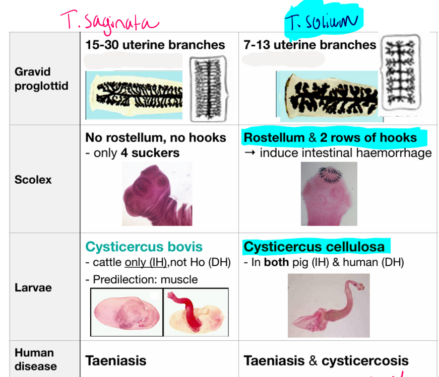
Location: adults in SI of humans. Larval stage (Cysticercus cellulosae) in striated muscle, subcutaneous tissue, eye, CNS/brain.
FH: Humans
IH: pigs
Geo: common in latin america, Asia, sub-saharan africa. RARE in Europe. Long-lived worms (25 years).
Life cycle: similar to T.saginata.
gravid proglottids released → pigs eat eggs → oncosphere into into cysticercus cellulosae (metacestode) in 2-3m → humans eat raw/undercooked pork → adult develops in intestine.
Humans can become accidental IH by eating eggs directly or autoinfect themselves (fecal-oral).
Pathogenesis/CS - in humans:
1) Taeniasis (adult in intestine) - usually mild, abd.pain, nausea, appetite loss, asymptomatic. But person continuously sheds eggs.
2) Cysticercosis (larvae in tissue)
Neurocysticercosis (larvae in CNS/brain) - epilepsy/seizures, headache, nausea/vomit, dizzy, mental disorders, loss of consciousness, death.
ocular type - eye pain, retinal detachment, vission loss
muscle/subcutaneous type - painless movable nodules, often asymptomatic
25% of cysticercosis infections are acquired by autoinfection.
Pigs: Most are asymptomatic. But heavy → weak, muscle pain/stiff, red. movement. possible neurological.
Diagnosis: eggs/proglottids in feces - not detectable until after 2-3months for Taeniasis. In cysticercosis - biopsy, CT/MRI for brain lesions, opthalmoscopy, serology/PCR
Treatment: praziquantel. for ocular/superficial cysts - surgical removal. No immunization.
Prevention: proper sanitation, cook pork, good hygiene to prevent autoinfection.
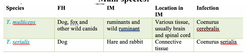
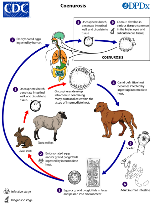
13.Coenurosis
Family: Taeniidae
Genus: Taenia
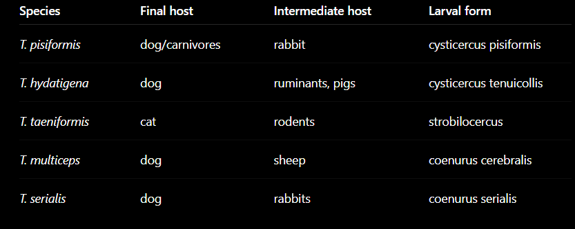
Species: T.multiceps, T.serialis
coenurosis = disease by coenurus larva.
Morphology:
coenurus: large, fluid-filled cyst, with many invaginated scolices.
Adult worm: lives in SI of canids, can read up to 5m.
FH: dogs/canids. Usually asymptomatic
IH: sheep (CNS infection), Rabbit (subcutaneous/CT infection), humans (accidental host)
Epidem: Humans infected by eating eggs from dog feces, contaminated water, veggies, soil. human cases are rare. No real endemic areas.
Life cycle: Dogs shed eggs in feces → sheep eat eggs → oncosphere hatches → into intestine, migrates via blood → larva develops into coenurus cerebralis (mainly brain/CNS) → dog infected by eating tissue/organs with these cysts → protoscolices evaginate → adult develops in intestine.
Similar in serialis, just that coenurus serialis infect connective tissue.
T.Multiceps - Mainly CNS - Brain
T.serialis - Connective/subcutaneous tissues
Patho: Disease depends on size of cyst & location. Main damage is pressure on tissues, CNS compression
CS:
Human Coenurosis - Depends on cyst location.
Brain: headache, seizure, ataxia
spinal cord: arachnoiditis (inflammation of arachnoid - meninges around cord)
eye: blindness/reduced
skin/subcutaneous tissue: painless movable nodules
FH - usually asymptomatic, extremely heavy infections in small dogs can cause blockage of intestine.
IH:
Rabbits: mostly painless subcutaneous nodules (neck, trunk, limbs ex.), neck cysts can affect swallowing + moving.
Sheep: early signs are isolation from flock, slow reactions, change in behavior. Progressive CNS signs are circling, ataxia, stumbling, blind, changed head posture, paralysis. More acute in young. Migrating larvae can cause meningoencephalitis, purulent tracks in brain.
Diagnosis: FH - eggs in feces, copro-Ag by ELISA. IH - Necropsy, CT-MRI, Biopsy.
Treatment: Surgical cyst removal, praziquantel, albendazole
Prevention: Proper meat disposal, hygiene, clean veggies, remove dog feces, proper cooking/freezing of meat
14.Cysticercosis of the Herbivores, Omnivores and rabbits
Family: Taeniidae
genera: Taenia
Cysticercosis - disease caused by cysticercus larval stage (Metacestode)
Species:
T.Hydatigenea
FH: Dogs, fox, wild canids
IH: domestic & wild ru, pigs
Location in IH: liver, peritoneal, abd.cavity
Larval stage: Cysticercus tenuicollis
T.pisiformis
FH: dogs, fox, wild canids
IH: rabbits, hares, rodents
Location in IH: liver, peritoneum
Larval stage: Cysticercus pisiformis
T.Ovis
FH: dogs, fox, wild canids
IH: sheep, goat
Location in IH: muscle
Cysticercus ovis
T.krabbei
FH - same
IH: Reindeer
location in IH: muscle
cysticercus tarandi
T.taeniaformis
FH: cat, lynx, other felids
IH: rodent
Location in IH: Liver
strobilocercus fasciolaris
Morphology:
Adults: scolex with 4 suckers, rostellum, many hooks (20-50 depending on species).
Larva: cysticercus - fluid-filled bladder, contains ONE invaginated scolex.
GEO: worldwide (cosmopolitan).
Location: Adults live in SI of FH, Larvae develop in tissue/organs of IH
FH: dog, car
IH: herbivore, ombivore, rabbits
Life cycle: Eggs released in feces of FH → IH eats eggs → oncosphere hatches → penetrates intestinal wall → migrates to tissue → develops into cysticercus → FH eats infected tissue/organ → adult develops in intestine.
Patho/CS: Usually asymptomatic in FH.
IH: damage depends on migration route, organs affected
Lesions:
Ex. Liver migration → liver parenchymal damage, hemorrhage, inflammation. CS - digestive disorders, painful/sensitive liver, abd./peritoneal pain on palpation.
Diagnosis: FH - proglottids/eggs in feces, IH-mainly PM inspection.
Treatment: Praziquantel, fenbendazole. IH - usually no treatment.
Prevention: prevent car from eating raw infected organs, proper disposal of carcass/offal, deworm dogs, good hygiene/pasture.
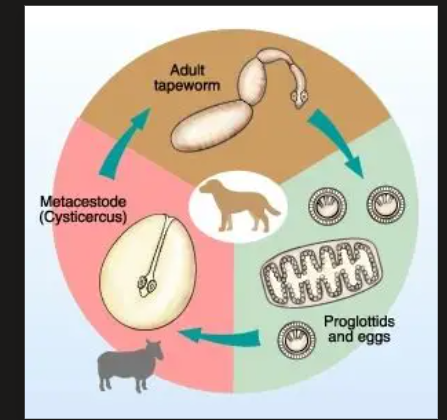
15.Echinococcosis (Hydatidosis) + Alveococcosis
Echinococcus - under fam: Taeniidae, order: Cyclophyllidea.
Main species: E.granulosus & E.multilocuaris
Other: E.equinesus, E.ortoleppi, vogeli, felidis, Shiquicus (all in liver)
1) Echinococcosis - E.granulosus
Generally tiny tapeworms of car, causing hydatid cyst disease. Main organs affected in humans are liver & lungs. Humans = accidental IH.
Morphology:
Granulosus is the smallest tapeworm (6mm), scolex has rostelleum with double row of hooks, 4 suckers. 3-5 proglottids.
Terminal proglottids measures less than half of length of whole worm.
FH: Car (dog, fox/wild)
IH: ru, pigs, deer, camels, humans
Transmission: Humans get infected by eating eggs from dog feces - contamianted food, water, soil. Eggs survive in soil for up to 1 year.
Location: SI in FH, Lungs&Liver in IH. Worldwide.
Life cycle: Dog shed eggs in feces → IH eat eggs → oncosphere into intestine → travels to liver → Lungs by blood → forms hydatid cyst → dog eats infected organs → adult tapeworm develops in intestine.
Only one gravid proglottid is shed each week.
Hydatic cyst - Grows slowly, can become very large, contains protoscolices, daughter cysts that can fill the interior of lung.
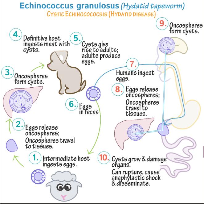
Patho/Clinical signs: Often asymptomatic for years, before the enlarging cysts cause symptoms in the affected organs.
Liver involvement: abd. pain, hepatomegaly (mass), biliary obstruction
Lung involvement: cough, chest pain, coughing up blood (hemoptysis)
complication: cyst rupture → Anaphylactic shock (fever, urticaria, eosinophilia, cyst dissemination).
Other organs can also be involved
Diagnosis: USG/CT, serology
Treatment: surgery, Albendazole
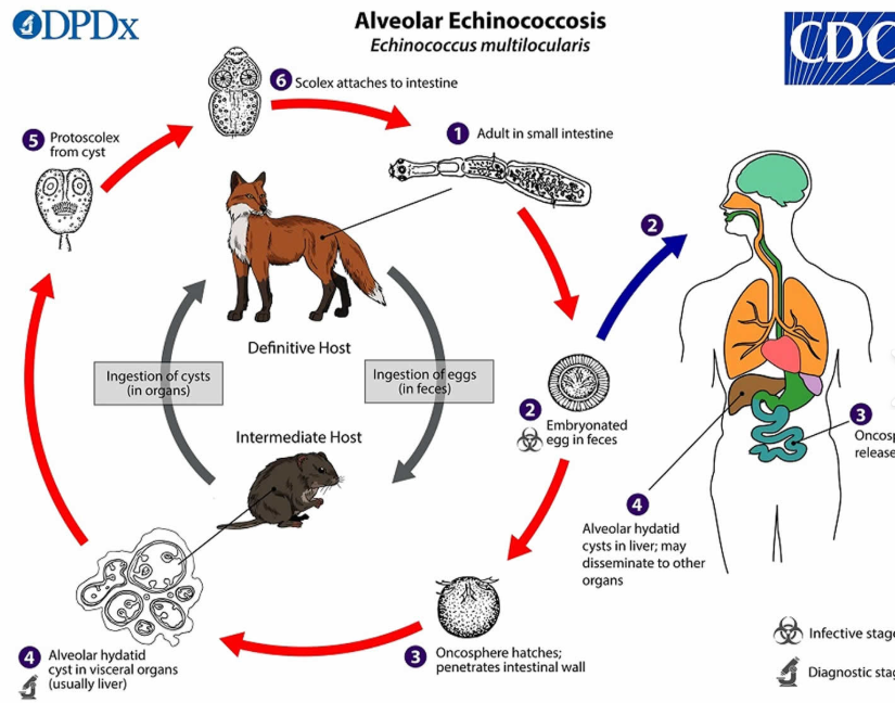
2) Alveococcosis - Echinococcus multilocularis
Difference: From the oncosphere, it creates an alveolar/multilocular cysts with outward budding → behaves like a malignant tumor! As it can invade surrounding tissues. Numerous protoscolices develop within these.
very small tapeworm (2-4mm). But generally similar to E.granulosis
FH: red fox (mainly) + other car
IH: small rodents, human, pigs, ru
main target organ: Liver
Patho: Slowly invasive liver lesion, that can spread to lungs & brain. (cause alveolar disease).
More aggressive & dangerous than E.granulosus
Abd.pain, biliary obstruction + sometimes metastatic lesions in brain/lungs.
Diagnosis: PM by intestinal scraping technique or sedimentation, counting. Detecting coproantigens, PCR
Treatment: surgery if possible, chemo-therapy
Long-term: albendazole, mebendazole
In car: praziquantel
16.Cestodosis in carnivores
Adult tapeworms in dogs/cats usually cause mild/no disease.
Diagnosis mostly by: Finding proglottids or finding eggs in feces
IH - usually more severely affected
Order: Cyclophyllidae, Fam: Dipylidiidae, Genus: Dipylidium
Species: D.caninum
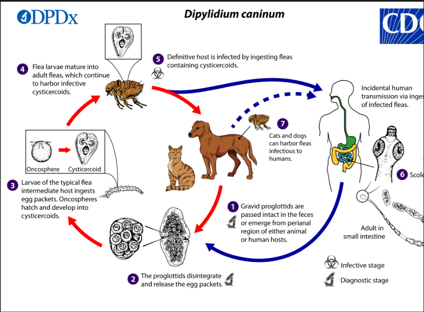
1) Dipylidium Caninum (Flea tapeworm)
Common tapeworm of dogs/cats - Zoonotic
Transmission through fleas (eating them!) or proglottids may creep around near dog anus. Occurrence varies - 8% in dogs, 3% cats in SK.
Morphology: Small/medium, scolex - 4 suckers, rostellium with 3-4 rows of hooks, eggs grouped in capsules. Proglottids resemble - cucumber seeds, motile (moving). Double set of reprod. glands.
Location: Small intestine. Worldwide.
FH: dog/cat/man (mostly children)
IH: fleas, dog lice
LC: Proglottids leave anus/feces → flea larva eats eggs → cysticercoid larva develops inside flea in haemocoel/body cavity → dog/cat swallows infected flea → adult tapeworm develops in SI.
Patho/CS: usually asymptomatic in dog/cat
possible signs: anal itching (scooting), weight loss, dull/bristly fur. RARE - nervous signs like vomit/convulsions due to toxin/metabolite
Characteristic: Moving proglottids around anus “
Diagnosis: Finding proglottids visually, eggs usually not found in feces
Treatment: Praziquantel, epsiprantel. Flea control!
fam: mesocerstoididae, Genus: mesocestoidius
Main species: M.lineatus, M.corti
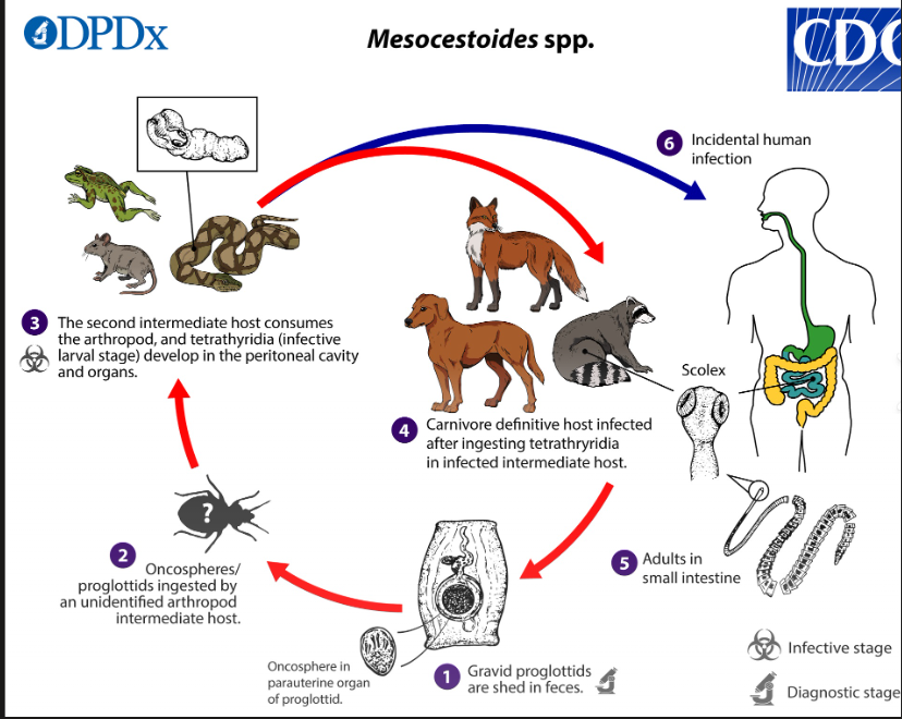
2) Mesocestoides spp.
carnivores, zoonotic, wild car - common reservoirs
Morphology: small/medium, scolex with 4 suckers, NO hooks. Eggs in parauterine organ. One set of genital organs.
Location: SI. Lung & Liver for IH. Wordlwide.
FH: car/humans (Zoonotic)
IH: 1st: oribatid mites, 2nd: small mammals/reptiles.
LC: proglottids shed with many eggs inside parauterine gland → mites eat proglottid/infective egg → oncosphere hatch in intestine → body cavity - develops into cysticercoids → small vertebrates eat the mites → cysticercoid released in the gut → migrate - lung, liver → tetrahyridium (can asexually reproduce by longitudinal division) → FH eats them → evaginates and attach to SI → adult. Humans can be accidental host by eating undercooked meat.
Larval stage: Tetrahyridium - develops in liver/lungs, can reproduce asexually, cause severe disease.
Patho/CS: in weak infection = asymptomatic, humans = severe diarrhea.
Severe: enteritis, vomiting, anorexia, resp. signs
Tetrahyridia in dog/cat can cause peritonitis, pleuritis, ascites
Diagnosis: proglottids & eggs in feces
Treatment: praziquantel, niclosamide
prevention by rodent control, insecticide, sanitation
3) Taenia spp. - of fam: taeniidaee.
Taeniformis - strobilocercus/Cysticercus fasciolaris
17.Equine Cestodosis
Horse tapeworm infection.
Caused by tapeworms in the family: Anoplocephalidae. Under order Cyclophyllidea.
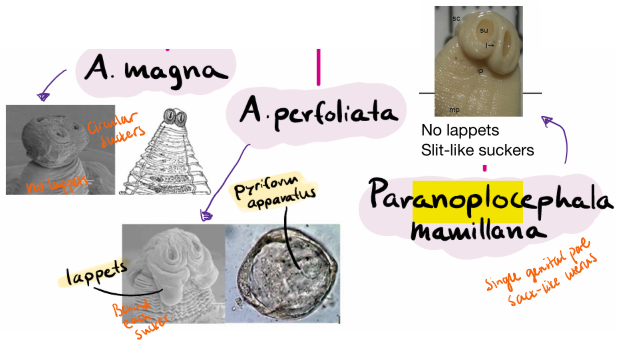
Genus: Anoplocephala & Paranoplocephala
Note: Anoplocephalidae - no dg. system, nutrients through tegument, protonephridial system - flame cells, hermaphroditic (all cestoda are)
Main species:
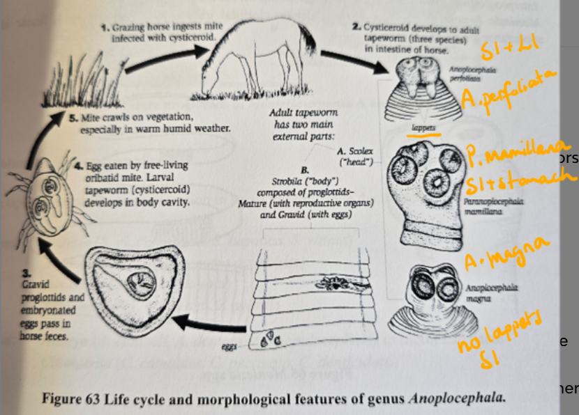
A.magna - jejunum
A.perfoliata - Ileocecal junction, SI + LI
Paranoplocephala mamillana - SI
Morphology:
scolex has 4 large suckers (No rostellum)
proglottids - wide & short, with a signel set of reproductive organs (perfoliata)
Eggs are similar to moniezia - polyedral shape with pyriform apparatus, with the embryo (ocosphere), hard shells
Perfoliata: Lappets behind suckers!
Location: ileocecal junction & SI of horse
Epidemiology: Most common species in horse. Usually affected in pasture. All age categories are susceptible. Lowest occurrence in sping, while highest in winter.
FH: horse
IH: Orbatid mites - Cysticerkoid stage (infective stage)
LC: indirect via the mites on pasture → horse eats infected mite (with cysticercoid) while grazing → develops to adult in intestine → gravid proglottids + embryonated eggs pass in horse feces → eggs eaten by mites.
Patho/CS: Mostly Mild infection without CS
In severe case: enteritis & colic, death. Obstruction & Perforation in both tapeworms.
A.perfoliata: Ulcers in the junction.
PM: small ulcers, edema, partial blockage of opening
Magna: cattarhal - hemorrhagic enteritis
P.mamillana - less patho, mild edema/infl.
Diagnosis: Fecal egg detection is unreliable (intermitted shedding), better is ELISA serology + PCR - determines presence, intensity of attack.
Treatment: Ivermectin, praziquantel + moxidectin
18.Cestodosis of ruminants
Order: Cyclophyllidea
Fam: anoplocephalidae
Genus: Moniezia, avitellina, Stilesia, thysanosoma, thysaniezia
All are tapeworms of ruminants.
IH: Oribatid mites (Ingest eggs → cysticercoid stage)
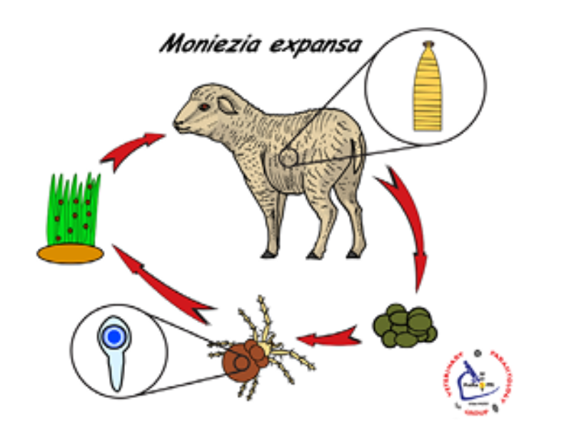
Genus MONIEZIA
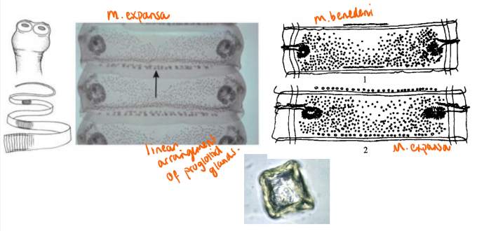
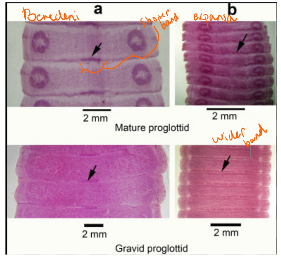
Main species: M.expansa (small ru) & M. benedeni (large ru)
Location: SI, Worldwide.
Morphology:
General for anoplocephalidae: Oval head, no rostellum but 4 suckers. proglottids - wide and short. Testes are many, ovary is flabelliform. Eggs - often has pyriform apparatus. Larvocyst = cysticercoid in arthropods - oribatid mites.
Moniezia - large strobila.
M. expansa: interproglottidal glands = wide band extending across most of proglottid. 6meters long proglottids.
M. benedeni: glands = narrow band - clustered arrangement or rosettes - glands are grouped in bunches rather than in a line, linear. 4m long but wider than expansa.
Epidemiology: More common in young, lighter infection in older. Peak infection in spring-summer (mite activity). Heavy pasture exposure = higher risk. Can survive for months in environment, some survive cold winter.
LC: Indirect, via mites - infective after 4 months → ingestion on pasture → adults in intestine, releases head and suckers attach to mucous wall. Producing proglottids, releasing eggs. After 3 months - spontaneously leaves the host.
Patho/CS:
Heavy infection: diarrhea - painful, enteritis, weight loss/poor growth, weak, possible intestinal irritation/obstruction.
Lambs can get weak, hard to stand up, bending their pack, nerve disorders due to toxins from moniezia.
light&moderate infection - can bypass without CS.
Diagnosis: Fecal exam (eggs/proglottids) - macro/microscopic
Treatment: Praziquantel. Others like albendazole can be more effective in some breeds but can also give resistance.
Other Important (Less common) Ru cestodes:
Less/non-pathogenic, difficult to distinugish from moniezia through fecal analysis. same LC as moniezia, alike.
1) Avitellina centripunctata - small ru
In SI, usually low patho, mild enteritis/asymptomatic
eggs - stored in parauterine capsules without pyriform apparatus!
2) Stilesia Hepatica - small ru
In bile ducts (liver), usually non-patho, found at slaughter (liver lesions)
eggs in parauterine organs.
3) Stilesia Globipunctata - small ru
SI, more pathogenic, cause intestinal inflammation, nodules, mucosal damage
4) Thysanosoma Actinoides - small, sometimes cattle
Bile + pancreatic ducts + intestine, has papillary projections at proglottids edge. eggs - no apparatus.
can block ducts → icterus, digestive disorders, weight loss
5) Thysaniezia giardi - small ru
SI, rare, low prevalence worldwide
eggs in parauterine capsules, without apparatus
19.Cestodoses of Poultry
1) Fam: Davaineidae - Genus: Davainea
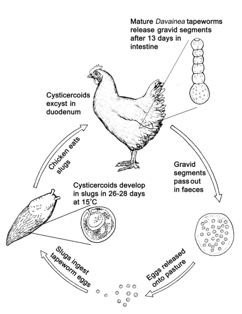
Species: Davainea Proglottina
Morphology:
general: S/M, “dwarf tapeworm”, retractable rostelleum armed with numerous hummer-shaped hooks. 4 suckers. single genital organs, uterus is replaced by egg capsules
D.proglottina - one of most common cestodes in poultry & pigeons. So small, that they are overlooked in PM examinations.
only has 4-9 proglottids, head is with many small hooks, suckers bear 4-5 rows of hooklets.
Location: Duodenum
FH: Chicken, pigeon (adult parasites) - Adult can live up to 3 years in FH.
IH: landsnail/slugs - various gastropod mollusks (cysticercoid)
Geo: Worldwide distribution - esp. in extensive poultry rearing (free-range/outdoor systems - slugs winter). In SK - 6%.
Patho/CS: most pathogenic poultry tapeworm.
cause severe hemorrhagic enteritis, necrosis, weight loss, possible death
Nervous disorders + duodenitis
Chronic - enteritis, weak + wasting
2) Fam: Davaineidae - Genus: Raillietina
All with rostellum with hooks.
Species:
R.tetragona - small scolex, oval suckers, spines, eggs in capsules
R.echinobotrida - spherical suckers, spines
R.cesticillus - broad restellum, suckers without spines, capsules with 1 egg
Location: SI, worldwide - occurrence of ish 20% in SK.
IH: Beetles, houseflies and ants
LC: same as Davainea
Patho/CS: non-specific - weak, poor growth, diarrhea
severe: Inflammation + degeneration of villi → can cause caseous nodules in intestinal wall at site of attachment + hyperplastic enteritis (thickening + inflamed intestinal wall)
Treatment: can be dewormed with flubendazole in feed. Also mebend/febendazole, praziquantel.
3) Fam: Hymenolepididae - Genus: Hymenolepis
Main species: H.lanceolata (most patho) & H.carioca
SI, scolex of 4 suckers + rostellum with hooks. one set of genital organs like the others. FH - water poultry/chicken.
IH: aquatic invertebrates - small crustaceans for lanceolata, dung beetle for carioca. Signs - enteritis + NS, diarrhea, poor growth. Mostly asymptomatic in adults.
4) fam: Dilepididae - Genus: Amoebotaenia & Choanotaenia
Main species: Choanotaeina infundibulum & Amoebotaenia cuneata
S/M, scolex has rostellum with rows of hooks. small head. Double set of genital organs opening laterally. eggs in capsules.
SI. IH: house fly, dung beetle, grass-hopper.
Cause enteritis, similar to others.
For all:
Generally parasite of intestine, causing intestinal issues.
Diagnosis: Generally fecal exam (eggs, proglottids) & PM examination (adults in intestine -Raillietina)
Treatment: Praziquantel.
Same LC: LC: Indirect. Gravid proglottids shed in FH feces → eaten by IH → develop to cysticercoid → FH eats IH → adult.
other: diphyllobothrium dendriticum (fish eating birds) - but not one of the main classical.
20.Cestodosis in Rodents
Order: Cyclophyllidea
Fam: Hymenolepididae. Genus: Hymenolepis/Rodentolepis
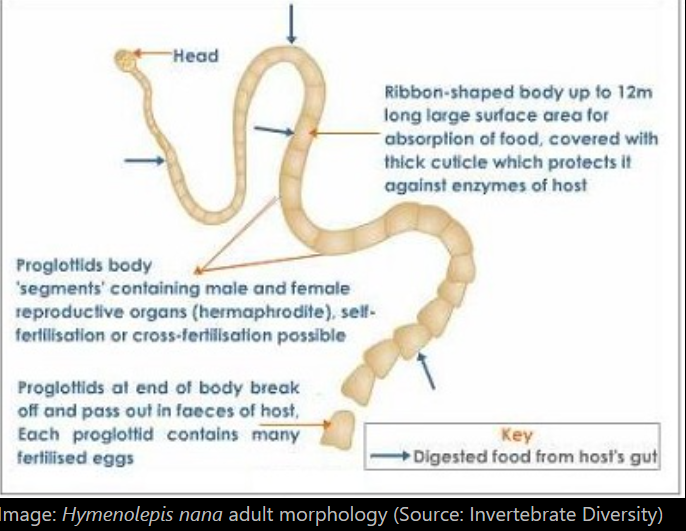
Main species: Hymenolepis nana (more common in man), H.diminuta (less common in humans but similar), H.microstoma (primarily of rodents)
Morphology:
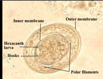
H.nana: S, 4 suckers on scolex, retractable rostellum with single circle of ish 30 hooks. Long and slender neck, wider segments than longer. Each - contain 3 testes. genital pores unilateral.
H.diminuta: longer, NO rostellum or hooks! but has 4 suckers. genital pores on left margin, 3 testes. Indirect LC with arthropods (grass-hopper, butterfly ex.). Does not have polar filaments in eggs - and are also more spherical.
Location: SI
FH: rodent, humans (Zoonotic)
IH: invertebrates, larval + adult beetles
Worldwide.
Epidem: Most common tapeworms of humans (esp. children). Transmission by eating infected beetle, contaminated food or by direct fecal/oral contact. Rats are heavily infected and spread in human population, esp. in areas of low hygiene.
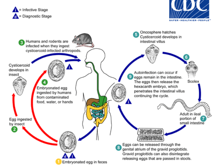
LC: Indirect with IH, direct & Autoinfection
Direct: Eggs passed in feces (infective) → host eats eggs → oncosphere released in intestine → villus → cysticercoid → villus rupture → returns to lumen → attach to mucosa → adult tapeworm
Indirect: eggs in feces → IH eats eggs → larva develops into cysticercoid inside IH → then rodent or human may eat infected insect → cysticercoid in intestine, adult develops.
Autoinfection: eggs release their hexacanth embryo in the intestine (instead of leaving the body) → goes into villus → new cysticercoids develop → new adults form. Allows for infection to last for years.
Patho/CS:
H.nana - in Humans → often asymptomatic but heavy infections (children commonly) can cause GIT issues, abd.pain (crampy), diarrhea, anorexia, vomit, anal/nasal itching. verminous intoxication.
Mice: growth issue, weight loss.
Diagnosis: Eggs in feces - they have polar filaments (H.nana). Diminuta does not!
Treatment: Praziquantel, niclozamide. Control after treeatment for 3 months, family or group retreatment recommneded. prevention - rat control.
21.Morphology, Classification and Life cycle of PSEUDOPHYLLIDEA
Class: Cestoda (tapeworms)
Subclass: Eucestoda
Order: Pseudophyllidea
Fam:
Diphyllobothridae - diphyllobothrium, spirometra
Bothriocephalidae - bothriocephalus
Ligulidae - ligula
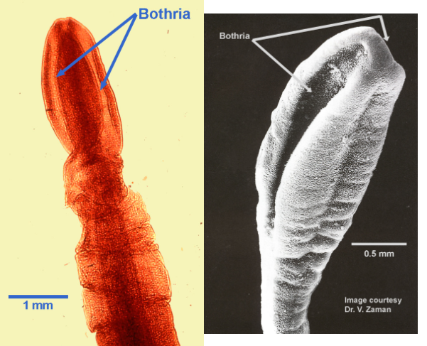
Morphology:
Scolex - 2 bothria (instead of 4 suckers)
Bothria = longitudinal sucking grooves
Body: Very long, short neck. Proglottids are short, broad/wide
Female: open reproductive system, vagina runs backwards from genital pore → joins oviduct + vitelline duct → forms ootype → uterus goes from it and stops in opening (uterine pore/ovipore) - outlet of eggs.
Uterus is long, colied and sac-like (NOT branched). Scattered vitelline glands.
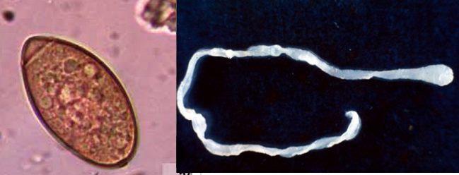
Eggs: operculated, released continuosly, needs some time before the hexacanth larvae develop in the water.
Larva inside the egg = coracidium (ciliated larva)
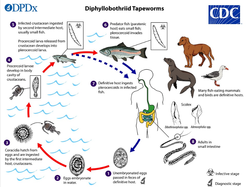
Life cycle: Indirect, 2 IH. Sometimes also Paratenic host.
Eggs leave with feces into water → embryonates and hatches into coracidium (free-swimming) → first IH eats coracidium (copepods/small crustacean) → looses cilia, develops into procercoid → 2nd IH (fish) eats the 1st IH → develops into plerocercoid (infective for FH) → FH eats fish → adult tapeworm in intestine.

22.Diphyllobothriosis, sparganosis, Ligulosis
class: cestoda, order: pseudophyllidea.
They all: 2 bothria on scolex, operculated eggs, coracidium larva, aquatic LC.
Similar LC: eggs in water → embryonates → coracidium → 1st IH (copepod/crustacean) → procercoid → 2nd IH → plerocercoid → FH eats IH → adult tapeworm develops.
Large fish may eat smaller infected fish → larvae can migrate into muscles (Diphyllo).
All depend on water, aquatic hosts + food chain transmission!
Location: intestine , Diphyllo (SI), spiro (adult in intestine, larvae in tissues), ligula (adult in intestine, larva in fish body cavity)
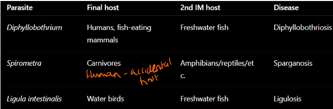
1) Diphyllobothriosis:
Fam: Diphyllobothriidae
Genus: Diphyllobothrium
Main species: D.latum (largest human tapeworm) & D.dendriticum
Morphology: Long tapeworm, scolex - 2 bothria, can grow over 10m.
FH: Fish-eating mammals & humans (Zoonosis)
IH: 1st: copepod crustacean, 2nd: freshwater fish
Geography: Scandinavia, russia, japan, north america
Transmission/Epidem: Humans infected by eating raw fish, undercooked. Eggs released daily in feces.
LC: 5-6w for infection to be detectable.
Patho/CS:
Usually asymptomatic
Possible signs - non-specific: Abd.pain, nausea, vomit, diarrhea
complications: Intestinal obstruction, gall bladder disease
IMPORTANT - Worm eats Vitamin B12 → causes macrocytic hypochromic anemia (larger cells + less colored) + malignant anemia
Blood findings: Thrombocytopenia, leukopenia
Diagnosis: fecal flotation & sedimentation
Treatment: praziquantel & niclosamide. Prevention - cook/freeze fish
2) Sparganosis - Spirometra mansoni
By: spirometra genus, fam: diphyllobothriidae
Morphology: Adults resemble diphyllobothrium.
Plerocercoid larva is called = Sparganum (Spargana)
Typical: White, ribbon-like, crinkled
Eggs have pointed ends - does rarely mature into adult in human body, so eggs are rarely found in human feces!
FH: car, humans as accidental host (Zoonotic)
IH: 1st:copepods, 2nd: amphibians, reptile, bird, mammals
Geo: asia, south america
Transmission: humans infected by drinking water, eating raw frogs/snakes, applying frog flesh to wounds/eyes (traditional medicine)
Patho/CS:
in animals: usually asymptomatic, small effect in intestines of dog/cat
In humans: spargana migrate through tissues → subcutaneous nodules, inflammation, edema, urticara, eosinophilia
Can migrate to brain, eye, skin, visceral organs
Brain → headache, seizure, NS
Diagnosis: Biopsy/specimen detection, ELISA
Treatment: surgical removal. Drugs usually ineffective - meben/albendazole, praziquantel.
3) Ligulosis - Ligula intestinalis
Fam: Ligulidae
Up to 1m long, in intestines. FH - fish eating water birds, IH: 1st: cyclopods, 2nd: freshwater fish.
Patho/CS:
infected fish: cannot dive, swim near surface, circling, easier prey for bird
Other signs: loss of appetite, enlarged abdomen/body cavity
Diagnosis: necropsy
treatment: Medicated food, preventive deworming of fish.
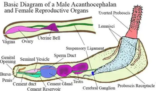
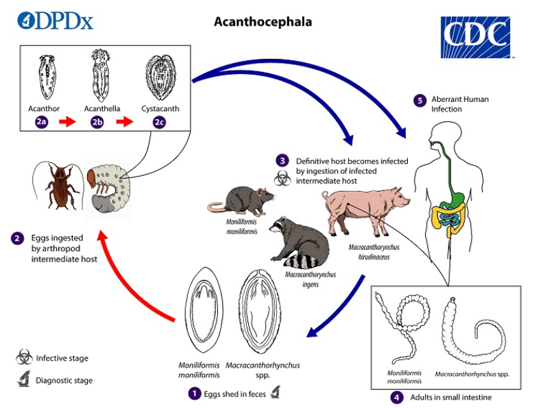
23.ACANTHOCEPHALA, morphology, classification, and life cycle + macracanthorhynchosis of pigs
Acanthocephala worms = Thorny-headed/Spiny-headed worms.
Mainly parasites of: fish, birds, rodents, pigs (sometimes humans)
Location: mainly Intestine
Morphology: Cylindical, females larger than male (separate sexes), light color
Proboscis - retractable in a sac, covered in hooks/spines/thorns. - for attachment to intestinal wall
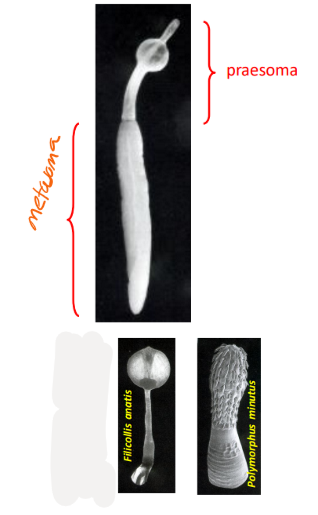
Body parts:
Anterior (presoma): proboscis + receptacle, neck, lemnisci + lacunae (moving/secretory), cerebral ganglion
Posteror (metasoma): contains reproductive organs. Genitals are stored in body cavity - pseudocel, defined by ligament sacs.
NO digestive system. Absorbs nutrient through surface - thick syncytial tegument. Glycocalyx on body surface protects host immunity.
Excretory system: most use diffusion! - but some, like oligoacanthorhynchidae members → 2 protonephridial organs
all systems (excretory/nervous/circulatory) = reduced
IH: arthropods, esp. insects. Sometimes also paratenic host-fish
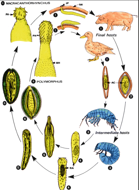
LC: indirect. Eggs in feces (with acanthor) → IH eats eggs → acanthor into preacanthella → acanthella → cystacanth (infective) → FH eats the IH → juveniles attach to SI wall → matures into adults
Important families:
Miniliformidae → moniliformis
oligacanthorhynchidae → macracanthorhynchus
Polymorphidae → filicollis, polymorphus
Macracanthorhynchosis in pigs
species: macracanthorhynchus hirudinaceus
Large, slightly ping, wrinkled, retractable proboscis with rows of hooks. In SI of pigs (FH). Beetles as IH. Worldwide.
LC: Eggs in pig feces (with acanthor) → beetles eat eggs (→ acanthella → cystacanth) → pig eats beetle → larva attach to SI, mature
Patho/CS: usually signs only in heavy infection. Bloody diarrhea, anorexia, cramps.
Diagnosis: fecal examination, PM. Treatment with thiabendazole. - ivermectin.
24.Polymorphosis and filicollosis of aquatic birds
Fam: polymorphidae, order: Polymorphida (under acanthocephala)
“thorny-headed/spiny worms”
general of the acanthocephala - mainly in freshwater fish&birds, but also rodent, pig, man. In intestines. Cylindrical body, 1mm-60cm. Females are longer + heavier. Fixation organ - proboscis, covered in thorns. Glycocalix - surface protection against host-immune system. neck, lemnischi - pair on sides of the octopus vagina (secretory + moving of proboscis).
main species: Filicollis anatis, Polymorphus minutus, P.rubra
Morphology: thick, orange color when fresh. Anterior end - proboscis with spines and hooks.
Location: SI
FH: aquatic birds, P.minutus also chickens
IH: shrimps, crustaceans
Geo: Worldwide
LC: Eggs with fully developed acanthor shed in feces of birds → eggs eaten by IH → acanthella → cystacanth → FH eats IH → juveniles attach to SI wall → mature, produce eggs.
Patho/CS: cause mechanical damage to intestinal wall, local inflammation, tissue proliferation + nodule formation, digestion issues, diarrhea, anorexia, decr. growth, death (young esp.)
Dg: Sedimentation, necropsy
Treatment: Bitinol