L99: Circulatory Disorders and Hepatic Infiltrations
1/91
There's no tags or description
Looks like no tags are added yet.
Name | Mastery | Learn | Test | Matching | Spaced |
|---|
No study sessions yet.
92 Terms
what is passive congestion characterized by?
reduced hepatic outflow due to cardiac dysfunction
what is passive congestion caused by?
right-sided heart failure which produces elevated pressure in the caudal vena cava that extends to the hepatic vein and its tributaries
what does high pressure in hepatic veins lead to?
centrilobular congestion of sinusoids
what are common causes of right sided heart failure resulting in hepatic congestion?
valvular endocardiosis of the tricuspid valve (old dogs)
canine heartworm
what is the pathway that leads to passive congestion?
congestion
hypoxia
centrilobular degeneration
atrophy and loss of hepatocytes
besides congestion, what else can chronic hypoxic injury lead to?
steatosis (fatty degeneration)
what do the combined changes in passive congestion lead to?
enhanced lobular pattern (nutmeg liver)
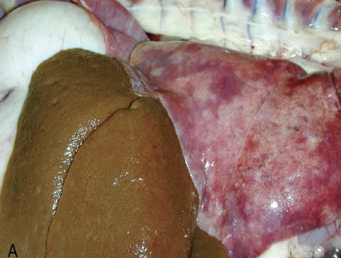
how does passive congestion appear grossly?
lobes of liver are enlarged with rounded edges
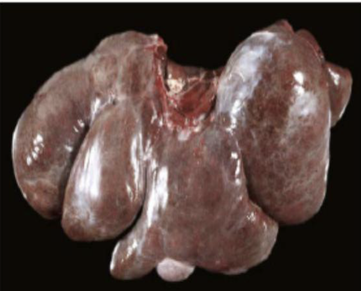
what does the image show?
passive congestion
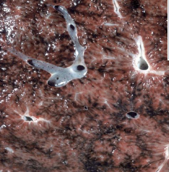
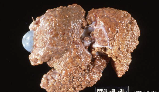
what does the image show?
enhanced lobular pattern from passive congestion
MCQ: a seven year old male lab presents with right-sided congestive heart failure and is diagnosed with chronic canine heartworm disease. What lesion would be expected in the liver of this dog?
chronic passive congestion
what is congenital portosystemic shunt?
abnormal vascular structure that allows portal blood to bypass the liver and drain directly into the systemic circulation
in which circulatory disorder will animals appear stunted?
congenital portosystemic shunt
what signs will animals with congenital portosystemic shunt develop?
signs of hepatic encephalopathy due to hyperammonemia
how does the liver appear grossly in animals affected by congenital portal systemic shunt?
liver is small with single anomalous vessels connecting portal circulation with systemic circulation
how does the liver appear histologically in animals affected by congenital portal systemic shunt?
lobular atrophy
portal miniaturization with small or absent portal veins
reduplication of arterioles
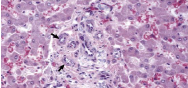
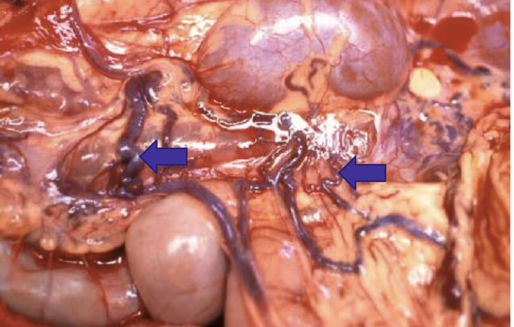
what does the image show?
Congenital portal systemic shunt
what are the types of PSS?
intrahepatic shunts
extrahepatic shunts
what causes intrahepatic shunts?
failure of closure of the ductus venosus (fetal vessel)
what animal is intrahepatic shunts common in?
large breed dogs
what are the causes of extrahepatic shunts?
portal vein to caudal vena cava anastomosis
portal vein to azygos vein anastomosis
what animal are extrahepatic shunts common in?
small breed dogs and cats
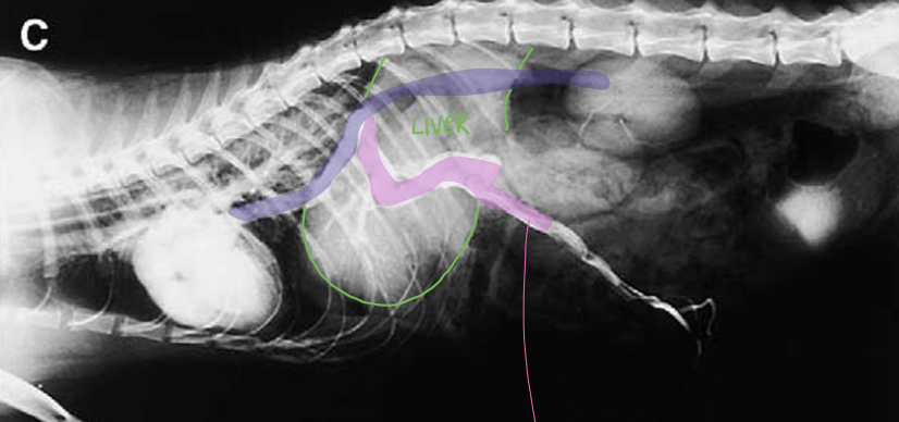
what does the image show?
congenital intrahepatic portosystemic shunts
what can result in dogs from congenital PSS?
ammonimum biurate crystalluria
what does the image show?
urinary bladder with ammonium biurate crystals
what else can congenital portal vein hypoplasia be called?
hepatic microvascular dysplasia
what animals does congenital portal vein hypoplasia occur in?
dogs and sometimes cats
what breeds does congenital portal vein hypoplasia appear in?
yorkshire terrier
maltese
cairn terriers
tibetan spaniels
shih-tzus
havanese
what does congenital portal vein hypoplasia result in?
diminished hepatic perfusion and portal hypertension
what signs do animals affected by congenital portal vein hypoplasia typically have?
microhepatica
ascites
what test is conducted to differentiate PSS from congenital portal vein hypoplasia?
radiology
what are the causes of portal hypertension?
thrombosis
occulusion within portal vein or hepatic outflow
intrahepatic causes
what are intrahepatic cause of portal hypertension?
fibrosis
nodular regeneration
lobular remodeling
veno-occlusive disease
microvascular disease
sinusoidal amyloidosis
what can persistent portal hypertension lead to?
ascites
development of acquired portosystemic shunts
what does the image show?
acquired portosystemic shunts from portal hypertension
what are examples of hepatocellular infiltrations?
amyloid
copper
iron
bile pigments
lysosomal storage disease
glycogen
lipids
what are the main mechanisms of abnormal intracellular accumulations?
inadequate removal and degradation
excessive production of an endogenous substance
deposition of an abnormal exogenous material
what are the four pathways in abnormal cellular infiltrations?
defect in metabolism
defect in protein folding or transport
lack of an enzyme resulting in failure to degrade a substance
ingestion or inhalation of indigestible materials (respiratory not hepatic)
amyloidosis
extracellular deposition of abnormal proteinaceous substance in tissues
what type of disorder is amyloidosis?
protein misfolding disorder
what two forms of protein can result from amyloidosis?
amyloid light chain protein (AL)
amyloid associated protein (AA)
amyloid light chain protein (AL)
derived from abnormal plasma cells secreting light chain fragments into circulation
amyloid associated protein (AA)
secreted in liver in response to cytokines
which form of protein from amyloidosis is most common?
amyloid associated protein (AA)
what is amyloidosis commonly a result of?
secondary (reactive) amyloidosis due to prolonged systemic inflammation (amyloid AA)
what breeds is amyloidosis seen in?
shar-pei dogs
abyssian cats
siamese cats
how do amyloidosis in the liver appear grossly?
livers are enlarged with rounded edges, friable, and pale
what are severely affected livers from amyloidosis susceptible to?
fracture and hemorrhage
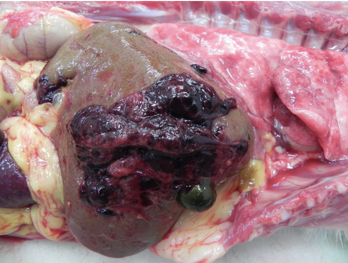
what does the image show?
amyloidosis
where does amyloid deposit?
space of disse
portal tracts
within and around blood vessels
where does amyloid deposition start?
space of disse and extends into the sinusoids
what can severe deposition of amyloid cause?
pressure atrophy and necrosis of hepatocytes
what stain is used for amyloid?
congo red
what does the image show?
amyloidosis
what does accumulation of copper lead to?
production of reactive oxygen species causing oxidative injury to mitochondria
what breeds do we see disorders of copper metabolism?
bedlington terriers
labrador retrievers
what is canine copper associated hepatopathy the common cause of?
chronic hepatitis in dogs
where does copper accumulate in the liver?
centrilobular regions and within kupffer cells
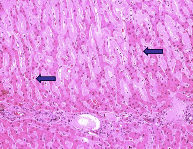
what does the image show?
canine copper-associated hepatopathy
what animal is copper storage poorly regulated in?
sheep; more prone to copper toxicosis
what can exacerbate copper toxicosis in sheep?
low dietary molybdenum and sulfur
what can trigger copper release in sheep?
stress or illness
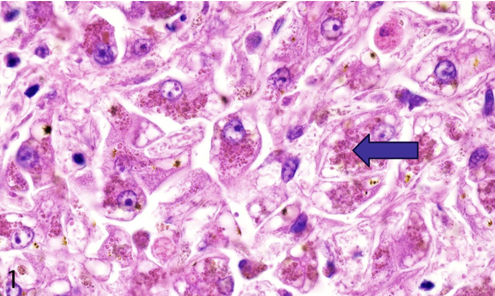
what does the image show?
copper accumulation
what does diagnosis of hepatocellular copper accumulation require?
liver biopsy
what stain is used to identify granules of copper?
rhodanine special stain
iron storage disease (hemochomatosis)
abnormally increased amount of iron storage within the liver
when does bile pigment accumulate in the liver?
during cholestatic disease and contributes to hepatoceullular injury
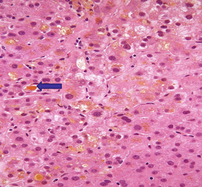
what does the image show?
bile pigment acummulation
what causes lysosomal storage diseases?
missing enzyme that are inherited as autosomal recessive disorders
what does accumulation of lysosomes result in?
abnormally increased amount of iron storage within the liver
MCQ: the bedlington terrier carries a genetic mutation that predisposes this breed to which of the following liver diseases?
copper-associated hepatopathy
what are the two types of canine degenerative vacuolar hepatopathy?
glycogen type
lipid type
what is glycogen-type VH associated with?
stress
cushing’s disease
genetic storage disease
glucocorticoid administration
what is lipid-type VH associated with?
hypoxia
certain toxins
hypothyroidism
diabetes
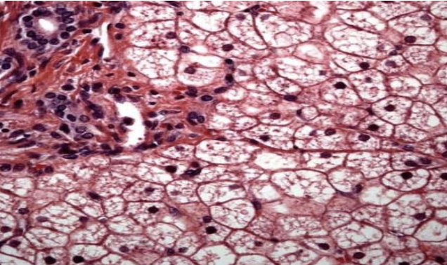
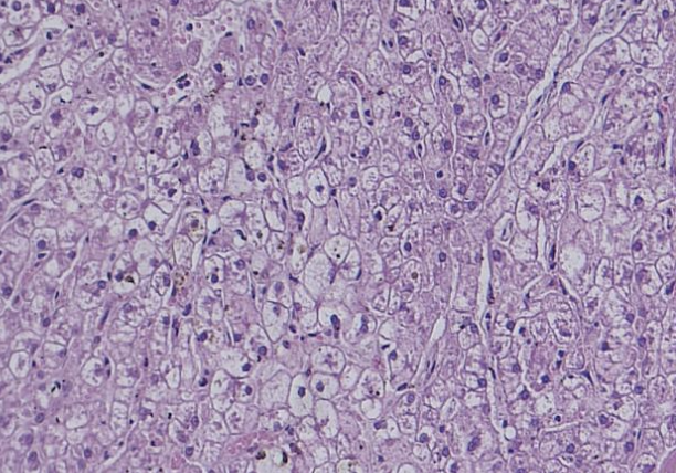
what does the image show?
glycogen type VH
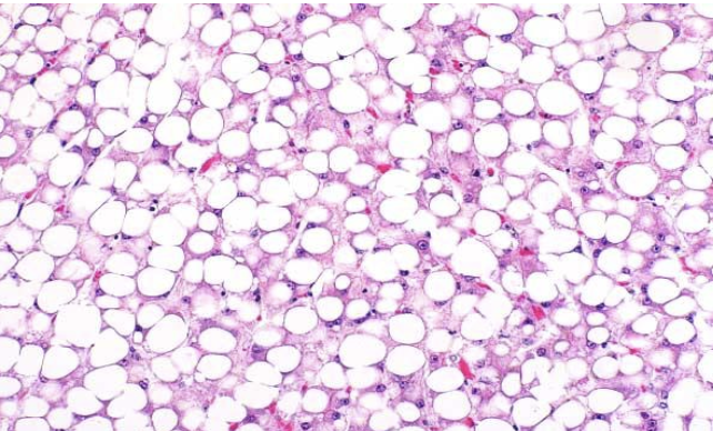
what does the image show?
lipid type VH
what can severe cases of VH result in?
progessive injury leading to end-stage liver
why is liver biopsy pursued in patients suspected of canine degenerative VH?
due to unexplained increase in serum ALP and mildly elevated ALT
why is there increased ALP in patients with canine degenerative VH?
severe swelling of hepatocytes leads to blockage of bile canaliculi and intrahepatic cholestasis
how does the liver appear with hepatocellular glycogen accumulation?
liver is enlarged and pale with enhanced lobules
what does diagnosis fo hepatocellular glycogen accumulation require?
liver biopsy
what does the image show?
canine steroid induced hepatopathy
what does treatment of hepatocellular glycogen accumulation require?
identification of underlying cause
what does the image show?
hepatocellular glycogen accumulation
bovine fatty liver disease
common in dairy cows in late gestation or peak lactation, especially after any period of inappetence or anorexia
when does hepatic lipidosis of ponies, mini horses, and donkeys occur?
occurs in overweight pregnant or lactating mares after a period of stress or anorexia
feline hepatic lipidosis
occurs in obese cats after a period of anorexia; frequently develop hepatic failure, icterus, and hepatic encephalopathy
what type of hepatocellular accumulation is characterized by discrete vacuoles that push the nucleus to the periphery?
lipid VH
what type of hepatocellular accumulation occurs when the liver is enlarged, yellowish, with an enhanced lobular pattern. Small pieces can float in water if severe
hepatic lipidosis
what kind of hepatocellular accumulation has Indiscrete intracytoplasmic vacuoles. Can be induced by prolonged treatment with steroids in dogs
hepatocellular glycogen accumulation
what kind of hepatocellular accumulation occurs as brown granules after staining with rhodanine special stain. Can cause oxidative injury to hepatocytes
hepatocellular copper accumulation
what kind of hepatocellular accumulation is associated with cholestasis?
bile pigment