CSN 251 Lab practical #1
1/207
There's no tags or description
Looks like no tags are added yet.
Name | Mastery | Learn | Test | Matching | Spaced |
|---|
No study sessions yet.
208 Terms
differential media
show differences between microbial colonies based on their cultural characteristics (what they look like when growing in colonies)
ubiquitous
they can be found in every habitat on earth
microorganisms
the most numerous and most diverse life forms on earth
symbiotic relationship
close interaction between species in which one species lives in or on the other
commensals
microbes that inhabit our bodies in ways that may benefit both them and us
opportunists
Microbes that cause disease in the right circumstances. When they have the opportunity to cause a disease they will often do so.
fomite
a person or object that mechanically carries microbes from one person or place to another
culture medium
used as a nutrient source to allow microbes to grow
liquid culture medium
liquid at room temperature
solid culture medium
solid at room temperature; typically made of agar
petri plates
agar solid media in round plastic culture plates with loose fitting lids
inoculating
introducing microorganisms into a culture where they can grow and reproduce
general purpose media
allow the growth of many microorganisms that do not need special growth factors
enrichment media
contain special growth factors required by some microorganisms to grow; i.e. blood agar
growth factors
stimulate the growth and division of cells
selective media
inhibit the growth of some groups of microorganisms and select for the growth of other microorganisms
hemolysis
lysis of blood cells
tryptic soy agar
general or all purpose culture media plate (a)
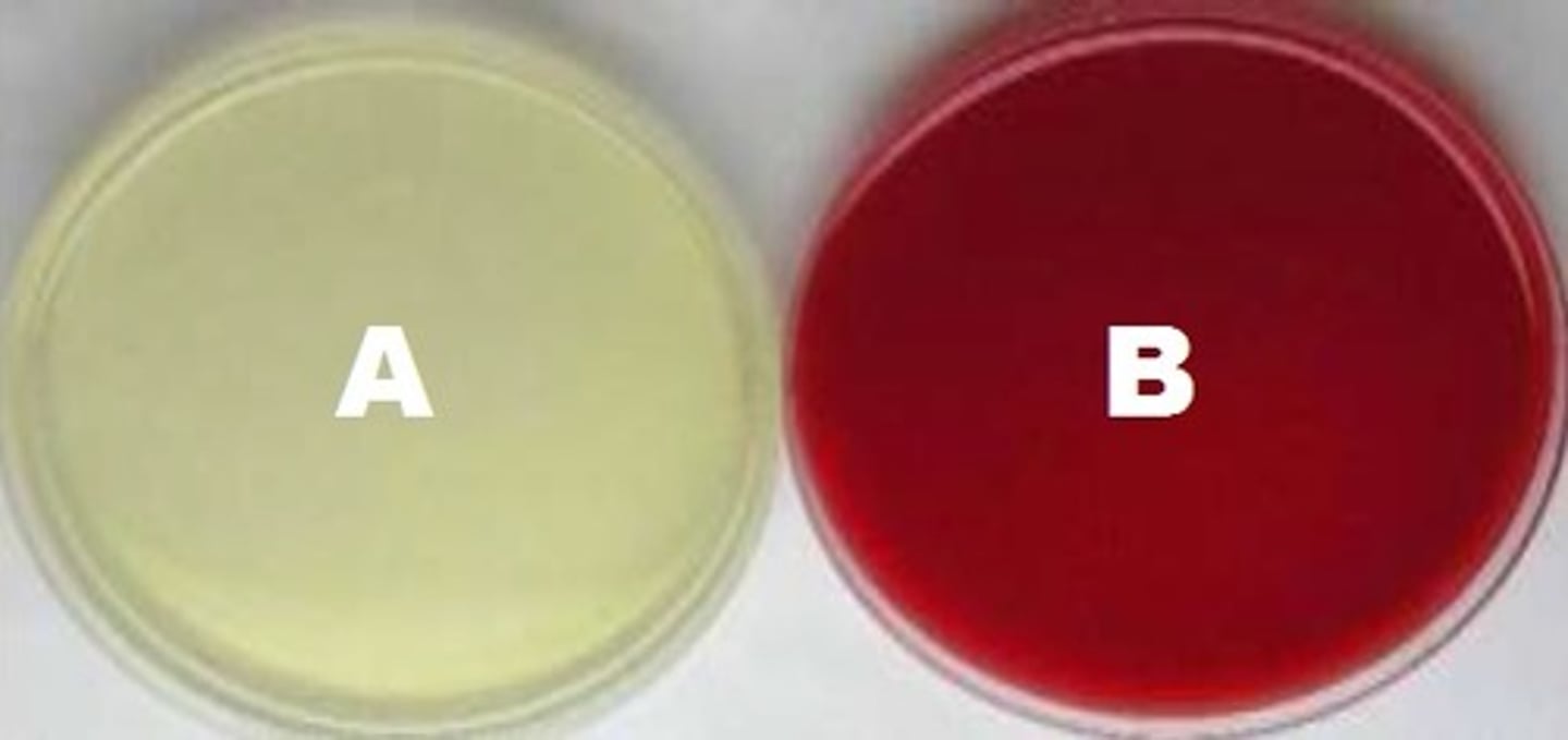
sheep blood agar
enrichment and differential media plate (b)

colony
clones from one bacterium
TSA
tryptic soy agar
SDA
Sabouraud's agar
BAP
blood agar plate
colonies
individual clustered groups of visible growth
cultural characteristics
size, shape and color can help in the identification of unknown microorganisms
Environmental samples
TSA and SDA
alpha hemolysis
partial hemolysis of red blood cells
gamma hemolysis
not hemolytic
normal microbiota
microbes normally present in and on the human body
beta hemolysis
complete hemolysis
light microscopy
brightfield microscopy produces an image by transmitting light through a specimen
ocular lens
microscope part (A)

objectives
microscope part (B)

arm
microscope part (C)

condenser lens and adjustment knob
microscope part (D)

coarse adjustment knob
microscope part (E)

fine adjustment knob
microscope part (F)

lamp switch
microscope part (G)

base
microscope part (H)
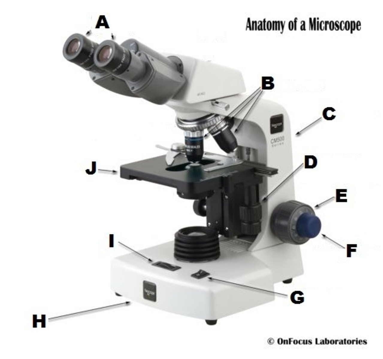
rheostat
microscope part (I)

mechanical stage
microscope part (J)

4x
scanning objective
10x
low power objective
40x
high dry objective
100x
oil immersion objective
domains
tree of life classifications
Archaea, Bacteria, Eukarya
What are the three domains?
protist, fungi, plants, animals
Domain Eukarya subdivided kingdoms
prokaryotic cells
relatively small and simple in structure and have generally have no true nucleus
eukaryotic cells
typically larger and more complex than prokaryotic cells. They contain a nucleus and other membrane bound organelles
unicellular
made of a single cell
multicellular
consisting of many cells
colonial and filamentous forms
protists that are intermediate kinds of organization
yeasts
Kingdom Fungi's unicellular microscopic members
molds
Kingdom Fungi's multicellular but are microscopic and macroscopic
protozoa
protist
locomotion
movement
algal protists
unicellular, free-living eukaryotes, although some __________ are filamentous and colonial
producers
photosynthetic phytoplankton and consumers
heterotrophic decomposers
use organics for carbon and energy
mycelium
mass of hyphae
budding
Asexual reproduction in which a part of the parent organism pinches off and forms a new organism
spores
Asexual reproductive or resting cell capable of developing into a new organism without fusion with another cell, in contrast to a gamete
Aseptic technique
working with microbial cultures that insures the environment, personnel, and the microbial cultures are NOT contaminated
pathogens
microbes that cause disease
Work Area Disinfection
The work area (lab bench) is treated with disinfectant twice when working in the lab. The first disinfection is performed when first entering the lab prior to any materials being placed on the bench. The disinfection will destroy most vegetative cells and viruses but generally will not destroy endospores. Therefore it is not a method of sterilization. The physical process of scrubbing the bench removes microorganisms. The work area is also disinfected after work is complete.
Bacteriological Loops and Needles
The transfers of culture material are achieved by using inoculating loops and needles which are sterilized before and after coming in contact with the culture. The incinerator is used to sterilize the loop or needle by placing in the incinerator opening until they glow orange. The loop or needle must be cooled prior to use which is done by holding for at least 5 seconds. The loop or needle is sterilized again after transferring cultures and before returning to the bench to prevent contamination of the environment. Do not store the loop or needle in the incinerator as the loop becomes so hot the rubber holder may melt. Return the loop or needle to the bench top location.
Culture Tube Handling, Flaming, and Inoculation
Hold culture tubes in your non-dominant hand. Prior to inserting a sterile, cooled loop or needle into the culture, the cap is removed, and the mouth is held next to the incinerator opening. Pass the culture next to the hot opening several times to produce convection currents to prevent microbes from the air from entering the culture. Hold the broth tube at an angle but still upright to minimize the chance of airborne contamination. When finished inoculating, flame the culture mouth again using the same procedure, and replace the cap on the tube. The broth tube is placed in a test tube rack and not laid on the bench top.
Petri Plate Inoculations
The plate lid is used as a shield to prevent airborne contamination by raising the cover and holding it diagonally over the plate. The loop with the inoculum is streaked over the agar surface gently to as the agar can be torn or gouged. The cover is replaced and loop is flamed.
Hand Washing (degerming).
Hands are washed as the last act before students leave the lab and after the bench top disinfection. Students also wash hands if they come into contact with bacterial cultures
Minimize Potential of Contamination
Do not perform transfers of microbes over your books and papers. Place materials safely away from the culture material
Suspend Bacteria in a Broth
Mix broth cultures gently prior to obtaining inoculum by drumming your
fingers on the tube or using a vortex mixer. When using a vortex mixer, start the process slowly. Do not allow broth to get into the cap, lose control of the tube, or allow broth to come out of the tube
Disposal of Used Materials
Materials should be discarded in the appropriate areas for eventual autoclaving (steam sterilization) or incineration (burning).
mixed culture
A culture with many different species of bacteria
Isolation
the separation of different species in a sample or culture medium from each other
pure culture
Isolated bacteria can be placed in new culture medium to have only one species of bacteria
streak plate and pour plate
two common methods of isolation
streak plate method
a bacteriological loop is used to obtain a sample or culture medium using aseptic technique. The loop is then streaked across a nutrient agar surface in a specific manner. As the streaking progresses the bacterial cells are spread out (thinned out), farther and farther apart from each other. The separated cells reproduce and form a colony separated from other colonies. The colony consists of identical cells, which are called clones, because they are all descended from a single, original cell. If the original sample contained more than one type of bacteria, they should be isolated into separate single colonies
pour plate method
a loop is used similarly to sample material. Serial transfers are made to additional tubes so that each transfer results in a greater dilution of the bacteria present in the original medium or sample. If the dilution has been adequate, bacteria will be separated sufficiently in the solidified agar, so that individual colonies will form
morphology
bacteria in a variety of shapes
arrangements
association with each other
cocci
spherical bacteria
bacilli
Rod shaped bacteria
spirilla
spiral shaped bacteria
vibrios
slightly curved rods
coccobacilli
short rods
spirochetes
flexible spirals
pleophorphism
variety of shapes seen in a given sample
diplococcus
pair of cocci
diplobacillus
rod-shaped double bacillus
streptococcus
chain of cocci
streptobacillus
chain of rods
tetrad
a group of four cells
sarcina
cube shaped arrangement of cells
filamentous
thread-like and branching pattern of growth
negative stain
useful in observing morphology, size, and arrangement of bacterial cells
chomogen (nigrosine)
acidic (gives up a hydrogen ion) and carries a negative charge
smear prep
To view bacteria microscopically, the bacteria are applied and adhered to a microscope slide so they can be colored by chemical reagents called stains. The application of bacteria to a slide for the purpose of staining is called a smear. Smears can be made from bacteria growing in liquid and solid culture media.
Gram stain
crystal violet, iodine, alcohol, safranin
differential stain
uses various dyes to differentiate two large groups of bacteria based on their cell walls (peptidoglycan layer)