Chapter 11: Functional Organization of Nervous Tissue
1/38
There's no tags or description
Looks like no tags are added yet.
Name | Mastery | Learn | Test | Matching | Spaced | Call with Kai |
|---|
No analytics yet
Send a link to your students to track their progress
39 Terms
what are the four functions of the nervous system
•Maintaining homeostasis: regulates and coordinate activities to maintain balance
•Receiving sensory input: monitor internal and external stimuli
•Integrating information: brain and spinal cord process sensory input and initiate responses
•Controlling muscles and glands
•Establishing and maintaining mental activity: consciousness, thinking, memory, emotion
what is the divisions of Peripheral Nervous system?
•Sensory (afferent): transmits action potentials from receptors toward the C N S.
•Sensory receptors:
•Can be neuron endings or specialized cells that detect external and internal stimuli
•send input along nerves to brain or spinal cord.
•Motor (efferent): transmits action potentials from C N S to effectors (muscles, glands).
what is the Motor Division of Peripheral Nervous system?
•Somatic nervous system
•from CNS to skeletal muscles
•voluntary
single neuron system
what is the Motor Division of Peripheral nervous systems (part 2 hint: ANS)
•Autonomic nervous system (A N S): from C N S to smooth muscle, cardiac muscle and certain glands
•Subconscious or involuntary control
•Two neuron system: first from C N S to ganglion; second from ganglion to effector.
•Divisions of A N S
•Sympathetic
•Parasympathetic
Enteric
The Cells of the Nervous System are? (hint: 4 major parts)
•Neurons are electrically excitable cells of the nervous system.
•Three major parts:
•Neuron cell body or soma
•Dendrites
•dendritic spines
•Axons:
•Trigger zone = axon hillock + initial segement
what are the four types of neurons?
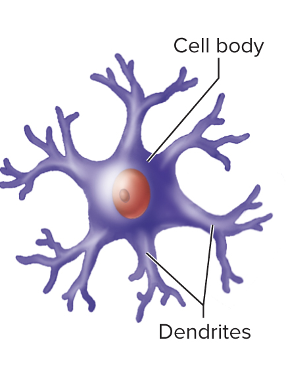
•Multipolar:
•most neurons in C N S
•Motor neurons
•Bipolar:
•sensory in retina of the eye and nasal cavity
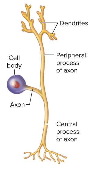
•Pseudo- unipolar:
•single process that divides into two branches
•Part that extends to the periphery has dendrite-like sensory receptors
•Anaxonic:
•no axons, only dendrites
•found in brain and retina where they only communicate using graded potentials
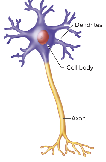
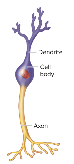
what is this type of neuron and do you know?
Multipolar: many dandrites
what are the action potential types of neurons?
•Sensory or afferent
•action potentials toward CNS
•Motor or efferent
•action potentials away from CNS
•Interneurons:
•within C N S from one neuron to another
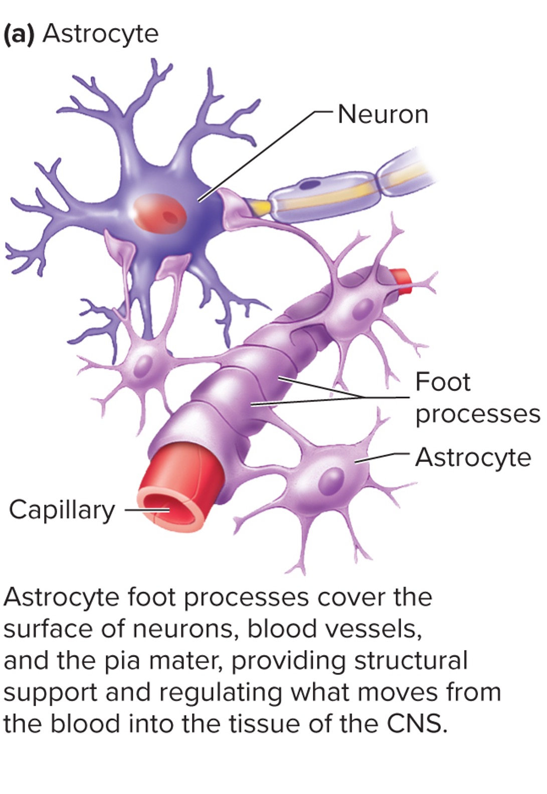
what are the glial cells Astrocytes?
•cover the surfaces of neurons, blood vessels, and the pia mater
•Regulate extracellular brain fluid composition
•form blood-brain barrier to regulate what substances reach the CNS from the blood
•Release chemicals to promote development of synapses and help regulate synaptic activity
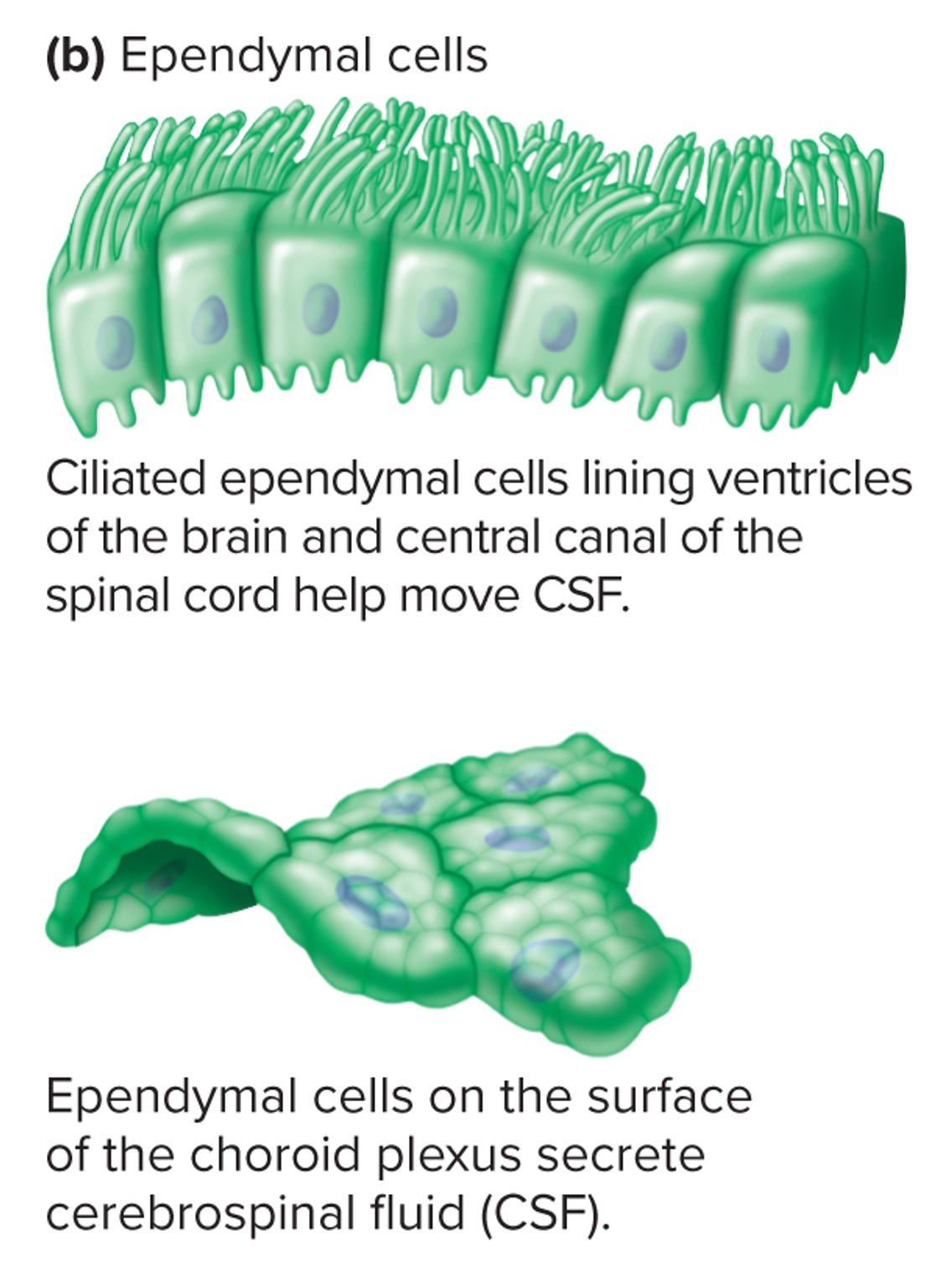
What are the glial cells Ependymal Cells?
•Line brain ventricles and spinal cord central canal.
•Specialized versions of ependymal form choroid plexuses.
•Choroid plexus: Secrete cerebrospinal fluid (CSF)
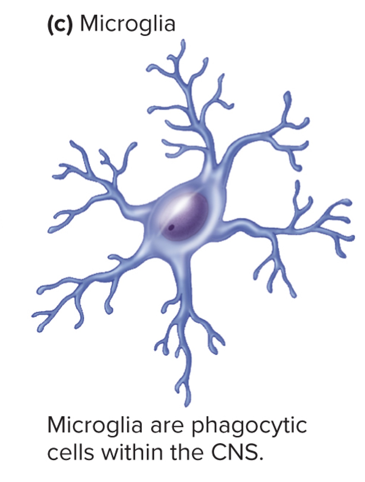
What are the glial cells Microglia ?
•specialized CNS macrophages
•Respond to inflammation, phagocytize necrotic tissue, microorganisms, and foreign substances that invade the CNS.
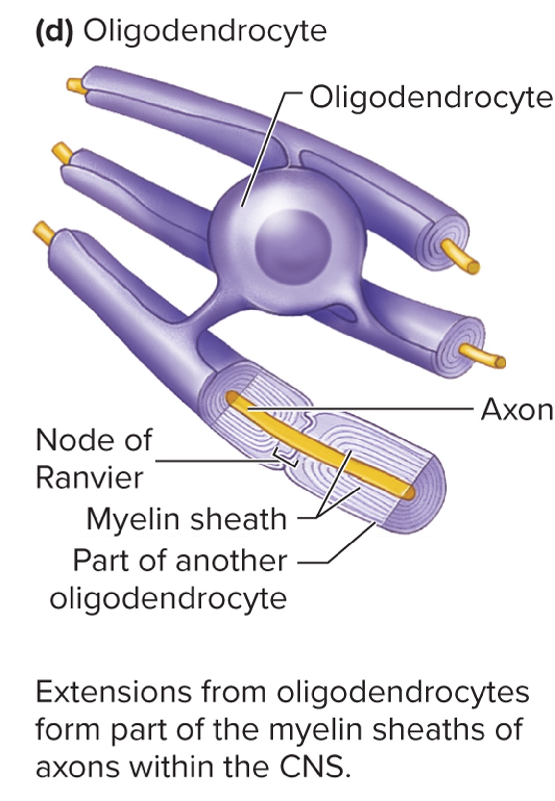
What are the Gilal cells Oligodendrocytes?
•form insulating myelin sheaths by wrapping cytoplasmic extensions around axons
•A single oligodendrocyte can form myelin sheaths around portions of several axons
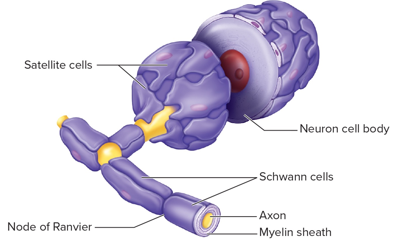
What are the Glial Cells of the Peripheral Nervous System? (hint only 2)
•Schwann cells:
•wrap around portion of only one axon to form myelin sheath
•Satellite cells:
•surround neuron cell bodies in sensory and autonomic ganglia
•provide support, nutrients, and protection from heavy-metal poisons.
what are Myelinated axons?
•Myelin protects and insulates axons from one another, speeds transmission, functions in repair of axons
•Not continuous; gaps called nodes of Ranvier
•Found in white matter
what are Unmyelinated axon?
•found in invaginations of Schwann cells or oligodendrocytes
•Not wrapped around the axon
•gray matter
what are the Gray matter organization of a nervous tissue?
•unmyelinated axons
•cell bodies
•dendrites
•Integrative functions
•The cortex of the brain is gray matter
what are the White matter organization of a nervous tissue?
•myelinated axons
•Propagate action potentials
what are the Central nervous system organization of a nervous tissue?
•clusters of neuron cell bodies are nuclei
bundles of myelinated axons arenerve tract
what are the peripheral nervous system organization of a nervous tissue?
•clusters of cell bodies are ganglia
•bundles of axons with their connective tissue sheaths are nerves
what is a Nerve and the two types of nerves?
•Nerve: a bundle of axons (neurons) outside the brain and spinal cord.
•Cranial nerves: originate from the brain; 12 pairs.
Spinal nerves: originate from spinal cord; 31 pairs.what
what is a Ganglion?
collection of neuron cell bodies outside the brain and spinal cord.
what is a Plexus?
•extensive network of axons, and sometimes neuron cell bodies, located outside CNS.
what is a Glial cells?
supportive cells with many functions.
What type of neuron is this, and how do you know?
Bipolar neuron: has a dendrite and an axon
what is this type of neuron and do you know?
Pseudo- unipolar: No dendrites but axons
what is this type of neuron and do you know?
anaxonic neuron: No axon but many dendrites
What are the types of electrical signals?
•Cells produce electrical signals called action potentials.
•Membrane potential is the result of ionic concentration differences across the plasma membrane and the permeability of the membrane.
what is the Ionic concentration differences across the plasma membrane?
•Ion concentrations across a membrane are a result of two processes:
•the Na+/K+ pump
•membrane permeability.
•Outside the cell = high concentrations of Na+ and Cl-
•Inside the cell = high concentrations of K+ and proteins
•Na+ and K+ have steep concentration gradients
what are the permeability characteristics of the plasma membrane?
•Gated ion channels open and close in response to a stimulus
•When they open, they change the permeability of the cell membrane
what does a sodium-potassium pump consist of?
•Neurons expend A T P to maintain the uneven distribution of ions across the membrane.
•The pump actively pumps 3 Na+ out of the cell and 2 K+ into the cell for each ATP used
what are the five gated ion channels we see?
•Gated ion channels
•open and close because of some sort of stimulus.
•When open, they change the permeability of the cell membrane
•Ligand-gated
•open or close in response to ligand
•Voltage-gated
•open or close in response to specific, small voltage changes across the cell membrane
•At rest, membrane is negative on the inside relative to the outside
•When cell is stimulated, that relative charge changes and voltage-gated ion channels either open or close.
•Touch receptors
•respond to mechanical stimulation
•Temperature receptors
•respond to temperature changes
how do we establishing the resting membrane potential?
•Opposite charges across the membrane, so membrane is polarized.
•Potential difference: unequal distribution of charge exists between the immediate inside and immediate outside of the plasma membrane: −70 to −90 mV.
•The resting membrane potential exists in an unstimulated (resting) cell, due to:
•Permeability characteristics of membrane.
•Differences in ion concentrations on each side of membrane.
when we are depolarization the resting membrane what are we doing? (hint: 6 answers)
•Depolarization
•inside of cell becomes more positive
•for example, from −70mV to −55mV
•Factors leading to depolarization:
•Sodium ions
•Most common way neurons become depolarized
•If gated sodium channels open, sodium diffuses into cell down its concentration gradient, and inside of cell becomes more positive
•Factors leading to depolarization:
•Calcium ions
•Calcium entry also causes depolarization, as seen in cardiac muscle
•Potassium ions.
•Normally, potassium diffuses out of the cell, but changes in the extracellular concentration of potassium can affect the resting membrane potential
•If extracellular potassium levels increase, intracellular stays inside cell (less concentration gradient), and membrane becomes depolarized
what is Hyperpolarization and how do we hyperpolarize a resting membrane potential?
•Hyperpolarization:
•inside of cell becomes more negative
•for example, from −70mV to −90mV
•Two major ways to hyperpolarize:
•Potassium ions
•Most common way neurons become hyperpolarized.
•If gated potassium channels open, potassium diffuses out of cell down its concentration gradient, and inside of cell becomes more negative
•Chloride ions
•Chloride is in higher concentration outside cell, so opening of ligand-gated chloride channels causes chloride to diffuse into cell.
•Adding negative charges into the cell hyperpolarizes it
•Mechanism for some inhibitory neurotransmitters
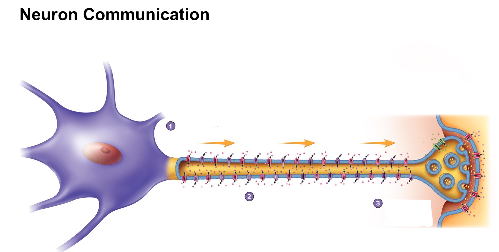
what are the three steps for neuro communication?
Generation of axon potential
action potential propagation along the axon
communication with target cells at the synapse
What is graded potentials and what are the results?
•A relatively small change in the membrane potential in a localized area
•Magnitude varies from small to large depending on stimulus strength or frequency
•Can be hyperpolarizing or depolarizing
•Can summate or add onto each other to reach threshold
•Threshold is the minimum membrane potential needed to generate an action potential.
•Spread (are conducted) over the plasma membrane in a decrementalfashion.
• rapidly decrease in magnitude as they spread over the surface of the plasma membrane.
•Result from:
•Ligands binding to receptors
•Changes in charge across membrane
•Mechanical stimulation
•Temperature changes
•Spontaneous change in permeability
what are the 5 steps of the graded potentials?
A small volume of Na+ entering the cell body causes a slight depolarization.
2 and 3. As greater volumes of Na+ enter the cell body, greater degrees of depolarization occur.
4.A stimulus is applied to a cell causing a small depolarization.
5.When a second depolarizing stimulus is applied before the first disappears, the second stimulus is added to the first to result in an even larger depolarization.
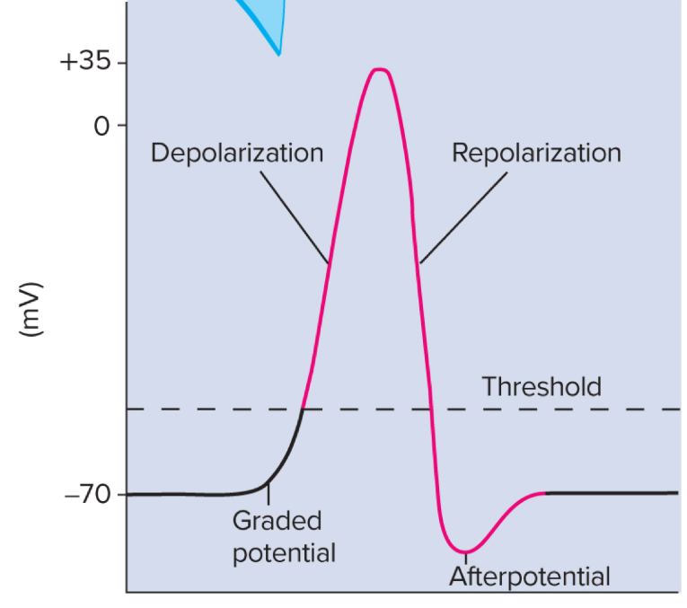
what is happening during an Action Potentials graded?
•Graded potentials summate at trigger zone, reaching threshold
•Four phases of action potentials:
•Depolarization
•Repolarization
•Afterpotential
•Return to resting potential
•All-or-none principle
•No matter how strong the stimulus, as long as it is greater than threshold, then an action potential will occur
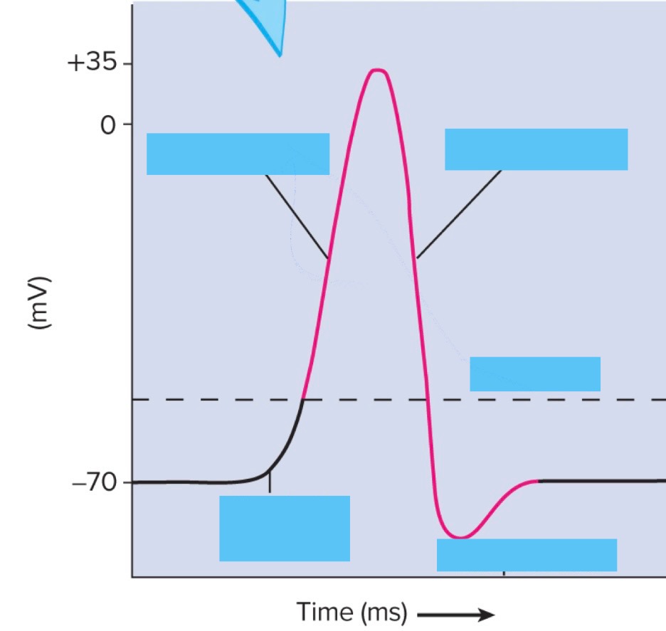
Fill in the graph for Action Potentials?