Neuro Week 4 - Early Development of the Nervous System
1/39
There's no tags or description
Looks like no tags are added yet.
Name | Mastery | Learn | Test | Matching | Spaced |
|---|
No study sessions yet.
40 Terms
what are the 3 germ layers of the embryo in early development?
ectoderm, mesoderm, endoderm
what and where is the notochord?
notochord is a long rod-like midline structure that develops ventral to the neural tube. It is transient, determining position of the nervous system and is
required for early neural differentiation
in between the mesoderm, ventral to the ectoderm and neural plate. formed by the invagination (folding in itself) of the mesoderm
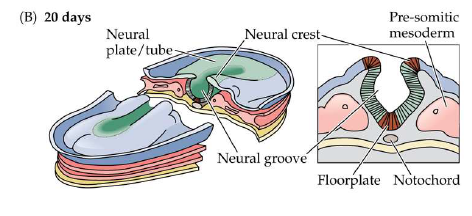
18 days
process of neurulation
development of the neural tube which leads to the development of the brain and spinal cord
As neurulation proceeds, the neural plate begins to fold at the midline (adjacent to the notochord), forming the neural groove and, ultimately, the neural tube.
Neural plate → neural groove → neural tube
20 days
what are the 3 primary swellings (brain vesicles) of the brain (ED)
prosencephalon (forebrain) (pro → forward)
mesencephalon (midbrain) (mes → middle)
rhombencephalon (hindbrain) (rhom → rhombus/behind)
what are the 5 secondary brain vesicles?
Telencephalon (cerebrum)
Diencephalon (thalamus, hypo)
Mesencephalon (midbrain)
Metencephalon (pons, cerebellum)
Myencephalon (medulla)
tell daniel messi met mya
what happens to the space that is present in the neural tube after brain development?
becomes our vesicles
what happens at 24 weeks for brain development?
The fetal brain and spinal cord are clearly differentiated by the end of the second trimester
Thalamic nuclei and basal ganglia are differentiated
Sulci and gyri of the cerebral cortex are beginning to emerge, and will continue developing
what are neural tube defects?
congenital conditions that occur when portions of the neural tube does not develop properly
what is spinal bifida?
Results in missing backbone that normally protects
the spinal cord and nerves
Different types, ranging in severity
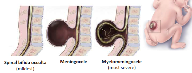
what are the severity levels of spinal bifida?
Spinal bifida occulta: mildest type; small gap in spine, but no opening or fluid-filled sac and spinal cord and nerves are usually unaffected. May not even be discovered until late childhood or adulthood. Most common.
Meningocele: a sac of fluid comes through an opening in the back, but the spinal cord and nerves are not contained within it. Usually little to no nerve damage. Least common.
Myelomeningocele: Parts of the spinal cord and nerves are contained within the sac of fluid and damaged, resulting in moderate to severe disability.
Sometimes surgery prior to birth or within 72-hrs can be performed to correct
what is anencephaly?
a fatal neural tube defect in which the baby is missing significant portions of the brain and skull
To prevent neural tube defects, which month of pregnancy would be most critical to ensure adequate folic acid levels? Explain your reasoning. [3pts]
the first month of pregnancy would be most critical to prevent neural tube defects. These congenital defects occur when the neural tube fails to close completely during early development. Since the neural tube is suppose to close by Week 4. and preventative measures needs to happen within this window, otherwise its too late.
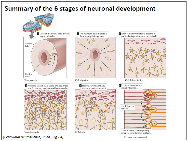
what are the six steps of brain development as a sequence of distinct cellular stages:
Neurogenesis: mitotic division of nonneuronal cells to produce neurons
Cell migration: the movements of cells to establish distinct nerve cell populations (brain nuclei, layers of cortex, etc)
Differentiation: the transformation of precursor cells into distinctive types of neurons and glial cells
Synaptogenesis: the establishment of synaptic connections, as axons and dendrites grow
Neuronal cell death: the selective death of many nerve cells
Synapse rearrangement: the loss of some synapses and development of others, to refine synaptic connections
do neurons divide?
no, only precursor cells
where do neurons and glial cells originate?
ventricular zone
what happens in the marginal zone?
Precursor cells start to migrate out to the marginal zone to become neurons, but some precursor cells are sent back down to the ventricular zone to divide again
where are radial glial cells generated?
they are generated before neurogenesis and extend from the inner to outer surfaces of the brain (like spokes on a wheel).
what is synaptogenesis?
the establishment of synaptic connections as axons and dendrites grow
what are three parts of a growing neuron at the synapse?
Growth cone: growing tip of an axon or dendrites
Filopodia (singular: filopodium): very fine, tubular outgrowths from the growth cone
Cell adhesion molecules (CAMs): protein found on the surface of a cell that guides
cell migration and/or axonal pathfinding
Some CAMs attract certain growth cones (chemoattractant) and some repel certain growth cones (chemorepellent)
when does myelination occur?
Some myelination occurs during gestation (cranial and spinal nerves become myelinated ~24 weeks), but the most extensive phase of myelination occurs shortly after birth and extends into young adulthood
explain apoptosis in ED
naturally occurring cell death
start off with a much greater number of cells in ED and loose them over the period of incubation or gestation
synapse rearrangement
the loss of some synapses and development of others, to
refine synaptic connections
Lissencephaly
rare gene-linked brain malformation resulting in the absence of convolutions in cortex (no sulci/gyri)
large ventricles
incorrect forming of white matter
what is histology
the scientific study of the composition of tissues at a microscopic level
structure → function
How to visualize cells
Combines specialized staining procedures to help visualize
cells
• Count cells and measure density in brain regions [Nissl]
• Examine the morphology of individual neurons [Golgi]
• Map expression of cellular products [IHC]
• Trace interconnections between neurons[Tract tracers]
Steps of histological processing (ADD)
prepare brain for cutting
trans cardiac prefusion (add formaldehyde in place of blood, fixation to remove skull, immerse in 30% in sucrose solution for 48 hours
Cut the brain into thin slices
• Brain mounted and frozen on a microtome
• Sharpe knife cuts into ~50nm thin sections
• Kept in order and placed into wells with solution
mount sections onto slides
run staining prodecure (nissl) - dehydrate tissue with ethanol → Soak in cresyl violet → Rehydrate tissue → Final clearing rinse
add coverslip to slides → now ready for viewing under microscope
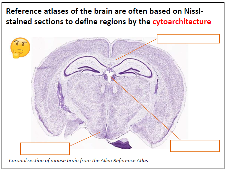
Nissl Stain
outlines all cell bodies in the tissue by staining RNA, and is useful for defining cytoarchitecture
does not stain dendrites and axons
what is a limitation of Nissl stains
detailed morphology of the neurons is not stained
cytoarchitecture
the study of the structural organization and arrangement of
cells within tissues, and they vary from brain region to brain region
label
insert completed image
corpus callosum
third ventricle
hypothalamus
Golgi staining
useful for examining the precise shape and structure of individual neurons
how much of the brain does golgi staining stain?
5-10% (not a limitation!)
what do Immunohistochemistry (IHC) and immunofluorescence do?
both use antibodies to target cells with a specific protein
Antibodies seek out and attach themselves to target protein
• Reveals a distribution of only those neurons that make the
target protein
add c-fos flashcard
How do you categorize neural cell types?
shape
genes
electrical properties
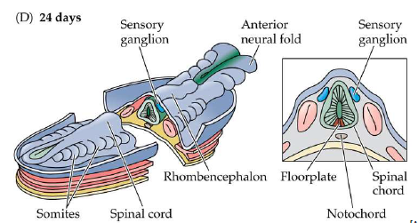
somites
form in the the mesoderm around the neural tube, that develop into the axial musculature and skeleton
what does the neural tube turn into?
the forebrain, midbrain, hindbrain, and spinal cord (the ones close to the somites)
what happens at 4 weeks (24 days) of germination?
the neural tube adjacent to the somites → spinal cord, and the neural crest → sensory and autonomic ganglia
anterior ends of neural plate (anterior neural folds) grow together at the midline and continue to expand, eventually giving rise to the brain.
By 4-weeks the neural tube will be fully closed
where is the ventricular zone?
the thickness of the neural tube
what are the two layers that early neural tube have
ventricular + marginal and later in development the wall thickens and forms an intermediate layer