lab 5
1/63
There's no tags or description
Looks like no tags are added yet.
Name | Mastery | Learn | Test | Matching | Spaced | Call with Kai |
|---|
No analytics yet
Send a link to your students to track their progress
64 Terms
what are the 5 vital signs?
heart rate
body temperature
respiratory rate
blood pressure
oxygen saturation
what is the resting heart rate physically determined by?
a combination of the rate at which the SA node produces action potentials and the effects of innervation by the parasympathetic nervous system (rest and digest)
on average, how many action potentials does the SA node produce per minute?
100 APs per minute = 100 beats per minute
at rest, the parasympathetic neurons send a constant stream of signals to SA node that decrease AP rate to what?
an average of 75 APs per minute = 75 beats per minute
what is the average heart rate and the range?
average is 75bpm, range is 60-100bpm
what is bradycardia?
this is when the resting heart rate is lower than 60bpm
what is tachycardia?
this is when a resting heart rate is higher than 100bpm
how does heart rate increase from exercise? in terms of SNS and SA node
the SA node gets stimulated by SNS and the endocrine system as it releases epinephrine from adrenal glands. these both act on SA node to increase heart rate by increasing the amount of APs per minute.
how do we calculate the maximum heart rate HRmax of an individual?
we do HRmax = 220 - age
heart sounds relate to what?
the closure of atrioventricular valves as systole begins = LUB
the closure of the semilunar valves as ventricular diastole begins = DUB
listening to heart sounds can provide what information?
information on the heart’s condition and function
diagnose heart murmurs that are a result from malfunctioning valves, holes in interventricular or interatrial septa, or other cardiac abnormalities
when listening for heart sounds, where should you place the stethoscope to hear LUB/S1?
apex of the heart, left of sternum/under breast
occurs from turbulent blood flow caused by closure of mitral and tricuspid valves
when listening for heart sounds, where should you place the stethoscope to hear DUB/S2?
2-3 cm underneath the clavicle, left of sternum
occurs from turbulent blood flow caused by the closure of the pulmonary and aortic semilunar valves
the contraction of cardiac muscle is initiated by what?
the conduction system of the heart
what is the conduction system composed of?
non-contractile cardiac muscle cells that have been modified to generate and spread electrical impulses.
in which atrium or ventricle can we find the SA node in?
the right atrium
how do the impulses of the SA node spread throughout the heart?
see notes
what are the 3 distinguishable waves in an ECG?
P wave
QRS wave
T wave
what does ECG mean?
electrocardiogram
what are the 4 segments/intervals of an ECG?
P-Q interval
S-T interval/segment
T-P interval
R-R interval
what is the P wave?
this is atrial depolarization. SA node fires and depolarization spreads from SA node to throughout atria
what is the QRS wave?
this is ventricular depolarization. the depolarization wave spreads from AV node to the rest of the conduction system and through the ventricles.w
what is the T wave?
ventricular repolarization
what is the P-Q interval?
the start of the P wave until the start of Q wave. this represents the atria contracting and the APs spreading through the AV node
what is the S-T interval?
the end of S wave to the start of T wave. the ventricles are depolarized and contracted. blood is ejected from the ventricles into the large arteries (stroke volume)
what is the T-P interval?
this is the period of time between cardiac cycles when heart is at rest. both atria and ventricles are at rest.
what is the R-R interval?
represents the time it takes for 1 full cardiac cycle since it’s in between two R waves
what is an ECG composed of?
all of the action potentials generated by nodal and contractile cells at a given time.
what is an ECG used to assess for?
to see if the heart’s rhythm and electrical conduction is normal for a person
what are arrhythmias?
these are irregular rhythms caused by abnormal impulse formation or conduction
what are conduction delays?
these are slower than normal electrical transmission through the heart’s conduction pathways.
what is heart block?
this is a conduction delay that produces “dropped” beats because some atrial impulses (the P waves) fail to conduct through the AV node/bundle branches/pukinje fibers. this results in P waves that don’t follow QRS complexes = atria contract more than ventricles = an extra P wave
what is fibrillation?
this is uncoordinated signalling and contraction either in the atria or the ventricles. basically, a bunch of rapid irregular waves where you can’t even identify any of the waves. this is fatal if left untreated since ventricles can no longer coordinate a proper pump for blood flow
is bradycardia unhealthy?
not necessarily! this can also be a sign of excellent cardiovascular fitness. high performance athletes tend to have resting heart rates below 60bpm!
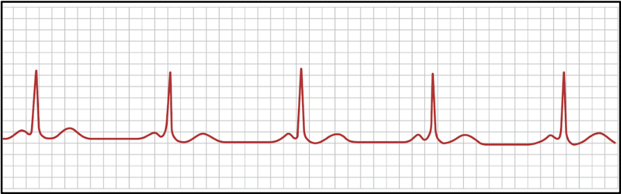
what is this
bradycardia
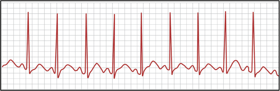
what is this
tachycardia
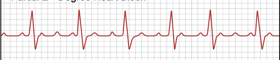
what is this
heart block
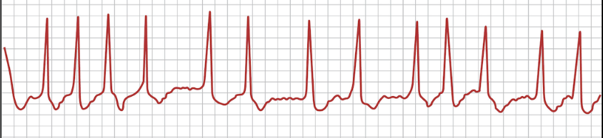
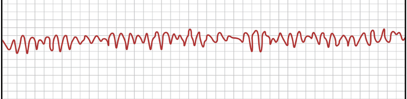
what is this
atrial fibrillation
what is this
ventricular fibrillation
how do we calculate heart rate from an electrocardiogram?
take chart speed and divide it by the distance between two QRS waves
take that value and multiply by 60
final value units should be beats/min
what is blood pressure used to assess for?
cardiovascular health
why is it important to maintain blood pressure?
it’s important for the proper functioning of the heart and all of the organs it supplies blood to
what is perfusion?
blood flow removing wastes
what will happen if mean arterial pressure MAP is too low?
there won’t be enough perfusion which can damage our tissues
what does blood pressure represent physiologically?
the force that blood puts on the arterial walls during contraction—systole—and relaxation—diastole—of the ventricles
where is blood pressure generally measured?
in the arteries
TRUE OR FALSE: since the heart alternates in contracting and relaxing, the blood pressure rises and falls, which is why you must take two blood pressure readings
TRUE! these are systolic and diastolic pressure
what is systolic pressure?
the pressure in the large arteries of the heart at the peak of ventricular ejection
what is diastolic pressure?
this is the pressure of large arteries during ventricular relaxation
what is hypertension?
high blood pressure. typically a value of 140/90 or higher when measured manually and 135/85 for digital instruments
what instruments do we use to measure blood pressure?
a stethoscope and a sphygmomanometer
TRUE OR FALSE: some drugs like caffeine and nicotine can cause long-term increases to blood pressure.
FALSE! they cause temporary SHORT-term increases to blood pressure. this is called acute hypertension
what is chronic hypertension?
this is when high blood pressure lasts for much longer periods and is usually associated with aging → our arteries become less elastic as we age
can also be from…
diet rich in fat or excess salt
lack of exercise
stress
some drugs and alcohol intake
what is the auscultatory method?
listening to the sounds of the body by using a stethoscope
what is blood pressure measured using?
the brachial artery since this artery is similar height/position as the large arteries exiting heart
what are the sounds of korotkoff?
this is the resumption of blood flow into the brachial artery
when using the sphygmomanometer, we inflate the cuff until what
until 160mmHg
when using the sphygmomanometer, we keep the cuff inflated for no more than?
no more than 1 minute
how do we calculate pulse pressure?
pulse pressure = systolic pressure - diastolic pressure
how do we calculate for mean arterial pressure MAP?
MAP = (1/3)(pulse pressure) + diastolic pressure
what is MAP?
the pressure of blood being delivered to organs.
what happens if MAP is too low?
if this is too low, blood may not reach the organ or may pass to slowly to have proper exchange of oxygen and nutrients and removal of wastes
what if MAP is too high?
the blood may pass through the organ too quick, not exchanging properly. it can also cause damage to the vessels of the organ
what values should MAP be?
between 70mmHg and 110mmHg