1.02 Asynchronous
1/74
There's no tags or description
Looks like no tags are added yet.
Name | Mastery | Learn | Test | Matching | Spaced | Call with Kai |
|---|
No analytics yet
Send a link to your students to track their progress
75 Terms
What are the two ways each vertebra articulates with the upper and lower vertebrae?
Each vertebra articulates in two ways:
JOINT 1: Bodies of the vertebrae articulate with the body of the upper and lower vertebra via an intervertebral disc.
JOINT 2: The articular processes of each vertebra articulate with the upper and lower vertebrae articular processes
Describe the functional and structural classification of the articulations of vertebral bodies.
Articulations of vertebral bodies are functionally amphiarthrodial and structurally cartilaginous symphysis joints.
How much movement occurs between individual vertebrae and what is the total range of movement for the vertebral column?
The individual vertebrae move only slightly on each other. However, when this slight movement occurs in all joints of the vertebral column, the total range of movement is very considerable

What is the Anterior Longitudinal Ligament (ALL)?
The Anterior Longitudinal Ligament (ALL) is a broad and strong band of fibers.
Where does the Anterior Longitudinal Ligament (ALL) extend?
The ALL extends along the anterior surfaces of the bodies of the vertebrae, from the occipital bone to the sacrum.
What are the functions of the Anterior Longitudinal Ligament (ALL)?
The ALL limits backward bending and supports the anterior convexity in the lumbosacral area.
Where is the Posterior Longitudinal Ligament (PLL) situated and where does it extend?
The Posterior Longitudinal Ligament (PLL) is situated within the vertebral canal and extends along the posterior surfaces of the bodies of the vertebrae, from the body of the axis, to the sacrum.
Does the Posterior Longitudinal Ligament (PLL) attach directly to the vertebral body, and what does it cover?
The posterior longitudinal ligament (PLL) doesn’t directly attach to the vertebral body itself.
Instead, it lies over a network (plexus) of arteries, veins, and lymphatic vessels.
These vessels pass through nutrient foramina (tiny openings in the bone) to reach the cancellous (spongy) bone inside the vertebral body.
What is the tensile strength of the Posterior Longitudinal Ligament (PLL) and what is its effect on forward bending?
The PLL has a relatively low tensile strength and does not significantly restrict forward bending
What is an intervertebral disc?
An intervertebral disc is a fibrocartilaginous structure that intervenes between the bodies of the adjacent vertebrae and binds them together.

What percentage of the vertebral column's length (height) does the intervertebral disc account for?
20-25% of the length (height) of the vertebral column.
What are the two main parts of an intervertebral disc?
An intervertebral disc is made of 2 parts:
The nucleus pulposus
The anulus fibrosus
What can result from degenerative changes in the anulus fibrosus?
Can lead to herniation of the nucleus pulposus
What can postero-lateral herniation of the nucleus pulposus impinge upon?
Postero-lateral herniation can impinge on the roots of a spinal nerve in the intervertebral foramen
What type of joints are the articular processes (JOINT 2) of the vertebrae?
The articular processes articulate via synovial planar joints called zygapophysial (facet) joints

To which variety of joints do zygapophysial (facet) joints belong, and what type of motion do they allow?
The zygapophysial (facet) joints belong to the diarthrodial (synovial) variety and are Planar joints allowing slide and glide motion
What envelops zygapophysial (facet) joints?
Zygapophysial (facet) joints are enveloped by capsules lined by synovial membranes
How do the articular capsules of zygapophysial joints vary across regions of the vertebral column?
The articular capsules are more extended and looser in the cervical than in the thoracic and lumbar regions
Name the ligaments that connect to the laminae, spinous, and transverse processes (associated with JOINT 2).
The Ligamenta Flava
The Supraspinous Ligament
The Interspinous Ligaments
The Intertransverse Ligaments

Describe the Ligamenta Flava, including their attachment points and function.
Attachment Point: The Ligamenta Flava connect the laminae of adjacent vertebrae, from the axis to the first segment (vertebra) of the sacrum.
Function: Their marked elasticity serves to preserve the upright posture, and to assist the vertebral column in resuming it after flexion.

Describe the Supraspinous Ligament, including its attachment points and its upward continuation
Attachment Point: A strong fibrous cord, which connects together the apices of the spinous processes from the seventh cervical vertebra to the sacrum.
It is continued upward to the external occipital protuberance as the ligamentum nuchae.

Describe the Interspinous Ligaments
The Interspinous Ligaments are thin and membranous and connect adjoining spinous processes and extend from the root to the apex of each process.

Describe the Intertransverse Ligaments.
The Intertransverse Ligaments are interposed between the transverse processes.
What is the Ligamentum Nuchae?
The Ligamentum Nuchae is a fibrous membrane, which, in the neck, represents the supraspinous ligaments of the lower vertebrae.
Where does the Ligamentum Nuchae extend?
It extends from the external occipital protuberance to the spinous process of the seventh cervical vertebra.
Which cervical vertebra has the most prominent spinous process, and what is it called?
CVII (the 7th cervical vertebra) has the most prominent spinous process (not bifurcated) in the vertebral column, called vertebral prominent.
What are the two sets of craniovertebral joints?
Atlanto-occipital
Atlanto-axial joints
How many atlantoaxial joints are there, and what are their types?
There are three atlantoaxial joints: two lateral and one medial atlantoaxial joints.
Describe the lateral atlantoaxial joints
The lateral atlantoaxial joints are synovial gliding joints made between the lateral masses of the atlas and the superior articular processes of the axis.
Describe the medial atlantoaxial joint.
The medial atlantoaxial joint is a synovial pivot joint between the odontoid process of the axis and the ring formed by the anterior arch and the transverse ligament of the atlas
What movement does the medial atlantoaxial joint allow, and what is its axis of motion?
This joint allows the rotation of the atlas (and, with it, the skull) upon the axis. The axis of motion is vertical through the dens.
Approximately what percentage of cervical spine rotation occurs at the atlanto-axial joint?
Approximately 50% of rotation in the cervical spine occurs at the atlanto-axial joint
Name the supportive elements (ligaments/membranes) of the atlantoaxial joints.
Supportive elements include:
Anterior Atlantoaxial Ligament (membrane)
Posterior Atlantoaxial Ligament (membrane)
Describe the Anterior Atlantoaxial Ligament (membrane).
This ligament is a strong membrane, fixed, above, to the lower border of the anterior arch of the atlas; below, to the front of the body of the axis.
Describe the Posterior Atlantoaxial Ligament (membrane)
This ligament is a broad, thin membrane attached, above, to the lower border of the posterior arch of the atlas; below, to the upper edges of the laminae of the axis.
What does the Posterior Atlantoaxial Ligament (membrane) replace?
It supplies the place of the ligamentum flavum.
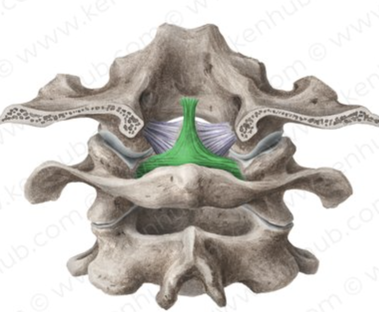
Describe the Transverse Ligament of the Atlas.
The Transverse Ligament of the Atlas is a thick, strong band, that arches across the ring of the atlas, and retains the odontoid process in contact with the anterior arch
Where is the Transverse Ligament of the Atlas attached?
It is firmly attached on either side to a small tubercle on the medial surface of the lateral mass of the atlas.
What fasciculi are associated with the Transverse Ligament of the Atlas, and what is the whole structure named?
As it crosses the odontoid process, a small fasciculus is prolonged upward (attached to the basilar part of the occipital bone), and another downward (fixed to the posterior surface of the body of the axis) from its superficial/posterior fibers.
Hence, the whole ligament is named the cruciate ligament of the atlas.
Name the ligaments connecting the Axis with the Occipital Bone.
Tectorial membrane
Alar ligaments
Describe the Tectorial Membrane
The Tectorial membrane is situated within the vertebral canal. - It covers the odontoid process
It’s a prolongation upward of the posterior longitudinal ligament of the vertebral column.
Describe the Alar Ligaments
Arise one on either side of the upper part of the odontoid process and, passing obliquely upward and lateral ward, are inserted into the medial sides of the condyles of the occipital bone.
What other ligament connects the vertebral column with the cranium, besides those directly uniting the atlas and axis to the skull?
The ligamentum nuchae must be regarded as one of the ligaments connecting the vertebral column with the cranium.
Describe the articulation of the atlas with the occipital bone.
The articulation of the atlas with the occipital bone consists of a pair of synovial condyloid (condylar) joints.
Name the ligaments connecting the atlas and occipital bone.
Articular capsules
The Anterior Atlantooccipital Membrane
The Posterior Atlantooccipital Membrane
Describe the articular capsules of the atlanto-occipital joint.
The articular capsules surround the condyles of the occipital bone and connect them with the superior articular surfaces of the lateral masses of the atlas. They are thin and loose.
Describe the Anterior Atlantooccipital Membrane.
The Anterior Atlantooccipital Membrane passes between the anterior margin of the foramen magnum above, and the upper border of the anterior arch of the atlas below.
Describe the Posterior Atlantooccipital Membrane.
The Posterior Atlantooccipital Membrane is connected above, to the posterior margin of the foramen magnum; below, to the upper border of the posterior arch of the atlas
What movements are permitted in the atlanto-occipital joint?
Flexion and Extension, which give rise to the ordinary forward and backward nodding of the head.
Slight lateral flexion (bending)
Rotation.
What are uncovertebral “joints” or clefts (of Luschka's joints)?
Uncovertebral “joints” or cleft (of Luschka's joints) are synovial planar joints formed between uncinate processes of the cervical vertebrae.
Where are uncovertebral joints located?
They are located in the cervical vertebrae between C3 and C7.
Describe the structure of uncovertebral joints.
2 lips project upward from the superior surface of the vertebral body below and one projects downward from the inferior surface of the vertebral body above.
What movements do uncovertebral joints allow and limit?
They allow for flexion and extension and limit lateral flexion in the cervical spine
What type of joints are the articulations of the head of the ribs (Costovertebral Joint)?
Articulations of the head of the ribs constitute a series of synovial planar (gliding) joints (diarthrodial joints).
How are costovertebral joints formed?
They are formed by the articulation of the head of the typical ribs with the facets on the sides of the bodies of the thoracic vertebrae and with the intervertebral discs between them.
Which ribs articulate with a single vertebra?
The 1st, 10th, 11th, and 12th ribs each articulate with a single vertebra.
Name the ligaments of the costovertebral joints.
Radiate ligament of head of rib
The Intra-articular ligament of head of rib
Describe the Radiate ligament of the head of the rib.
The Radiate ligament of the head of the rib connects the anterior part of the head of each rib with the side of the bodies of two vertebrae, and the intervertebral disc between them
Describe the Intra-articular ligament of the head of the rib.
The Intra-articular ligament of the head of the rib is situated in the joint’s interior. It attaches on one end to the crest separating the two articular facets on the head of the rib, and on the other end to the intervertebral disc.
What is the function of the Intra-articular ligament of the head of the rib?
It divides the joint into 2 cavities.
What are costotransverse joints and what type of joints are they?
Costotransverse joints are formed between the tubercle of the rib with the articular surface on the adjacent transverse process.
They are diarthrodial joints
Name the ligaments of the costotransverse joints.
The superior costotransverse ligament
The costotransverse ligament
The lateral costotransverse ligament
Describe the superior costotransverse ligament.
The superior costotransverse ligament is attached below to the upper border of the neck of the rib and to the transverse process immediately above.
Describe the costotransverse ligament.
The costotransverse ligament connects the rough surface on the back of the neck of the rib with the anterior surface of the adjacent transverse process.
Describe the lateral costotransverse ligament.
The lateral costotransverse ligament passes obliquely from the tip of the transverse process to the rough non-articular tubercle of the rib.
Summarize how each rib articulates posteriorly.
Each rib touches 2 vertebral bodies and 1 transverse process
Describe the articulations of the cartilages of the true ribs with the sternum (Sternocostal joints).
The articulations of the cartilages of the true ribs with the sternum are synovial joints, except for the 1st rib.
How does the first rib articulate with the sternum?
For the first rib, the cartilage is directly united with the sternum, and it is therefore a synchondrosis articulation.
Name the ligaments connecting the sternocostal joints
Anterior radiate sternocostal ligaments
Posterior radiate sternocostal ligaments
Describe the anterior and posterior radiate sternocostal ligaments.
These ligaments consist of broad and thin membranous bands that radiate from the front and back of the sternal ends of the cartilages of the true ribs to the anterior and posterior surfaces of the sternum.
What movements are permitted in the sternocostal joints?
Slight gliding movements are permitted in the sternocostal joints.
Describe costochondral articulations.
The lateral end of each costal cartilage is received into a depression in the sternal end of the rib, and the two are held together by the periosteum.
What type of joint are all costochondral articulations?
They are all synchondrosis types of the joint (structurally are synarthrosis)
What joint movements work together to produce "bucket handle" motion, and what is this motion?
Sternocostal + Costotransverse and Costovertebral joints work together to move the ribs in a superior and lateral direction.
This is known as “bucket handle” motion
What joint movement produces "pump handle" motion, and what is this motion?
Sternocostal alone pulls the sternum in a superior direction.
This is known as “pump handle” motion