L21: diagnostic imaging of thoracic limb small animal
1/103
There's no tags or description
Looks like no tags are added yet.
Name | Mastery | Learn | Test | Matching | Spaced | Call with Kai |
|---|
No analytics yet
Send a link to your students to track their progress
104 Terms
how should the external surface of the cortical bone be?
smoothly marginated except at the point of the muscle and tendon insertion
what coverse the bone surface in normal mature bone?
periosteum
periosteum
outer vascular connective tissue of bone that is connected to the underlying cortex
what is the opacity of the periosteum of normal mature bone on a radiograph?
soft tissue opacity
endosteum
lines inner surface of cortex of bone; made up of connective tissue
what is between the periosteum and endosteum layers?
cortical bone
what bone is denser for normal bones?
compact cortical bone denser than spongy cancellous bone
how would the cortices of bone appear if there was high stress and loading on the bone?
would appear thickened
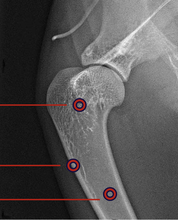
what is the top circling highlighting?
cancellous bone

what is the middle red circle highlighting?
cortex

what is the bottom red circle highlighting?
medulla
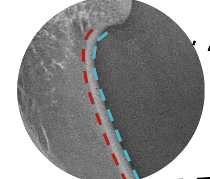
what layer of bone is the blue?
periosteum

what layer of bone is the red?
endosteum
how many radiographic views is required?
atleast 2. prefer 3
what are the radiographic views?
lateral
cranial/caudal
ventral or dorsal
if we are trying to image a lateral view of the scapula, what position will we put the animal in?
right or left lateral recumbency with the affected limb down
what view of the scapula is shown?
lateral view
what position will we put the animal in if we want to get a caudo-cranial view of the scapula?
dorsal recumbent position with forelegs extended cranially
what radiograph view of the scapula is shown?
caudo-cranial view
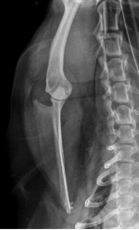
what position will we put the animal if we want to get a lateral view of the shoulder joint?
right or left lateral recumbency with the affected limb down (pulled cranially and ventrally)
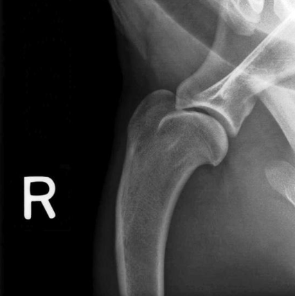
what radiograph view of the shoulder joint is shown?
lateral view
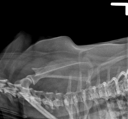
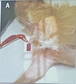
what position will we put the animal in to get a skyline view of the shoulder?
sternal recumbency with the head turned slightly away from the shoulder without rotating the thorax; elbow joint of affected leg will be flexed and film-screen cassette is placed in crook of elbow

what radiographic view of the shoulder is shown?
skyline view
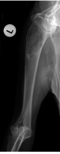
what position will we put the animal for a lateral view of the humerus?
right or left lateral recumbency with the affected limb down (pulled cranially and ventrally)
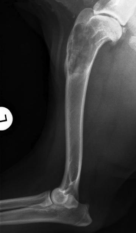
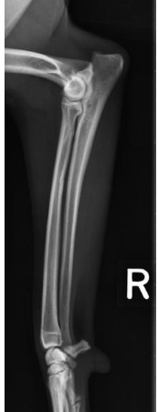
what radiographic view of the humerus is shown?
lateral view
what position will we put the animal in for a caudal-cranial view of the humerus?
dorsal recumbent position with the forelegs extended cranially
what radiographic view of the humerus is shown?
caudo-cranial view
what position will we put the animal in for a cranio-caudal view of the humerus?
dorsal recumbent position with the forelegs extended caudally
what radiographic view of the humerus is shown?
cranio-caudal view
what are the two ways the elbows can be flexed for a lateral view of the elbow joint?
flexed at 45 or 90 degrees
fully flexed
what position will we put the animal for a lateral view of the elbow joint?
right or left lateral recumbency with the affected limb pulled down cranially and ventrally
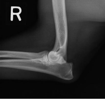
what radiographic view of the elbow is shown?
lateral view with elbow flexed 90 degrees
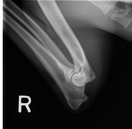
what radiographic view of the elbow is shown?
lateral view with elbow fully flexed
what structure of the elbow is focused on if we are doing a lateral view with the joint fully flexed?
olecranon process
what position do we put the animal in if we want a cranio-caudal view of the elbow?
sternal recumbency with the forelegs extended cranially
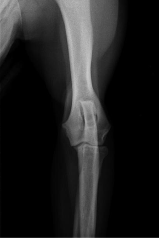
what radiographic view of the elbow is shown?
cranio-caudal view
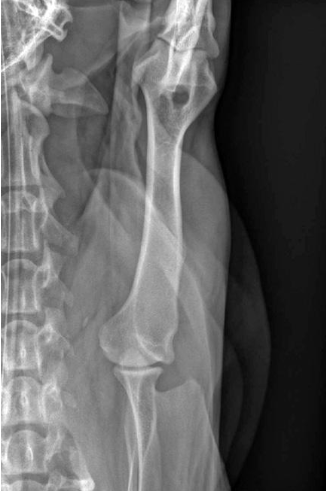
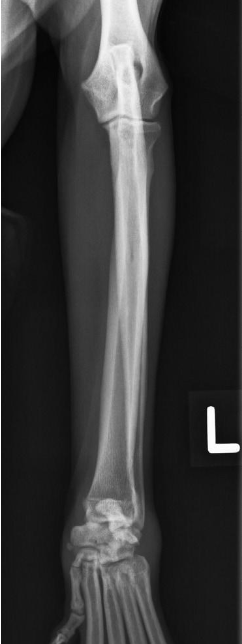
what position do we put the animal in for a lateral view of the radius and ulna?
lateral recumbency with the affected limb down (slightly flex the elbow and pull limb cranially)
what radiographic view of the radius and ulna is shown?
lateral view
what position do we put the animal in for a cranio-caudal view of the radius and ulna?
sternal recumbency with forelegs fully extended cranially and head elevated away from limb
what radiograph view of the radius and ulna is shown?
cranio-caudal view
what radiograph view of the carpal joint is shown?
lateral view
what radiograph view of hte carpal joint is shown?
lateral view with carpal joint flexed at 90 degrees
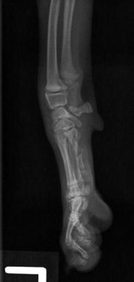
what position do we put the animal in for a dorso-palmar view of the carpal joint and digits?
sternal recumbency with affected forelegs extended cranially and head elevated away
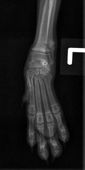
what radiograph view of the carpal joint and digits is shown?
dorso-palmar view
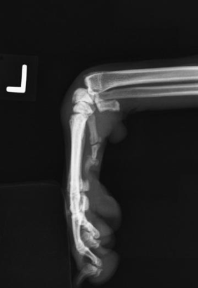
what digits are taped if we are doing a splayed lateral view of the digits?
digits 2 to 5
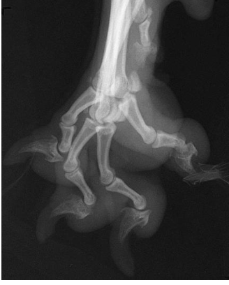
what radiographic view of the digits is shown?
splayed lateral view of the digits
MCQ: what view is this dog positioned for?
medio-lateral of the shoulder joint
MCQ: to aquire a skyline view of the shoulder, the x-ray beam direction is…
proximo cranial-disto cranial view

what is 1 (red box)?
spine of scapula

what is 2?
supraspinous fossa

what is 3?
infraspinous fossa

what is 4?
acromion
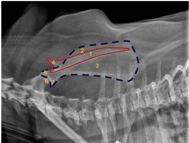
what is 5?
supraglenoid tubercule

what is 6?
glenoid cavity

what is 7?
infra-glenoid tuberosity

what is 1?
spine of scapula
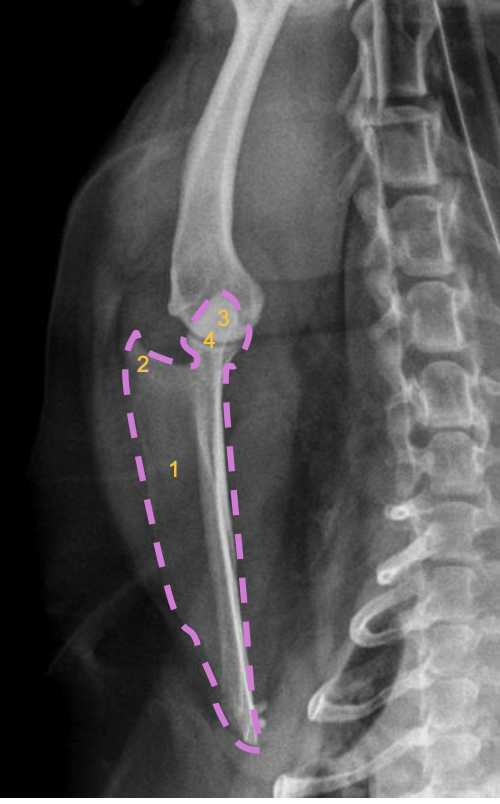
what is 2?
acromion

what is 3?
supraglenoid tubercule

what is 4?
glenoid cavity

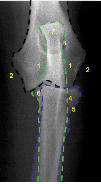
what is 11?
lateral epicondyle
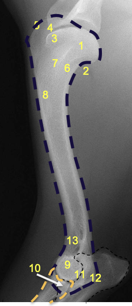
what is 12?
medial epicondyle
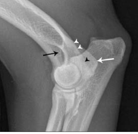
what are the white arrowheads pointing to?
lateral supracondylar crest

what is the black arrowhead pointing to?
caudal aspect of the lateral epicondyle
what is the red circle highlighting?
tricipital line present in the humerus
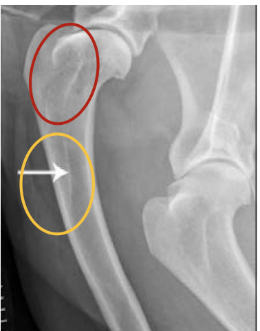
what is the orange circle highlighting?
deltoid tuberosity
where does the tricipital line of the humerus extend?
disto-cranially to merge with the deltoid tuberosity
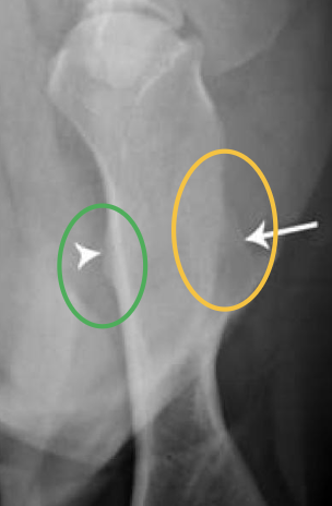
what is the green circle highlighting?
teres major
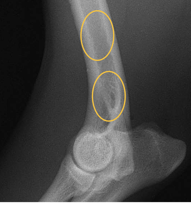
what are the orange circles showing?
mid-diaphyseal medullary region of the humerus (radioluscent)

what is the blue arrow pointing to?
greater tubercle of the humerus
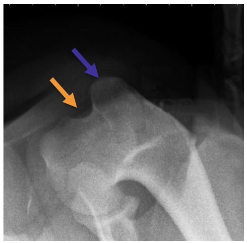
what is the orange arrow pointing to?
lesser tubercle of the humerus

what is the groove between the blue and orange arrows called?
intertubercular groove or bicipital groove

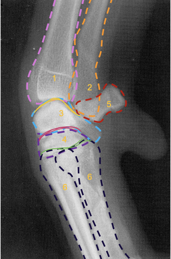
what is the pink line?
medial epicondyle of the humerus (makes 90 degree angle)

what is the purple line?
lateral condyle of humerus
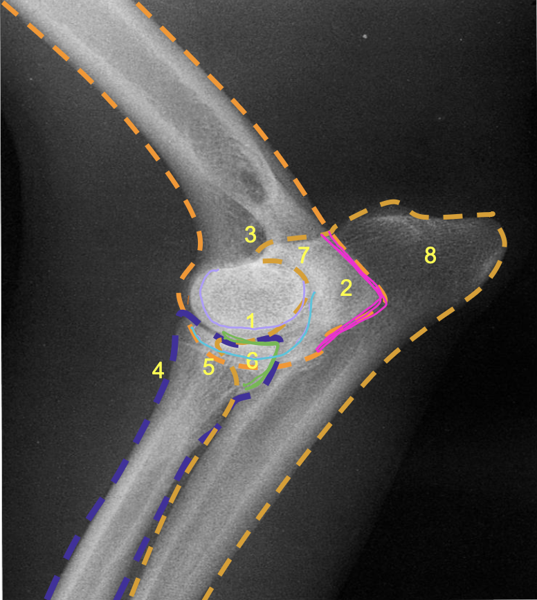
what is the light blue line?
medial condyle of humerus

what is the green line?
medial coronoid process of the ulna

what is 8?
olecranon process of the ulna

what is 7?
olecranon of ulna
what is 1?
condyles of humerus

what is 2?
epicondyles of humerus
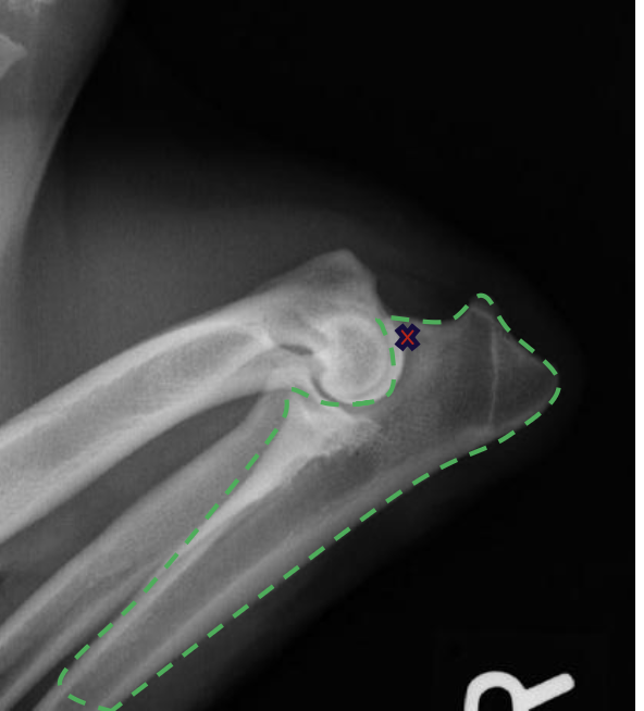
what is the red X marking?
anconeal process of the ulna
how does the olecranon process extend on a lateral view?
caudo-proximally
what is the relation of the medial condyle to the lateral condyle on a medio-lateral view of the elbow in a dog?
medial condyle is distal to the lateral condyle
where in the ulna is there a slight increase in opacity?
at the proximal extremity of the medullary cavity
where is the caudal ulnar cortex thicker?
in the proximal third of the diaphysis
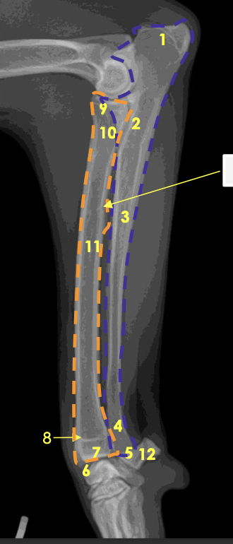
what is the arrow at 8 pointing to?
physeal scar on distal physis of the radius
how will the limbs of chondrodystrophic breeds appear on radiographs?
short with marked craniolateral bowing and curvature
how are the diaphyses of chondrodystrophic breeds appear on radiographs?
short and wide
proximally form large flared elongated articulations
what is circled in orange?
sesamoid bone in the tendon of m. abductor pollicis longus
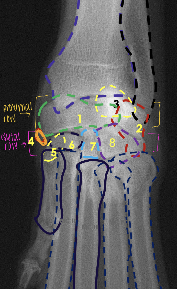
what is the yellow line?
radiocarpal joint

what is the berry red line?
intercarpal joint

what is the green line?
carpometacarpal joint
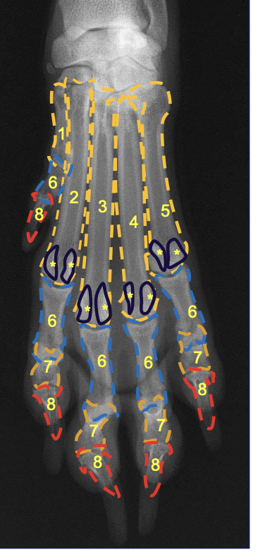
what is the navy blue circles with astericks?
proximal sesamoid bones of distal metacarpal bones
how does the long diaphyses of cats compare to dogs?
cats are more straight
how does the acromoin in cats compare to dogs?
pointed more cranially
how is the clavicle in the cat compared to the dog?
clavicle is well formed and developed
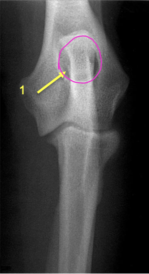
how does the olecranon process appear in cats?
more square
what is the pink circling?
supracondylar foramen in the cat
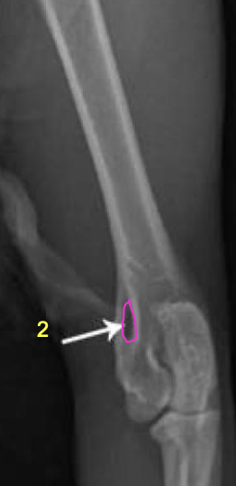
which species has the supratrochlear foramen at the elbow joint?
dogs
which species has the supracondylar foramen at the elbow joint?
cats?
what is the pink circling?
supratrochlear foramen