Chp 3
1/37
There's no tags or description
Looks like no tags are added yet.
Name | Mastery | Learn | Test | Matching | Spaced |
|---|
No study sessions yet.
38 Terms
Culture Media
Def: Nutrients that supports growth of microbes in the lab.
What does culture microbes involve? Why is this important?
Requires aseptic technique → prevents contamination.
Culture media = nutrients that support microbial growth.
Agar → solid surface in petri dishes.
Broths → liquid media in tubes.
Significance: Provides controlled conditions to isolate and study microbes.
Name and describe the Five I’s of microbiology.
Inoculation – introducing microbes into media.
Samples from body fluids, soil, water, food, surfaces.
Requires sterile instruments.
Purpose: Begin microbial growth for study.
Incubation – placing cultures in temperature-controlled environments.
Factors: temperature, oxygen/CO₂, humidity.
Example: human pathogens often incubated at 37°C.
Purpose: Promotes optimal microbial growth.
Isolation – separating individual species.
Produces colonies = visible mounds of identical cells.
Streak plate technique commonly used.
Purpose: Essential in medical microbiology to identify a pure pathogen.
Inspection – observing colony characteristics.
On solid media → size, shape, color, growth patterns.
On liquid media → turbidity, sediment, film.
Purpose: Provides first clues to species identity.
Identification – confirming which organism is present.
Methods: staining, biochemical tests (enzymes, metabolism), immunological tests (antibodies).
Purpose: Determines the microbe causing infection.
Describe the 3 physical states of media.
Liquid Media (Broths)
Water-based, flow freely.
Allow maximum population growth.
Purpose: Good for bulk growth, but colonies not separated.
Semisolid Media
Clot-like consistency (gelatin/agar).
Example: motility agar → determines if bacteria are motile.
Purpose: Tests movement, can differentiate species.
Solid Media (Agar plates/slants)
Firm surface at room temp; liquefies at 100°C.
Used for colony growth and isolation.
Purpose: Allows separation of pure colonies.
Name & describe the 3 types of bacterial cultures.
Pure — a single bacterial species
Mixed — two or more bacterial species (at least one is known)
Contaminated — unwanted microbe in pure or mixed cultures
Contaminated cultures are the result of ___________________.
Contaminated cultures are the result of unsterile lab practices.
Mixed Culture
Two or more bacterial species
Contaminated Culture
Unwanted microbes present
T or F: A pure culture is essential for reliable diagnostic results.
True
What is the purpose of microscopy?
Microbes are too small to be seen by the naked eye.
Microscope allows visualization of stained/unstained specimens.
Allows us to define cell details
Properties of Light Microscopy
(1) Magnification – enlargement of specimen.
Total magnification = ocular lens × objective lens.
Example: 10× ocular × 100× oil immersion objective = 1000×.
(2) Resolution (Resolving Power) – ability to distinguish two points as separate.
Human eye: 0.1 mm.
Bacteria: 0.5–5 μm.
Light microscopes can resolve objects ~0.2 μm apart.
Significance: High resolution is needed to see bacterial details.
(3) Contrast – difference between specimen and background.
Controlled by iris diaphragm and staining.
Too much light = low contrast.
Purpose: Improves visibility of structures.
Brightfield Microscopy
(*NTS: High-yield info)
Dark image against bright background.
Requires staining → cells usually dead.
Used in routine labs.
Darkfield Microscopy
(*NTS: High-yield info)
Light reflects off specimen against dark background.
Good for live, unstained microbes.
Capsule and external structures visible.
Phase Contrast Microscopy
Enhances contrast in unstained specimens.
Useful for observing internal structures in live cells.
Fluorescence
Uses UV light; specimens stained with fluorescent dyes.
Used in diagnostics (e.g., TB detection, antibody-tagging).
Conofocal Microscopy
3D imaging with laser scanning.
Used for biofilms and detailed cell studies.
Electron Microscopy
(*NTS: High-yield info)
Uses electron beams instead of light.
Highest magnification & resolution.
Transmission EM (TEM): electrons pass through specimen → internal structures.
Scanning EM (SEM): electrons bounce off surface → surface images (e.g., cilia, pili).
Culturing microbes is essential for identifying __________.
Culturing microbes is essential for identifying pathogens.
The properties/principles of microscopy (i.e. magnification, resolution, and contrast) are crucial for _________________.
The properties/principles of microscopy (i.e. magnification, resolution, and contrast) are crucial for visualization.
Why do we use diff types of microscopes?
Different types of microscopes reveal different aspects of microbes (e.g. live vs dead cells, surface vs internal structures)..
The _________ and ______ of a culture determines that accuracy of lab results.
The media type and purity of a culture determines that accuracy of lab results.
Culture
Growth of microorganisms in/on a nutrient medium.
The term “media” is plural of “________”
The term “media” is plural of “medium”
__________ is the introduction of microbes into or on media for microbial growth.
Inoculation is the introduction of microbes into or on media for microbial growth.
Sterile
Free of all life forms including spores and viruses
Any instrument used for sampling and inoculation must be _______.
Any instrument used for sampling and inoculation must be sterile.
Turbidity
Refers to appearance of microbes grown in liquid medium.
Cloudy sediment
Scum
Colour
Colonies
Visible masses of piled-up microbial cells grown on solid medium.
T or F: Water-based liquid media solutions do not solidify at room temperature.
True
Why is the practice of growing isolated bacterial colonies important in microbiology?
Colony
Growth of pure (meaning one species) of bacteria cells on agar plate.
Originates from one cell (or group of genetically identical cells)
can vary in shape, margin, colour, texture, and elevation, etc.
Identifying Microbes
What observations should you make?
What tests should you perform?
Observing
Observe bacterial colonies on media
Note bacterial cell shape, colour, & growth patterns
Testing
Staining
Microscope
Biochemical test (e.g. enzymes, metabolic, immunological testing, and antibody testing
Oil Immersion
Function: Increase resolution of specimen
How it works:
Some bacteria cannot be seen using a total magnification of 400x or less —> due to light refraction
Therefore, we use oil immersion to prevent refraction
A pure culture originates from a _____________ cell or a __________.
A pure culture originates from a single progenitor cell or a colony.
Contrast
Def: Degree of contrast
Identify the type of microscopy used in each of the slide images below.
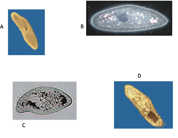
A — scanning electron microscope (SEM)
B — Darkfield microscopy
C — Brightfield microscopy
D — transmission electron microscopy