sound - chemoreception
1/10
There's no tags or description
Looks like no tags are added yet.
Name | Mastery | Learn | Test | Matching | Spaced | Call with Kai |
|---|
No analytics yet
Send a link to your students to track their progress
11 Terms
sound detection: what is sound?
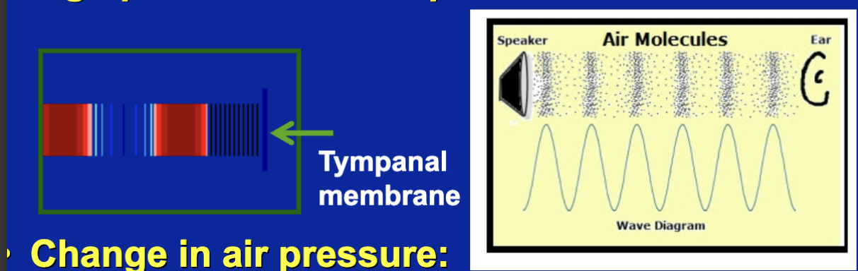
its changes in air pressure
airpressure is determend on how many molecules of air there are in space
changes in air pressure: frequency; cycles per second (hz: hertz)
changes in airpressure gives us sound waves
these sound waves hit our tympanic membranes in our ears causing it to flex back and forth
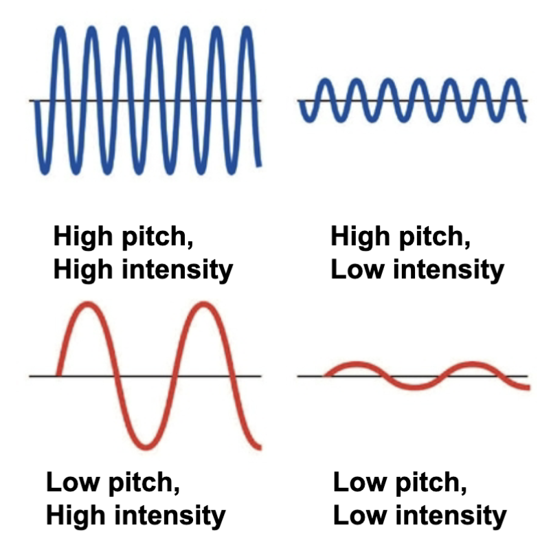
Pitch and Intensity
we can have increased and decreased pitch and intensities
pitch: frequency of pressure changes
intensity: amplitude of pressure changes
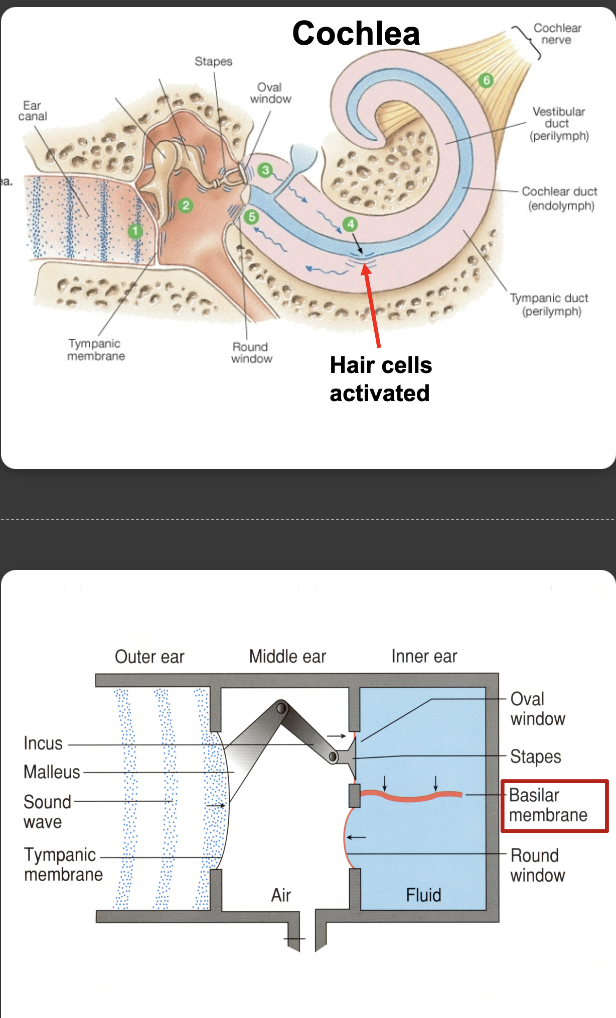
How sound waves travel through our ears
Sound waves strike the tympanic membrane and become vibrations
the sound wave energy is transfered to the 3 bones of the middle ear which vibrate them
the stapes is attached to the membrane of the oval window. vibrations of the oval window create fluid waves within the cochlea
the fluid waves push on the flexible membranes of the cochlear duct
energy from the waves is transfered across the cochlear duct into the tympanic duct and is disipated back into the middle ear at the round window
hair cells with in the cochlear duct create AP’s in the sensory neurons of the cochlear nerve
Sound Waves → Ear Canal → Tympanic Membrane (Eardrum) → Malleus → Incus → Stapes → Oval Window → Cochlea (Fluid Vibrations) → Basilar Membrane → Hair Cells → Electrical Signals to the Brain
Hearing
hair cells are located on basilar membrane in the cochlea
tips of hair cells are bent by basilar membrane movement
bending tips of hair cells causes AP firing
Organ of corti
where the production of hair cells are made (this is within the cochlea)
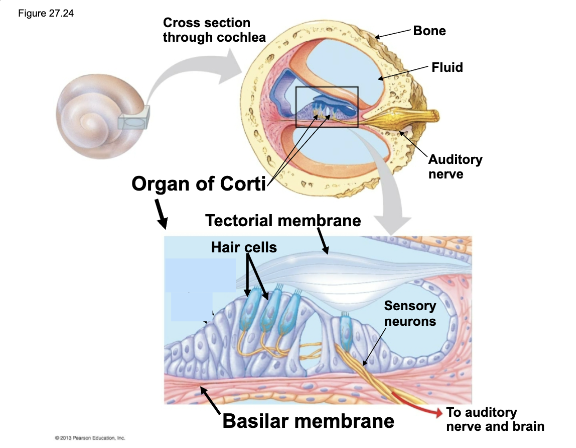
auditor system: hair cells
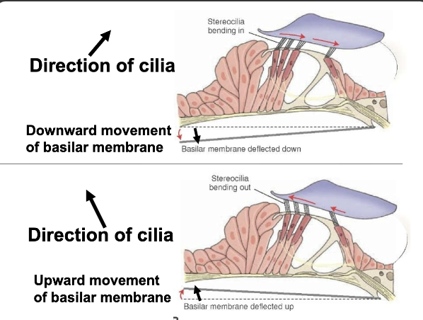
stereocillia: hair cells within the inner ear
these cillia move right and left
L direction of cilia: upward movement of basilar membrane
R direcetion of cilia: upward movement of basilar membrane
Hair cells modulate release of NT’s: change in AP frequency above spontaneous frequency
moving these hair cells causes more APs and releases more NTs
More NTs = more APs
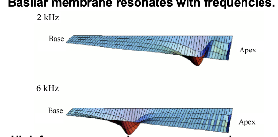
how do we mamals distingusih different sound frequencies (pitch)?
human hearing: 20-20000 hz
we have to look at the basal membrane and the hair cells
basilar membrane resonates with frequencies
the hair cells on top of the basilar membranes location codes for the frequencies
basilar membrane resonates with frequencies
High frequency sound waves move membrane closer to stapes; lower frequency sounds closer to distal end.
THUS: Hair cell location on membrane creates codes for sound pitch.
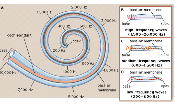
different regions of basilar membrane project to different areas of cortex: ionotopic representation
High frequency sound waves move membrane closer to stapes, lower frequency sounds closer to distal end
sensory coding for pitch discrimination
the map to show where on the cochlear the basal membrane flexes for certian frequencies
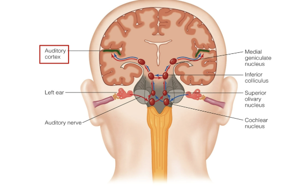
auditory pathways to the brain
information from the ear (the vibration energies - AP) go to the auditory complex via cochlear nerve
auditory structures are bilateral, messages can cross over between 2 sides
info from R/L can be sent to the R and L side of auditory cortex in the brain
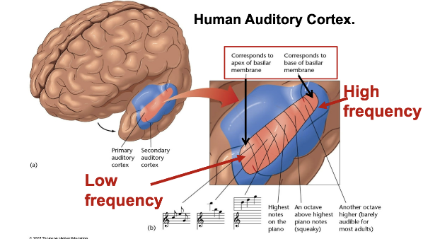
human auditory cortex
this cortex just like the cochlear map in our ears has a map telling it what frequencies are occurring
Only sense where you have a neural map
neurons, responding to particular frequencies, are arranged in a gradient, with cells responding to decreased frequency tones at one end and cells responding to increased frequency tones at the other end of auditory cortex