Anatomy Nervous system
1/72
There's no tags or description
Looks like no tags are added yet.
Name | Mastery | Learn | Test | Matching | Spaced | Call with Kai |
|---|
No analytics yet
Send a link to your students to track their progress
73 Terms
What is the Nervous system
the "master controller", controlling thoughts, emotions, sensations and movements (communication) at rapid rates, having 3 functions - to see something (glass of water) which is sent to brain, interprets information (thirsty for water), execution (lift glass of water)
What is central and peripheral nervous system
Central nervous system contains brain and spinal cord which are located dorsally, and CNS interprets information then processes it based on past experiences or reflexes
Peripheral nervous system are the nerves outside of CNS and has 2 divisions - sensory (afferent) division (carries information towards CNS) and motor (efferent) division (carries information away from CNS towards effector organs)
What is motor division
somatic nervous system (nerve fibres carry impulses towards skeletal muscles under voluntary or conscious control) and autonomic nervous system (controlled automatically like smooth muscles inside organs)
What are the 2 types of nervous tissue?
neurons (nerve cells) and neuroglia (support cells)
Neurons
functional unit of nervous system, uniquely shaped and is a large cells with long, spider-like appendages (dendrites) extending outward from central body which receive incoming signals (axons) and relay messages. Neurons have high metabolic rates (requiring fuel like glucose and oxygen or they can't survive) and be efficient for a lifetime
Neuroglia
support neurons and are much smaller and abundant than neurons.
4 types of neuroglia in CNS
Astrocytes cover capillaries with radiating processes and are shaped like stars, clinging to neurons. They make sure neurons are in contact with capillaries to have nutrient supply. They maintain homeostasis. Guide formation of junctions between neurons while maintaining chemical environment and cleaning excess potassium ions.
Microglia reside in CNS and maintain health of CNS by becoming macrophages, removing damaged cells or foreign invaders.
Ependymal cells are epithelial cells that line or cover body cavities like brain and spinal cord creating a semi-permeable barrier. Ciliated surface allow cushioning and protective layer in brain.
Oligodendrocytes produce insulated and protective myelin sheaths around thicker nerve fibres, which are the cells that are attacked by individuals with multiple sclerosis.
2 types of neuroglia in PNS
Satellite cells surrounding cell bodies of neurons and are similar to astrocytes (support cells)
Schwann cells surround thicker nerve fibres in periphery and have a regenerative role in PNS and also produce myelin.
4 classes of neurotransmitters
Acetylcholine - skeletal muscle contraction (excitatory affect)
Biogenic amines - reward pathway or emotional behaviour. Noradrenaline (responses like fight or flight), dopamine (enhanced by amphetamines), serotonin (feeling good), histamine (desire increases)
Amino acids - GABA, glutamate (enhanced by alcohol)
Peptides - endorphins (natural pain killers)
Demyelinating conditions in CNS and PNS
multiple sclerosis (CNS) and Guillain Barre Syndrome (PNS)
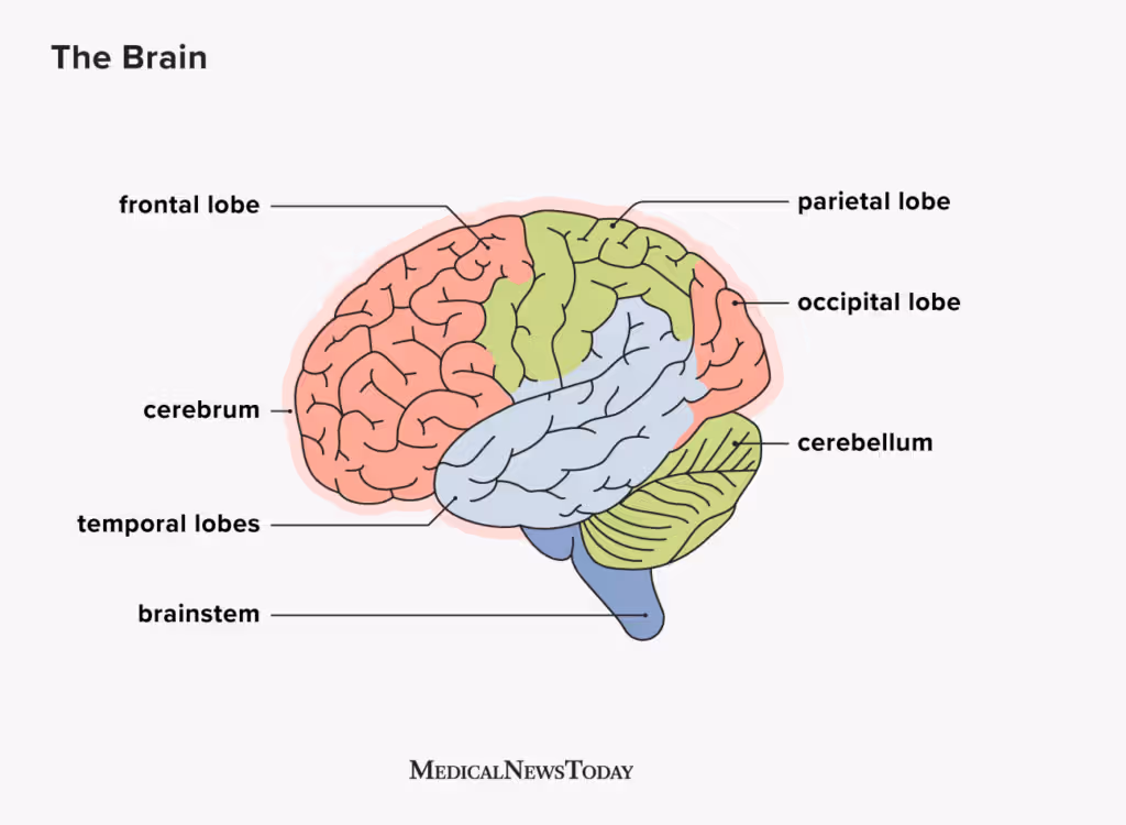
4 regions of brain -
Frontal lobe (frontal)
Diencephalon (deep, contains thalamus, hypothalamus, epithalamus)
Brainstem (midbrain, pons, medulla, oblongata)
Cerebellum (posterior)
Describe how brain looks
2 cerebral hemisphers separated left and right by longitudinal fissures. Hemispheres contain elevated ridges called gyrus which are separated by shallow grooves called sulcus, dividing 2 hemispheres into 5 lobes. Deeper grooves are called fissures (such as longitudinal fissure)
How much of brain mass does cerebrum, cerebrellum and brain stem take up each
Cerebrum takes around 83%, cerebrellum takes around 10% and brain stem takes the rest
Aphasia
speechlessness or partial loss of language skills because brain has been damaged in Broca's area
What are most association areas (motor and sensory)
multimodal association areas, which receive multiple senses and send output to multiple areas, and give meaning to the information received such as making memories, relating to previous memories, making decisions
Structure of diencephalon
thalamus is relay centre connecting hypothalamus and epithalamus, hypothalamus is anterior and helps homeostasis, epithalamus is posterior and helps melatonin
Structure of brainstem
Midbrain bridges diencephalon to pons, Pons bulges anteriorly, and inferior is medulla oblongata (autonomic reflexes in homeostasis like cardiovascular and respiratory centres regulating heart rate and breathing rate)
5 brain lobes
Frontal, parietal, occipital, temporal, insula (deep)
What is cerebral cortex
The conscious mind. Motor (directing movement), sensation (directing perception), association (integrate voluntary behaviours), visual and auditory
Motor areas (frontal) (4)
primary motor cortex (in precentral gyrus, voluntary movement of skeletal muscles), premotor cortex (anterior to primary motor, plans movement or complex tasks to produce accurate sequence of muscle movements like typing, coordination), Broca's area (sits anterior to inferior region of premotor cortex on left (dominant) hemisphere, directs muscles used for speech), frontal eye field (anterior to premotor cortex, eye movement)
Sensation areas (parietal) (2)
primary somatosensory cortex (postcentral gyrus, sensory information like touch, pain, pressure), somatosensory association cortex (posterior to primary somatosensory cortex, receives information from primary somatosensory cortex and processes the feeling (such as touching a coin in pocket))
Visual (occipital) (2)
Primary visual cortex (posterior tip of occipital lobe, receives visual information from eyes), visual association area (surrounds primary visual cortex, associates information of received image from eyes to past experiences)
Auditory (temporal) (4)
primary auditory cortex (close to ear, when hearing receptors become activated information is carried here to allow interpretation like volume, pitch and location of sound), auditory association area (posterior to primary auditory cortex, process what was heard and meaning like music or screaming), olfactory cortex (in temporal lobe, relays information on smell), Gustatory cortex (deep in insula close to lateral sulcus, relays information on taste)
Where are the sulcus and what do they separate
Frontal lobe separates from parietal through central sulcus. Precentral gyrus sits anteriorly housing motor cortex, postcentral gyrus sits posteriorly housing somatosensory cortex
Parietal and frontal separates from temporal through lateral sulcus. Insula is deep to lateral sulcus
Regions of cerebral hemispheres separated by tissue
Grey matter (superficial cortex), white matter (internal), basal nuclei (groups of neurons deep with cerebral cortex)
Grey matter
make up 40% of brain mass, short, non-myelinated neurons and cell bodies, which is where neurons communicate with cells (synapse)
White matter
myelinated and non-myelinated axons, connect grey matter to each other
Ventricles
hollow spaces (cavities) deep in brain filled with cerebrospinal fluid. Continuous and with central canal of spinal cord. Ependymal cells line these chambers. Chambers are paired and lateral (C-shaped), lying deep to each hemisphere. Thin membrane separates lateral ventricles called septum pellucidum. Third ventricle is in diencephalon, and communicate to lateral ventricles through interventricular foramen. Third is connected to fourth, which is continuous with central canal of spinal cord
Hydrocephalus
build up of CSF in brain ventricle due to poor circulation, increasing pressure. Can be seen in newborns where skull bones are not fused, causing head to expand.
Cerebrospinal fluid (CSF)
Provides cushioning and buoyancy to brain structures to prevent damage. Produced by choroid plexus (thin-walled capillaries hanging from ventricle roof). After production, CSF moves freely in ventricles with help from cilia, extending from ependymal cells, and enters subarachnoid space. Has a Blood brain barrier (BBB) which protects brain from blood born cells with near impermeable barrier is controlled by special features called tight junctions that lines capillaries. However, oxygen, carbon dioxide, alcohol, steroid hormones, nicotine, caffeine and glucose can pass through.
Label the parts
Lateralisation
both cerebral hemispheres are not completely identical in ability
Cerebral dominance
one hemisphere is more important for a task (e.g. language on left hemisphere)
Roles of left and right hemisphere
Left hemisphere role - maths, logic, memorisation, language (90% of population are left dominant, making them right handed)
Right hemisphere role - creativity, emotion, visual-spatial skills
Fibre tracts
white matter which cerebral hemispheres communicate through (only in CNS)
Different between tracts and nerves
Tract is only in CNS, Nerve is only in PNS
Types of fibre tracts (classified according to direction)
Association fibres - run horizontally within same hemisphere
Commissural fibres - run horizontally between hemispheres (corpus callosum)
Projection fibres - run longitudinally or vertically from brain to brain stem or spinal cord, and they decussate (cross into other hemisphere)
Basal nuclei
includes caudate nucleus, putamen, globus pallidus. Caudate nucleus and putamen create striation with fibres running through them creating striped pattern. Basal nuclei receive information from cerebral cortex via thalamus before projecting to motor and prefrontal cortex, and has role in emotions and cognition
Basal ganglia
group of deep nuclei within each hemisphere (white matter)
Caudate nuclei
tail shaped nucleus
Cerebellum
Sits under occipital lobe, bilaterally symmetrical separated by vermis, has many gyrus called folia (leaves), white matter looks like tree branches called Arbor vitae
Influences subconscious coordination of skeletal muscle contraction
Thalamus function
filled with egg-shaped nuclei, relay point for information projection
Hypothalamus function
sits inferior to thalamus extending from optic chiasma, contains important nuclei, control point for homeostasis, pituitary gland is connected
Epithalamus function
Roof of 3rd ventricle, contains pineal gland which produces melatonin
Midbrain
Cerebral peduncles (hold up cerebrum)
Corpora quadrigemina (superior and inferior paired nuclei)
Cerebral aqueduct (connect 3rd and 4th ventricle)
Substantia nigra (dopamine)
Pons
Anterior bulge (bridges midbrain, pons and medulla oblongata)
Conduction tracts (spinal cord connect to higher brain centre)
Medulla oblongata
Ridges - pyramids containing tracts from cerebral cortex
Cerebellar peduncles (fibre tracts connecting medulla to cerebellum)
Medulla oblongata function
Cardiovascular centre - heart rate and force of contraction
Vasomotor centre - blood vessel diameter
Respiratory centre - rate, depth of breathing
Hypothalamus can override these actions but medulla does them autonomically
4 layers for brain protection
Bone
Meninges
CSF
BBB (blood brain barrier)
3 layers of meninges (lie external to brain and spinal cord)
Dura mater (outer layer "tough mother")
Arachnoid mater (middle layer "spider-like" extensions)
Pia mater (inside layer delicate "gentle mother", CSF circulates, clings to brain tissue, delicate network of connective tissue)
Purpose of meninges
cover and protect CNS, protect blood vessels surrounding cerebrospinal fluid, form partitions (spaces) of skull
Spinal Cord
highway of conductions in both directions and major reflex centre, with reflexes initiated and completed at spinal cord. Spinal cord is also protected by 3 meningeal layers, bony vertebra and cerebrospinal fluid. Runs from foramen magnum (occipital bone) to L1/L2 (conus medullaris)
Epidural space
space between bony vertebra and spinal dura mater filled with fat padding (anaesthesia is administered here)
Types of paralysis
Spastic paralysis - Damage to upper motor neurons (within cortex), indicates muscles are paralysed but reflexes can occur in spinal cord
Flaccid paralysis - damage at spinal cord, synapses cannot occur, muscle atrophy (wasted) due to lack of stimulation
Spinal cord damage
Quadriplegia - C1-C8, damage to entire spinal cord, can lose sensory and motor function in all upper and lower limbs
Paraplegia - T1-L1, paralysis of lower limbs
Efferent corticospinal tract (cortex to spine)
originates in pyramidal cells (upper motor neurons) in cortex to medulla, fibres decussate at pyramidal cells, then goes down to spinal cord where it synapses with lower motor neuron in ventral horn before creating response in skeletal muscles.
3 steps to spinothalamic tract
activation of receptors sends information to dorsal horn in spinal cord where it decussates, then goes to thalamus, then goes to somatosensory cortex
Afferent spinothalamic tract (spinal cord to thalamus)
also has 3 steps, carries information about pain, temperature and crude touch, crosses at spinal cord.
3 steps to afferent pathways
activated receptors (caused by joint movement) send information to medulla where it decussates, medulla sends information to thalamus, thalamus sends information to somatosensory cortex
Afferent pathways
dorsal column, carries information about fine touch and pressure from proprioceptors in joints, crosses over (decussation) in medulla in brain stem, has a 3 step process
Nerve roots
inferior to conus medullaris is cauda equina
Filum terminale
anchors spinal cord to coccyx
How many pairs of spinal nerves in PNS, and what are important ones
31. Cervical and lumbar enlargements are rich in nerves to supply either upper or lower limbs.
Sensory cell bodies of spinal cord
Ventral median fissure - anterior, wider
Dorsal median sulcus - posterior, narrower
Central canal - contains CSF, runs through centre of spinal cord
Spinal Grey matter - found in centre of spinal cord
Spinal White matter - surrounds grey matter
Spinal nerve - mixed afferent (carry sensory information) and efferent (carry motor output) fibres. Formed by ventral and dorsal spine roots
Cervical spinal cord
runs from C1-C8, cervical enlargement serving upper limbs
Thoracic spinal cord
runs from T1-T12, no enlargements
Lumbar spinal cord
Cauda equina runs L2-L5, Conus medullaris is end of spinal cord from L1-L2, Filum terminale anchors spinal cord to coccyx, lumbar enlargement serves lower body and legs, subarachnoid space between L4-L5
Lumbar puncture
CSF sampling extracted from subarachnoid space to test for conditions like meningitis or meninges inflammation
Spinal nerve rootlets
emerge from spinal cord and fuse to become spinal roots
Dorsal vs ventral surface identification
dorsal root ganglion is enlarged and rich in cell bodies for sensory neurons
Sensory and Motor tract directions
Sensory tract (afferent) - ascending, dorsal column (tract in dorsal) - medial lemniscal (medulla ribbon) system, anterolateral system - spinothalamic tract, spinocerebellar tract
Motor tract (efferent) - descending, lateral corticospinal tract, rubrospinal tract
Spinal grey matter
butterfly-like appearance with grey commissure containing interneurons surrounding central canal. Motor output leads via ventral horns via autonomic or somatic neurons before merging into mixed spinal nerve. Dorsal horn (backdoor) contains sensory interneurons. Ventral horn (front door) contains motor neurons (information travels in the backdoor and out the front door).
Spinal white matter
myelinated and non-myelinated axons divided into columns (funiculi), with dorsal, ventral and central (location). Fibres run in 3 directions - ascending (afferent or sensory input), descending (efferent or motor output), transversely (commissural fibres across)