FILM SCREEN P1
1/145
There's no tags or description
Looks like no tags are added yet.
Name | Mastery | Learn | Test | Matching | Spaced | Call with Kai |
|---|
No study sessions yet.
146 Terms
darkroom;
this is the place where the film is
processed and made useful for viewing.
processing room
is perhaps
the most important single room
in the radiology department
latent image,
processing room is the place where the ___ placed on the film by
radiographic equipment, is
converted into a useful visible
image.
DARKROOM
specially designed and constructed
room
–Used for the proper storage and
maintenance of radiographic films, as well
as for the handling of films in the process of
loading and unloading cassettes and the
processing of the exposed radiographic
film.
Processing Tanks
Used on the wet side of the darkroom.
Contain developer, fixer, and wash water.
Allow manual processing of radiographic
films.
Proper temperature and time control are
essential to avoid image errors.
Warning Light Outside the Room
Located outside the darkroom entrance.
Indicates when the darkroom is in use.
Prevents accidental opening of the door
and film fogging.
Lockable Door
Ensures the darkroom is completely light-tight.
Prevents unauthorized access.
Helps maintain radiation department safety and workflow
control.
Film Bin
Located on the dry side.
Used for temporary storage of
unexposed and exposed films.
Protects films from light, heat,
and humidity.
Loading Bench
Found on the dry side.
Used for loading and
unloading films into
cassettes.
Must be kept clean and dry
to prevent artifacts.
Pass Box
A light-tight transfer
compartment.
Allows films to be passed
between the darkroom and
X-ray room.
Prevents light exposure
while maintaining workflow
efficiency.
Safelight
Provides safe illumination
during film handling.
Uses appropriate filters (e.g.,
GBX, Wratten 6B) depending on
film type.
4 feet
Safelight Positioned at least__ feet from
work areas to prevent fogging.
Darkrooms can be smaller if distributed all throughout the X-ray Department but should not be less than 8 feet along any wall. The size of the darkroom will depend on the overall size and activity of the department. Minimum of 10m sq
Ceiling height must be at least 2.5 - 3m
Darkrooms can be smaller if distributed all throughout the X-ray Department but should not be less than ___ feet along any wall. The size of the darkroom will depend on the overall size and activity of the department. Minimum of —- m sq
Ceiling height must be at least—- m
DARKROOM
Minimum area: 10 m2
Minimum wall length: 8 feet
Ceiling height: 2.5–3 m
DARKROOM
Minimum area: ___
Minimum wall length: ___
Ceiling height:___
1:2
RATIO OF darkroom to radiographic rooms
: Barium plaster
Alternative wall shielding material used in X-ray rooms
Lead ply sheet
Material used to line X-ray room doors
• 6 inches thick concrete
Standard concrete thickness for radiation protection
1.5 mm aluminum equivalent lead lining
Required wall lining equivalence for radiation shielding
5 mR
Stored radioactive materials should be located as
remotely as possible from the darkroom because even as little as__ mR total exposure from x- or gamma rays causes detectable fog.
Windows
should be avoided because they are
extremely difficult to render lightproof.
Plastic tiles)
Type of flooring recommended in darkrooms for durability and easy maintenance
Walls are generally light gray or ivory
color to provide reflection from
the “safe” lighting system.
Walls are generally ___or ___
color to provide reflection from
the “safe” lighting system.
Note that the darkroom walls do not
have to be painted black.
Note that the darkroom walls do not
have to be painted ___
40-60%
darkroom Relative humidity :
▸Low humidity- increase static marks
▸High humidity- increase fogging
▸Low humidity- increase ___
▸High humidity- increase ___
10
VENTILATION
Minimum of __ air changes per hour
18-20 degrees C
darkroom – Room temperature
Single Door Type
Double Door Type
Revolving Door Type
Maze Labyrinth Type
TYPES OF DARKROOM ENTRANCE
SINGLE DOOR
The simplest type of door.
• This type of entrance , although the
least expensive and requiring minimal
space , is also the greatest potential
source of light leaks into the darkroom.
DOUBLE-DOOR
• A less costly entrance is a in which access to
the darkroom is accomplished by opening an
outer door enabling you to enter a darkroom
anteroom.
An electrical system of locks is used to
prevent you from opening the inner door
leading into the darkroom itself until the
outer door is completely closed.
The locking devices are arranged to prevent
both doors from being opened at the same
time.
maze
•Because of the high cost
of the required floor
area, ___ are rarely
used today.
LABYRINTH/ MAZE TYPE DOOR
• Used in larger departments.
• This eliminates the need for doors
and the possibility of extraneous light
leaks but takes up considerable space
and increases the cost of
construction.
• The ideal darkroom entrance,
because of no door and air can freely
circulate in the room.
Safelight System.
•The special illumination for a radiographic darkroom is
called
1. Safelight
2. General
3. Radiographic
A properly designed darkroom should have three
types of illumination:
1 meter (3 feet)
Minimum working distance from the safelight
no
is “SAFE'' lighting totally safe?
Amber filter – blue-sensitive film
Red filter: green and blue-sensitive film
__ filter – blue-sensitive film
___ filter: green and blue-sensitive film
KodakWratten6-B safelight filter
specific brand for ‒ For Blue-sensitive Film
• Ortho G and GBX Filter
specific brand for ‒ Sensitive for Both Blue and Green
The standard safelight system used in many departments is a
7.5- 15-watt bulb with a Kodak Wratten 6B filter. (Red orange)
The standard safelight system used in many departments is a
____ watt bulb with a_____ filter. (Red orange)
Exposed radiographic film// Twice as sensitive
what is More sensitive to safelight illumination, exposed or unexposed film
latensification.
This increased sensitivity of film to exposure to safelight
illumination is called
post-exposure fog.
The effect on previously exposed x-ray film of an exposure to a safelight
is known as
Kodak Wratten 6B
: Older safelight filter used for blue-sensitive films
GBX
Modern safelight filter for green-sensitive (orthochromatic) films
Wratten 6-B
Safelight filter not suitable for orthochromatic-sensitive films
Low latensification, decreased contrast, gross fog
Negative effects of using Wratten 6-B on green-sensitive film
5-
minute
Maximum safelight exposure time for green-sensitive film using GBX without significant fog
is 120cm (4 feet)
Recommended minimum safelight distance from working surface
distance
or the bulb wattage or both.
correct intensity can be obtained by adjusting either
: 20 – 45 secs
‒ Maximum time a film can be exposed to safelight during
the procedure without causing any appreciable degree of
fogging.
Screen–film radiography
is an imaging
system in which X-ray photons interact
with intensifying screens, converting X-ray
energy into light, which then exposes a
radiographic film to create a latent image
that becomes visible after chemical
processing.
Image forming X-rays:
‒ x-rays that exit the patient and
interact with the image receptor
Exit X-ray beam:
X-rays that remain as the useful
beam exits the patient
Scattered X-rays, Image-forming X-rays
Components of the exit X-ray beam
IMAGE RECEPTOR
• The medium that converts the x-ray beam into
a visible image
‒ Base
‒ Emulsion
Radiographic film has two parts:
double
emulsion film
• The emulsion is coated on both sides:
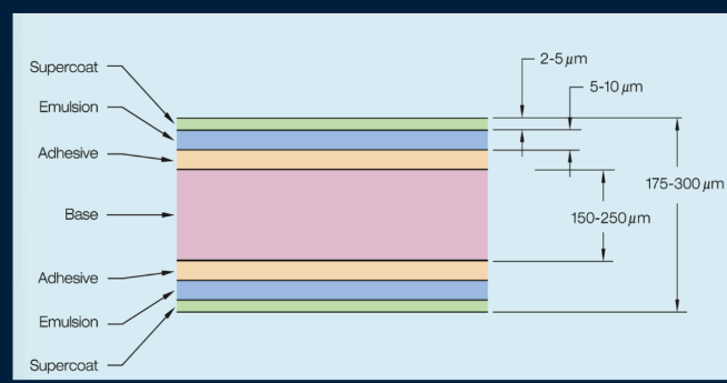
OVERCOAT/ SUPERCOAT
Protects emulsion from
scratches,
pressure, contamination during
handling, processing, and storage.
• Prevent damage to the sensitive
emulsion layer underneath
150 to 300 μm.
The thickness of radiographic
film is
approximately
ADHESIVE LAYER
Between the emulsion
and the base
BASE
Foundation of radiographic film
• Properties:
‒ Transparent and clear
‒ Provide a rigid structure
‒ Slight blue tint
‒ Flexible and sturdy
‒ Fracture resistant to allow easy
handling
‒ 150 to 300 μm thick,
base is how thick
polyester
base is made of
(Dimensional Stability)
Should be sturdy/ strong to maintain size and shape during use
and processing
(Uniform
Lucency)
Should be nearly transparent to light so there will be no unwanted
pattern or shading on the film caused by the base
1. Glass plate
2. Cellulose nitrate
3. Cellulose triacetate
4. Polyester
TYPES OF FILM BASE
Original base
‒ Plates coated with emulsion
‒ Difficult maintenance
‒ Exposure factors with plates
very high
‒ Origin of term “flat plate” of
abdomen
1. Glass plate
CELLULOSE NITRATE
• Replaced glass plates during
World War I glass shortage
CELLULOSE NITRATE
• Highly flammable film base
World War I
CELLULOSE NITRATE
• Replaced glass plates during ___
glass shortage
CELLULOSE TRIACETATE
• Safety base introduced in 1920s
• Less flammable than Cellulose Nitrate
• Disadvantages:
‒ Films would warp with age
‒ Not as resilient as today’s polyester films
POLYESTER
Base of today’s modern film
• Introduced in 1960s
• Introduced in 1960s
POLYESTER
• Introduced in ___
1920s
CELLULOSE TRIACETATE
• Safety base introduced in
Dye
Component added to radiographic film to reduce eyestrain and fatigue
Dimensional stability
Property that allows radiographic film to maintain size and shape during use and processing
EMULSION
The heart of radiographic film.
Gelatin
Component of emulsion that holds crystals
Silver halide crystals
Component of emulsion sensitive to X-rays
3 to 5 μm
Thickness of the emulsion
Binder
Component that holds the recording medium on the base and provides a form of material easier to work with
cows
The binder is a gelatin, which is extracted from
collagen, a protein found obtained from the
flatbones of
gelatin
The binder is a
Colloid
Substance which, in solution, fails to settle out but not dissolve; at higher temperatures it will and has some degree of porosity
Gelatin
Properties: clear & porous, provide mechanical support for silver halide crystals by holding them uniformly dispersed
SILVER HALIDE CRYSTALS
Active ingredient of emulsion
98% silver bromide; 2% silver iodide
composition of silver halide crystals in SF film
Tabular
Commonly used shape of silver halide crystals
1 μm
Approximate diameter of silver halide crystals
cubic form or cubic lattice.
Inner structure of silver halide crystals
Silver Nitrate (AgNO3)
Made by dissolving metallic silver (Ag) in nitric acid (HNO3) to form
Silver Bromide (AgBr)
Formed by mixing silver nitrate with potassium bromide (KBr)
Sensitivity Center/ sensitivity speck
Physical imperfection in the lattice of the emulsion layer
Silver Sulfide
Chemical contaminant responsible for the physical imperfection of the silver halide crystal
: ZBr = 35
Atomic number of Bromine in silver halide crystals
ZAg = 47
Atomic number of Silver in silver halide crystals