Abdomen lecture
1/469
There's no tags or description
Looks like no tags are added yet.
Name | Mastery | Learn | Test | Matching | Spaced | Call with Kai |
|---|
No analytics yet
Send a link to your students to track their progress
470 Terms
The hind gut structrues are supplied by the
IMA
Splenic flexure —> anus
The mid gut structures are supplied by
SMA
Mid duodenum —> approx splenic flexure
The foregut structures are supplied by the
celiac trunk
(oral cavity —> initial duodenum)
Body of the pancreas is supplied by the _________ which branches off the celiac trunk but the head and neck are supplied by branches from both the _____- and _____
Splenic artery
Gastroduodenal artery (common hepatic —> cel trunk)
SMA
The adrenal glands affect kidney function through the secretion of
aldosterone
Suprarenal medulla secretes
catecholamines (epinephrine and norepinephrine) fight or flight
Suprarenal cortex secretes
corticosteroids in response to stress, and androgens
How is the liver divided functionally?
Right and left based on blood supply and glandular secretions —> portal lobes (R and L )
What exits the porta hepatis?
Right and left hepatic ducts and lymph vessels
What enters the porta hepatis?
Portal vein, right and left hepatic arteries and nerve fibers
What bounds the abdomen superiorly? Inferiorly?
The diaphragm superiorly and the inguinal ligament and pelvic bones inferiorly
Aponeurosis
Flat, broad, tendon-like layers that serve as attachments for muscles
Fascia
A thin sheath of fibrous tissue enclosing a muscle or other organ
Peritoneum
Serous membrane that lines the abdominal cavity and its organs
Peritoneal ligament
A double layer of peritoneum that connects one organ to another organ or to the abdominal wall
Mesentery
A fold of the peritoneum that attaches the stomach, small intestine, pancreas, spleen and other organs to the posterior wall of the abdomen
Omentum
A double layered fatty sheath that supports and protects abdominal organs
Median plane
Divides the body into equal right and left halves
Transumbilical plane
This plane divides the body into upper and lower halves, across the middle of the abdomen
What are the 4 quadrants of the abdomen?
RUQ, LUQ, RLQ, LLQ
What are the 9 regions of the abdomen?
Right hypochondriac region // Epigastric region // Left hypochondriac region
Right lumbar region // Umbilical region // Left lumbar region
Right iliac region // Hypogastric region // Left iliac region
Liver
Gallbladder
Small intestine
Ascending/transverse colon
Right kidney
Right adrenal gland
Right ureter
These organs are found in what region of the abdomen?
Right hypochondriac region
Esophagus
Stomach
Liver
Pancreas
Small intestine
Transverse colon
R/L kidneys
R/L adrenal glands
R/L ureters
These organs are found in what region of the abdomen?
Epigastric region
Stomach
Tip of the liver
Tail of the pancreas
Small intestine
Transverse/descending colon
Spleen
Left kidney
Left adrenal gland
Left ureter
These organs are found in what region of the abdomen?
Left hypochondriac region
Liver
Gallbladder
Small intestine
Ascending colon
R kidney/ureter
These organs are found in what region of the abdomen?
Right lumbar region
Stomach
Pancreas
Small intestine
Transverse colon
R/L kidneys
R/L ureters
These organs are found in what region of the abdomen?
Umbilical region
Small intestine
Descending colon
L kidney
L ureter
These organs are found in what region of the abdomen?
Left lumbar region
Small intestine
Appendix
Cecum/ascending colon
R ovary/fallopian tube
These organs are found in what region of the abdomen?
Right iliac region
Small intestine
Sigmoid colon/rectum
R/L ovaries/fallopian tubed
Urinary bladder
Uterus
Vas deferens/seminal vesicles
Spermatic cord
Prostate
These organs are found in what region of the abdomen?
Hypogastric region
Small intestine
Descending/sigmoid colon
L ovary/fallopian tube
These organs are found in what region of the abdomen?
Left iliac region
What region of the abdomen is the appendix found in?
Right iliac region
What region of the abdomen is the prostate found in?
Hypogastric region
Thin cartilaginous extension off of the sternum that is easily palpated in the depression where the costal margins meet in the upper part of the anterior wall is identified as the _________
Xiphoid process
Where the xiphoid process meets the sternum
Xiphosternal junction
The xiphosternal junction is at which vertebral level?
T9
Small depression located in the upper-most midline abdomen in the infraxiphoid area
Epigastric fossa
Costal margin
The curved lower margin of the thoracic wall formed anteriorly by the cartilage of the 7th, 8th, 9th and 10th ribs and posteriorly by the cartilage of the 11th and 12th ribs
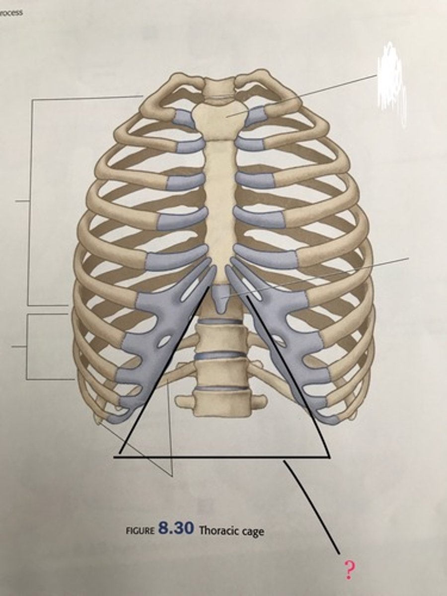
The cartilage of which ribs form the anterior portion of the costal margin?
7, 8, 9 and 10
The cartilage of which ribs form the posterior portion of the costal margin?
11 and 12
Why is the costal margin important?
The diaphragm connects to the costal margin. If there is something wrong with the costal margin, breathing can be interrupted
The superior portion of the diaphragm attaches to this area of the thorax
Costal margin
What is the Linea alba?
Fibrous band that extends from the symphysis pubis inferiorly to the xiphoid process superiorly and lies in the midline
What does the inferior portion of the linea alba connect to?
The symphysis pubis
What does the superior portion of the linea alba connect to?
Xiphoid process (linea alba)
This structure represents the fusion of the aponeuroses of the muscles of the anterior abdominal wall and is represented on the surface by a slight median groove
Linea alba
The linea alba is represented on the surface by a slight median groove. What is this median groove referred to as?
Rhaphe
You are scrubbing into your first surgery of your general surgery rotation. The head surgeon tells you to identify the linea alba when the patient is opened on the table. Explain where it is. What color is this structure?
You tell the surgeon that the linea alba is a fibrous band that extends form the pubic symphysis to the xiphoid process and lies in the midline of the abdomen. It is a pearly white color
Rhaphe
A suture/seam where two structures meet
Umbilicus
Puckered scar at the site of attachment of the umbilical cord in the fetus
The umbilicus lies in the linea alba and is ____ in position
Inconstant
What vertebral level is the umbilicus located at?
L4 (umb)
What vertebral level does the aorta bifurcate into the R and L common iliac arteries?
L4 (aorta)
What does it mean when we say the umbilicus is "inconstant in position"?
It can be found in slightly different spots on different people
The aorta bifurcates behind this structure
Umbilicus
Linea semilunaris
Lateral edge of the rectus abdominis semilunari
Where does the linea semilunaris cross the costal margin?
At the tip of the 9th costal cartilage
This bony structure of the pelvis can be felt along its entire length
Iliac crest
The iliac crest ends anteriorly at the _____
Anterior superior iliac spine (ASIS)
The highest point of the ASIS lies opposite ____
L5
A 56 year old male comes to the ER after a car accident with pelvic instability. You tell your student to check the stability of the patient's pelvis, and they tell you they won't be able to feel the iliac crest because the patient is extremely overweight. Is your student correct?
No they're wrong, throw a shoe at their face and then explain to them that the iliac crest can be palpated in its entirety regardless of the patient's weight
Diaphragm
Dome-shaped, musculotendinous partition separating the thoracic and and abdominal cavities
The diaphragm is made up of a central tendon with muscular attachments to the _____, ___ and ___
Xiphoid process, inferior thoracic cage and superior lumbar vertebrae
Which dome of the diphragm is higher?
Right
What are the 3 openings of the diaphragm, from anterior to posterior?
Caval opening (IVC)
Esophageal hiatus
Aortic hiatus
The IVC passes through the diaphragm through the _____
Caval opening
What does the caval opening allow for passage of?
IVC through the diaphragm
The IVC is adherent to the caval opening in the central tendon of the diaphragm. What happens when the diaphragm contracts during inhalation?
The IVC dilates, facilitating blood flow to the heart
In addition to the IVC, what else passes through the caval opening of the diaphragm?
The right phrenic nerve and some lymphatics
Oval aperture in the diaphragm that allows for the passage of the esophagus
Esophageal hiatus
In addition to the esophagus, what other structures pass through the esophageal hiatus?
Anterior and posterior vagal trunks
Esophageal branches of the left gastric vessels
Lymphatics
Which diaphragmatic aperture is the site of hiatal hernia?**
Esophageal hiatus (hernia)
Protrusion of a part of the stomach upward through the esophageal hiatus in the diaphragm**
Hiatal hernia
This is an opening posterior to the diaphragm through which the aorta passes
Aortic hiatus
Aortic hiatus
This aperture does not pierce the diaphragm, and therefore is not affected by movements during respirations
What structures pass through the aortic hiatus?
Aorta
Azygos vein
Thoracic duct
What groups of arteries supply the superior portion of the diaphragm?
Superior phrenic arteries (from thoracic aorta)
Musculophrenic and pericardiophrenic arteries (from the internal thoracic arteries)
What arteries supply the inferior surface of the diaphragm?
Inferior phrenic arteries (from the thoracic aorta)
Diaphragm
Phrenic = _____
On the superior portion of the diaphragm, the musculophrenic and pericardiophrenicn veins drain into the ______ vein
Internal thoracic vein
On the superior portion of the diaphragm, the superior phrenic vein drains into the _____
IVC
On the inferior surface of the diaphragm, the right inferior phrenic vein drains into the _____
IVC (rt phren drain)
On the inferior surface of the diaphragm, explain the venous drainage on the left side
The left inferior phrenic vein is doubled and drains into the IVC and left suprarenal vein
On the inferior surface of the diaphragm, the left inferior phrenic vein is doubled and drains into the ___ and ___
IVC and suprarenal vein
Which nerve roots provide motor supply to the diaphragm?**
C3, C4, and C5 keep the diaphragm alive! (phrenic nerve)
Centrally, the diaphragm's sensory innervation comes from _____**
The phrenic nerve (C3-C5)
Peripherally, the diaphragm's sensory innervation comes from _______
Intercostal nerves (T5-T11) and subcostal nerves (T12)
Fascia that is the fatty superficial layer continuous with the superficial fat of the rest of the body**
Camper's fascia
This abdominal fascia may become extremely thick in obese patients**
Camper's fascia (ob)
What is the first fascia encountered when cutting into the abdomen?**
Camper's fascia (1s)
Deep membranous layer of fascia of the abdomen. Fades out over the thoracic wall above and along the midaxillary line laterally. Inferiorly, it passes onto the front of the thigh where it fuses with the deep fascia**
Scarpa's fascia
Scarpa's fascia extends over the penis, where it helps to form _______**
Dartos fascia
Extension of scrapa's fascia into the perineum**
Colle's fascia
Fascia that lines the muscle is known as
Investing fascia
What is another name for the investing fascia?
Epimysium
Fascia that lines the organs of the abdomen. Names for the organ it lines
Endoabdominal fascia (transversalis)
The outer layer of the peritoneum that lines the interior of the abdominal wall/abdominal cavity**
Parietal peritoneum
There are 5 muscles of the abdominal wall, ____ flat muscles and ____ vertical muscles
3 flat, 2 vertical
All 3 flat muscles of the abdomen end anteriorly in a strong, sheet-like ______
Aponeurosis
The fibers of each of the 3 aponeuroses from the flat muscles of the abdomen interweave at the linea alba with the corresponding muscle of the opposite side, forming the ______**
Rectus sheath
Rectus sheath
Fibrous sheath formed by aponeuroses of abdominal muscles