3.6.4 Homeostasis
1/46
There's no tags or description
Looks like no tags are added yet.
Name | Mastery | Learn | Test | Matching | Spaced | Call with Kai |
|---|
No analytics yet
Send a link to your students to track their progress
47 Terms
what is homeostasis
● Maintenance of a stable internal environment within restricted limits
● By physiological control systems (normally involve negative feedback)
what are some examples of where homeostasis occurs in mammals
core temperature, blood pH, blood glucose concentration, blood water potential
Why is it important that we maintain a stable core temperature? (3)
- To provide an optimum temperature for enzyme activity
- If temperature is too high → Hydrogen bonds in tertiary structure of enzymes break, Enzymes denature; active sites change shape and substrates can’t bind, So fewer enzyme-substrate complexes
- Low temperatures will not provide sufficient kinetic energy for optimum enzyme activity, so fewer enzyme substrate complexes formed
Why is it important that we maintain a stable blood pH? (4)
- To provide an optimum pH for enzyme activity
- Above or below optimal pH, ionic / hydrogen bonds in tertiary structure break
- Enzymes denature; active sites change shape and substrates can’t bind
- So fewer enzyme substrate complexes
Why is it important that we maintain a stable blood glucose concentration? (2)
- To provide a sufficient substrate for respiration
- To release energy for metabolic processes that occur in the body
What happens if blood glucose concentration is too low (hypoglycaemia)
Not enough glucose (respiratory substrate) for respiration
So less ATP produced
Active transport etc. can’t happen → cell death
What happens if blood glucose concentration is too high (hyperglycaemia)
Water potential of blood decreases
Water lost from tissue to blood via osmosis
Kidneys can’t absorb all glucose → more water lost in urine causing dehydration
Why is it important that we maintain a stable water potential of blood within restricted limits? (2)
- So that excess water does not enter or leave body cells by osmosis
- Which will cause cells to burst or dehydrate and shrivel
What is a negative feedback system? (3)
if receptors detect change from optimum
effectors will respond to counteract change
Returning levels to normal
Explain the importance of conditions being controlled by separate mechanisms involving negative feedback
Departures in different directions from the original state can all be controlled / reversed
Giving a greater degree of control (over changes in internal environment)
Describe positive feedback
1. Receptors detect change from normal
2. Effectors respond to amplify change
3. Producing a greater deviation from normal
what type of feedback is the only type involved in homeostasis
negative
What are two factors that affect blood glucose concentration? (2)
- Consumption of carbohydrates (glucose is absorbed)
- Rate of respiration of glucose - this uses up glucose rapidly e.g. during exercise due to muscle contraction
What is the role of the liver in glycogenesis? (1)
Converts glucose to glycogen
What is the role of the liver in glycogenolysis? (1)
Converts glycogen to glucose
What is the role of the liver in gluconeogenesis? (1)
Converts amino acids and/or glycerol into glucose
what cells are involved in decreasing blood glucose concentration and how do they react
Beta cells in islets of Langerhans in pancreas detect blood glucose concentration is too high → secrete insulin
Describe the action of insulin (6)
1. Insulin attaches to specific receptors on the cell surface membrane of target cells, e.g., liver and muscle cells
2. This causes more glucose channel proteins to be incorporated into the cell surface membrane
3. This increases the membrane's permeability to glucose, allowing more glucose to enter cells by facilitated diffusion
4. Insulin also activates enzymes involved in the conversion of glucose to glycogen (glycogenesis)
5. This lowers the glucose concentration in the cells, creating a concentration gradient
6. So glucose enters cells by facilitated diffusion down a concentration gradient
7. Results in a decrease in blood glucose concentration
what cells are involved in increasing blood glucose concentration and how do they react
Alpha cells in islets of Langerhans in pancreas detect blood glucose concentration is too low → secrete glucagon
Fear / stress / exercise → adrenal glands secrete adrenaline:
Describe the action of glucagon in increasing blood glucose concentration (4)
1. Glucagon attaches to specific receptors on cell surface membranes of target cells eg. liver
2. Glucagon activates the enzymes that are involved in glycogenolysis (hydrolysis of glycogen to glucose)
3. It also activates enzymes that are involved in gluconeogenesis (the conversion of glycerol / amino acids into glucose)
4. This establishes a concentration gradient → glucose enters blood by facilitated diffusion
Describe the action of adrenaline in increasing blood glucose concentration (3)
1. Adrenaline attaches to specific receptors on cell surface membranes of target cells eg. liver
2. Adrenaline activates enzymes involved in glycogenolysis (hydrolysis of glycogen to glucose)
3. This establishes a concentration gradient → glucose enters blood by facilitated diffusion
what 2 negative feedback systems use the second messenger model
adrenaline and glucogon
Describe the secondary messenger model of adrenaline and glucagon action (5)
1. Adrenaline / glucagon ('first messenger') attaches to specific receptors on cell membrane
2. This activates enzyme adenylate cyclase (changes shape)
3. This enzyme converts many ATP to many cyclic AMP (cAMP)
4. cAMP acts as the second messenger that activates protein kinase enzymes
5. Protein kinases activates the enzymes that are required to break down glycogen to glucose
Advantages of the second messenger model
Amplifies signal from hormone
As each hormone can stimulate production of many molecules of second messenger (cAMP)
Which can in turn activate many enzymes for rapid increase in glucose
What is the cause of type 1 diabetes? (2)
- Occurs when β cells in islets of langerhans in pancreas produce insufficient insulin
Normally develops in childhood due to genetics causing an autoimmune response destroying β cells of Islets of Langerhans
Describe how of type I diabetes can be controlled (2)
Daily injections of insulin → blood glucose concentration monitored with biosensors; dose of insulin matched to glucose intake
Eat regularly and control carbohydrate intake eg. those that are broken down / absorbed slower → To avoid sudden rise in glucose
What is the cause of type 2 diabetes? (3)
- receptor (faulty) loses responsiveness to insulin (but insulin still produced)
- So fewer glucose transport proteins → less uptake of glucose → less conversion of glucose to glycogen
What is a significant risk factor for type 2 diabetes? (1)
Long term obesity
How can type 2 diabetes be controlled?
Not normally treated with insulin injections but may use drugs which target insulin receptors to increase their sensitivity → To increase glucose uptake by cells / tissues
Reduce sugar intake (carbohydrates) / low glycaemic index → less absorbed
Reduce fat intake → less glycerol converted to glucose
More (regular) exercise → uses glucose / fats by increasing respiration
Lose weight → increased sensitivity of receptors to insulin
how you can evaluate the positions of health advisers and the food industry in relation to the increased incidence of type II diabetes
Consider both sides:
Health advisers aim - reduce risk of type II diabetes due to health problems caused (eg. kidney failure) → So need to reduce obesity as it is a risk factor
Food industry aim - maximise profit
Draw the structure of a single nephron (11)
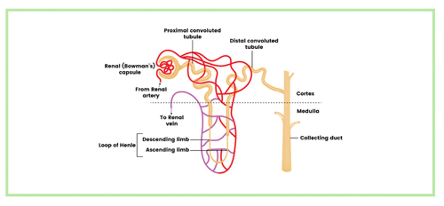
How is the glomerular filtrate formed? (5)
1. There is high hydrostatic pressure in the glomerulus
2. This is because the diameter of the afferent arteriole (in) is wider than that of the efferent arteriole (out)
3. Small substances, e.g., water, glucose, ions, and urea, are forced into the glomerular filtrate
4. These substances are then filtered through the pores between capillary endothelial cells, the capillary basement membrane, and podocytes
5. Large proteins and blood cells remain in the blood.
Describe how glucose is reabsorbed by the proximal convoluted tubule (4)
1. Sodium ions are actively transported out of epithelial cells into the capillary
2. Sodium ions then moves from the PCT into the epithelial cells by facilitated diffusion, down its concentration gradient
3. This brings glucose into the epithelial cells against its concentration gradient
4. Glucose moves into the capillary by facilitated diffusion down its concentration gradient
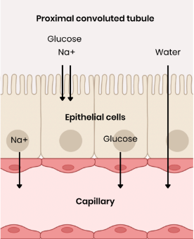
How is water reabsorbed by the proximal convoluted tubule? (2)
1. Glucose in the capillaries lowers water potential
2. Water moves by osmosis from a high water potential to a low water potential, down a water potential gradient into the capillary
What features of the cells in the PCT allow the rapid reabsorption of glucose into the blood
Microvilli / folded cell-surface membrane → provides a large surface area
Many channel / carrier proteins → for facilitated diffusion / co-transport
Many carrier proteins → for active transport
Many mitochondria → produce ATP for active transport
Many ribosomes → produce carrier / channel proteins
why glucose is found in the urine of an untreated diabetic person (2)
Blood glucose concentration is too high so not all glucose is reabsorbed at the PCT
As glucose carrier / cotransporter proteins are saturated / working at maximum rate
Explain the importance of maintaining a gradient of sodium ions in the medulla (concentration increases further down)
● So water potential decreases down the medulla (compared to filtrate in collecting duct)
● So a water potential gradient is maintained between the collecting duct and medulla
● To maximise reabsorption of water by osmosis from filtrate
What is the role of the loop of henle in maintaining the gradient of sodium ions in the ascending limb? (3)
1. Sodium ions are actively transported out of the ascending limb (so filtrate concentration decreases) and water remains as ascending limb is impermeable to water
2. This increases the concentration of sodium ions in medulla (surrounding tissue around the loop of henle)
3. Which lowers the water potential of the medulla
What is the role of the loop of henle in maintaining the gradient of sodium ions in the descending limb? (3)
1. Water moves out of the descending limb by osmosis
2. This allows water to be reabsorbed by capillaries (so filtrate concentration increases)
3. So sodium ions can diffuse back into the descending limb of the loop of henle
Suggest why animals needing to conserve water have long loops of Henle (thick medulla)
● More Na+ moved out → Na+ gradient is maintained for longer in medulla / higher Na+ concentration
● So water potential gradient is maintained for longer
● So more water can be reabsorbed from collecting duct by osmosis
Describe the reabsorption of water by the distal convoluted tubule and collecting ducts (3)
1. Water moves out of the distal convoluted tubule and collecting duct by osmosis, down a water potential gradient
2. This is controlled by ADH
3. Which increases the permeability of the collecting duct
What is meant by osmoregulation? (1)
The control of the water potential of blood via negative feedback
Describe the role of the hypothalamus in osmoregulation
Contains osmoreceptors which detect increase OR decrease in blood water potential
Produces more ADH when water potential is low OR less ADH when water potential is high
Describe the role of the posterior pituitary gland in osmoregulation
Secretes (more / less) ADH into blood due to signals from the hypothalamus
How does our body respond when the blood water potential is too high? (6)
osmoreceptors in the hypothalamus detect an increase in the water potential of the blood
nerve impulses are sent along sensory neurones to the posterior pituitary gland
this causes it to release less ADH into the blood, so aquaporins to leave the cell membrane and return to vesicles in the cytoplasm
\this decreases the water permeability of the collecting ducts
So less water is reabsorbed by osmosis from the DCT / collecting duct into the blood
So a larger volume of more diluted urine is produced
How does our body respond to a decrease in water potential? (5)
osmoreceptors in the hypothalamus detect an decrease in the water potential of the blood
nerve impulses are sent along sensory neurones to the posterior pituitary gland
this causes it to release more ADH into the blood
ADH binds to receptors on collecting duct and increasing permeability of cells to water (aquaporins join cell surface membrane)
So more water is reabsorbed by osmosis from the DCT / collecting duct into the blood
So a smaller volume of more concentrated urine is produced
What happens when the blood water potential has returned to normal
the posterior pituitary gland secretes the normal amount of ADH