19. Antibody Effector Functions

0.0(0)
studied byStudied by 0 people
0.0(0)
full-widthCall Kai
learnLearn
examPractice Test
spaced repetitionSpaced Repetition
heart puzzleMatch
flashcardsFlashcards
GameKnowt Play
Card Sorting

1/33

encourage image

There's no tags or description

Looks like no tags are added yet.

Study Analytics
Name
Mastery
Learn
Test
Matching
Spaced

No study sessions yet.

34 Terms

1
New cards

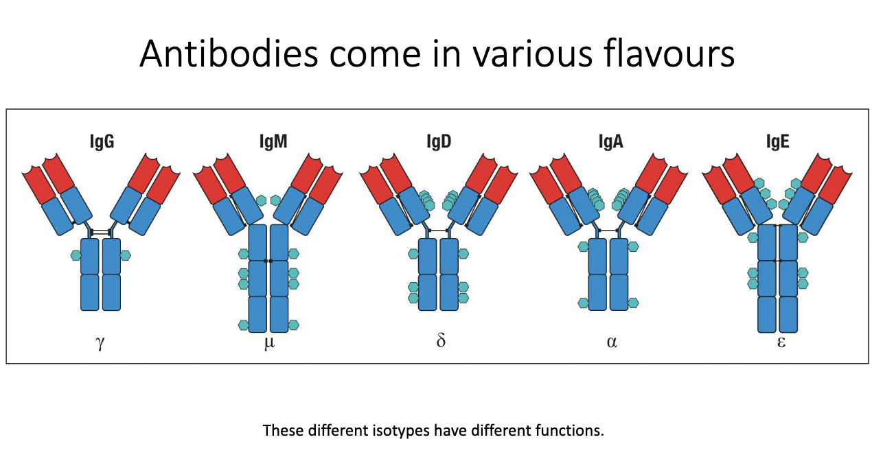
What are antibody isotypes, and why do they differ?

  • Five major isotypes: IgG, IgM, IgD, IgA, IgE.

  • Called isotypes/classes; each has distinct structure and function.

  • Structural differences include number/type of glycans and constant region features.

  • These structural variations determine their effector functions in immunity.

<ul><li><p class="isSelectedEnd"><span>Five major isotypes: </span><strong><span>IgG, IgM, IgD, IgA, IgE</span></strong><span>.</span></p></li><li><p class="isSelectedEnd"><span>Called </span><strong><span>isotypes/classes</span></strong><span>; each has distinct </span><strong><span>structure</span></strong><span> and </span><strong><span>function</span></strong><span>.</span></p></li><li><p class="isSelectedEnd"><span>Structural differences include </span><strong><span>number/type of glycans</span></strong><span> and </span><strong><span>constant region features</span></strong><span>.</span></p></li><li><p><span>These structural variations determine their </span><strong><span>effector functions</span></strong><span> in immunity.</span></p></li></ul><p></p>
2
New cards

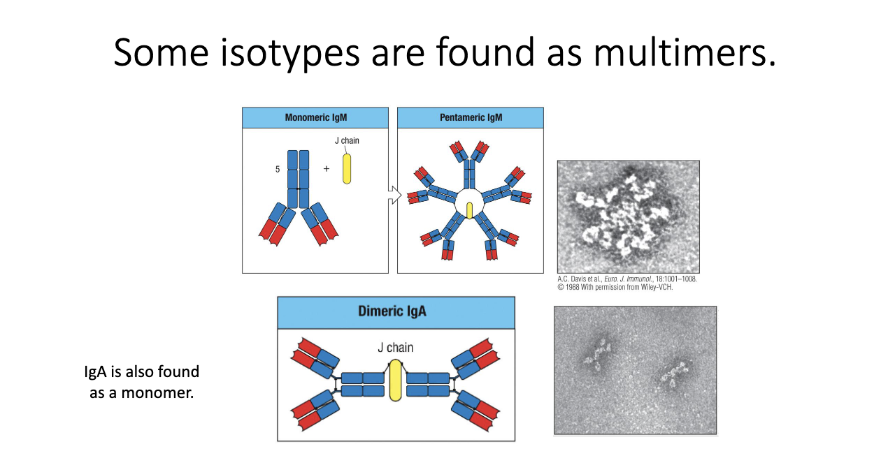
Why do IgM and IgA form multimers, and what does the J chain do?

  • IgM forms pentamers via the J chain → 5 antibodies linked.

  • IgA forms dimers (also J chain–dependent); found especially at mucosal surfaces (ex: intestines).

  • Purpose: increase avidity (total binding strength) to compensate for lower affinity monomers.

  • IgM: almost always pentameric.

  • IgA: can be monomeric or dimeric depending on tissue context.

<ul><li><p class="isSelectedEnd"><strong><span>IgM</span></strong><span> forms </span><strong><span>pentamers</span></strong><span> via the </span><strong><span>J chain</span></strong><span> → 5 antibodies linked.</span></p></li><li><p class="isSelectedEnd"><strong><span>IgA</span></strong><span> forms </span><strong><span>dimers</span></strong><span> (also J chain–dependent); found especially at </span><strong><span>mucosal surfaces </span></strong><span>(ex: intestines).</span></p></li><li><p class="isSelectedEnd"><span>Purpose: increase </span><strong><span>avidity</span></strong><span> (total binding strength) to compensate for </span><strong><span>lower affinity</span></strong><span> monomers.</span></p></li><li><p class="isSelectedEnd"><span>IgM: almost always pentameric.</span></p></li><li><p><span>IgA: can be </span><strong><span>monomeric or dimeric</span></strong><span> depending on tissue context.</span></p></li></ul><p></p>
3
New cards

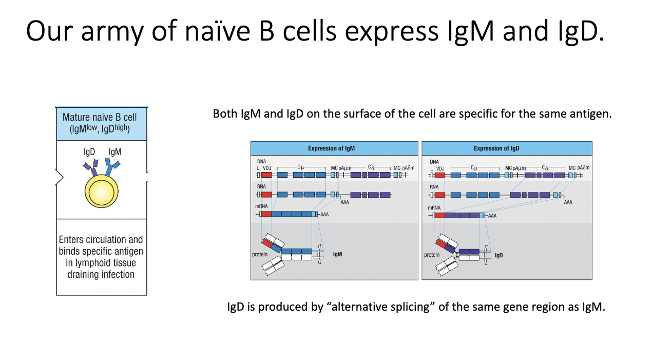
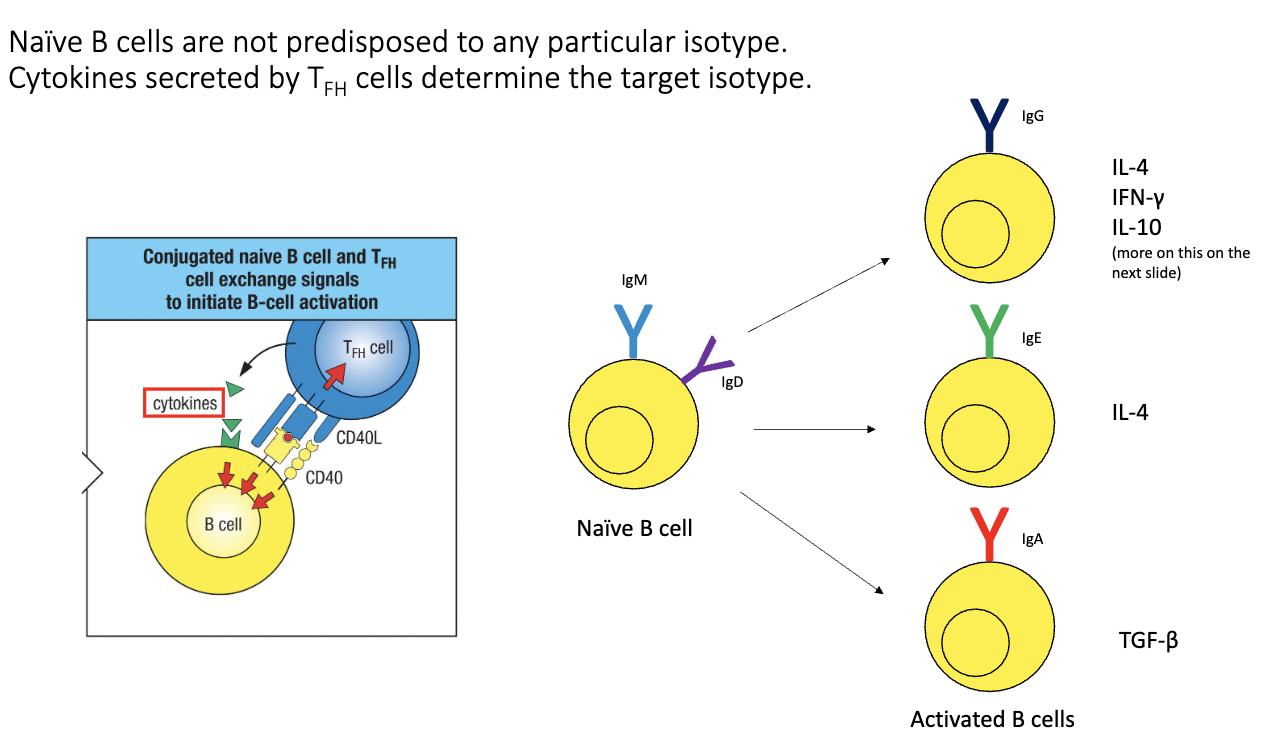
Why do naïve B cells express both IgM and IgD, and how is this achieved?

  • Naive B cells uniquely co-express IgM and IgD.

  • Both forms recognize the same antigen.

  • Achieved via alternative splicing of a shared transcript containing both constant regions.

  • Purpose of IgD is still unclear, but it is biologically required.

<ul><li><p class="isSelectedEnd"><span>Naive B cells uniquely co-express </span><strong><span>IgM and IgD</span></strong><span>.</span></p></li><li><p class="isSelectedEnd"><span>Both forms recognize the </span><strong><span>same antigen</span></strong><span>.</span></p></li><li><p class="isSelectedEnd"><span>Achieved via </span><strong><span>alternative splicing</span></strong><span> of a shared transcript containing both constant regions.</span></p></li><li><p><span>Purpose of IgD is still unclear, but it is biologically required.</span></p></li></ul><p></p>
4
New cards

How do B cells switch from IgM/IgD to other isotypes?

  • Driven by signals from TFH cells:

    1. Antigen-specific TCR–MHC II recognition.

    2. CD40–CD40L co-stimulation.

    3. Cytokines → determine isotype outcome.

  • Examples:

    • IL-4 → IgE

    • TGF-β → IgA

  • Dendritic cell → T cell → B cell signaling ensures isotype matches infection type.

<ul><li><p class="isSelectedEnd"><span>Driven by signals from </span><strong><span>TFH cells</span></strong><span>:</span></p><ol><li><p class="isSelectedEnd"><span>Antigen-specific </span><strong><span>TCR–MHC II</span></strong><span> recognition.</span></p></li><li><p class="isSelectedEnd"><strong><span>CD40–CD40L</span></strong><span> co-stimulation.</span></p></li><li><p class="isSelectedEnd"><strong><span>Cytokines</span></strong><span> → determine isotype outcome.</span></p></li></ol></li><li><p class="isSelectedEnd"><span>Examples:</span></p><ul><li><p class="isSelectedEnd"><strong><span>IL-4 → IgE</span></strong></p></li><li><p class="isSelectedEnd"><strong><span>TGF-β → IgA</span></strong></p></li></ul></li><li><p><span>Dendritic cell → T cell → B cell signaling ensures isotype matches infection type.</span></p></li></ul><p></p>
5
New cards

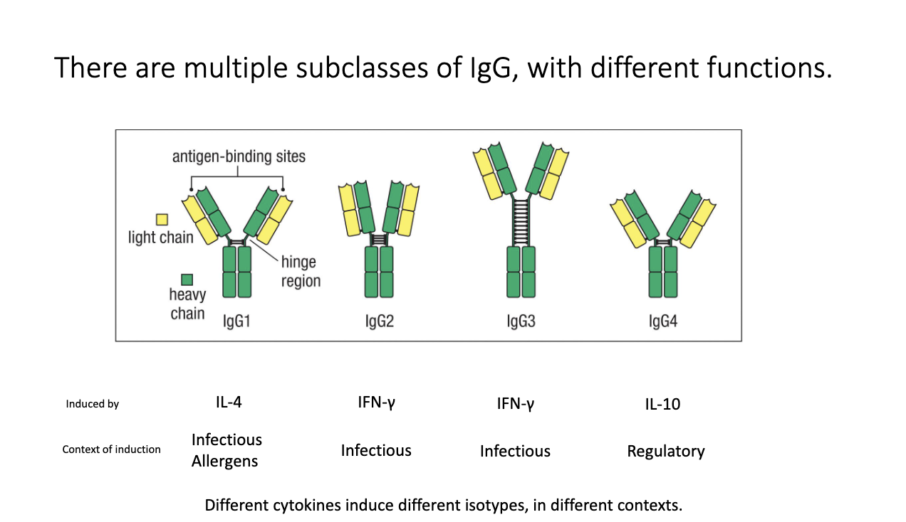
How do IgG subclasses differ, and what induces them?

  • Humans have four IgG subclasses: IgG1, IgG2, IgG3, IgG4.

  • Each has distinct hinge structures + effector functions.

  • Cytokine induction:

    • IL-4 → IgG1 (infectious and allergens)

    • IFN-γ → IgG2, IgG3 (infectious/anti-viral)

    • IL-10 → IgG4 (regulatory/anti-inflammatory)

  • Subclasses participate in different immune contexts.

<ul><li><p class="isSelectedEnd"><span>Humans have </span><strong><span>four</span></strong><span> IgG subclasses: </span><strong><span>IgG1, IgG2, IgG3, IgG4</span></strong><span>.</span></p></li><li><p class="isSelectedEnd"><span>Each has distinct hinge structures + effector functions.</span></p></li><li><p class="isSelectedEnd"><span>Cytokine induction:</span></p><ul><li><p class="isSelectedEnd"><strong><span>IL-4 → IgG1 </span></strong><span>(infectious and allergens)</span></p></li><li><p class="isSelectedEnd"><strong><span>IFN-γ → IgG2, IgG3</span></strong><span> (infectious/anti-viral)</span></p></li><li><p class="isSelectedEnd"><strong><span>IL-10 → IgG4</span></strong><span> (regulatory/anti-inflammatory)</span></p></li></ul></li><li><p><span>Subclasses participate in different immune contexts.</span></p></li></ul><p></p>
6
New cards

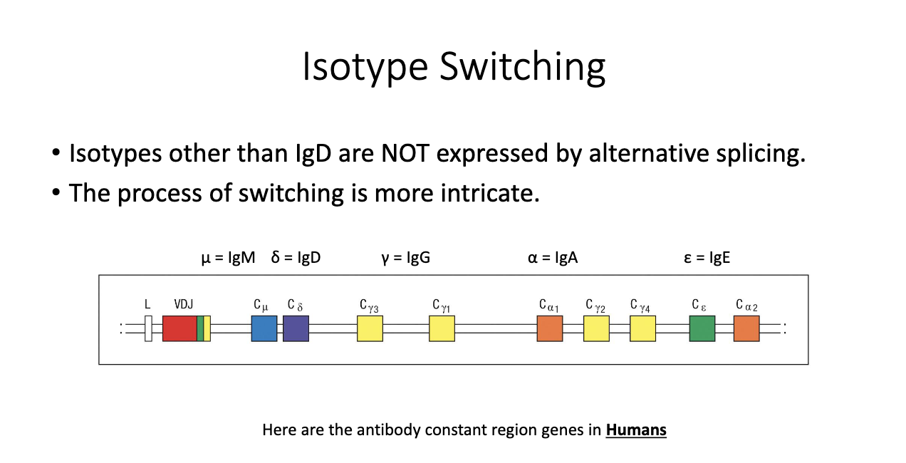
How are constant region genes arranged in the human genome, and why does it matter?

  • Genomic order (simplified): M → D → G3 → G1 → A1 → G2 → G4 → E → A2.

  • Result of gene duplication events during evolution.

  • Different isotypes evolved distinct roles despite shared ancestry.

  • Only IgM/IgD are close enough for alternative splicing; others require DNA recombination.

<ul><li><p class="isSelectedEnd"><span>Genomic order (simplified): </span><strong><span>M → D → G3 → G1 → A1 → G2 → G4 → E → A2</span></strong><span>.</span></p></li><li><p class="isSelectedEnd"><span>Result of </span><strong><span>gene duplication events</span></strong><span> during evolution.</span></p></li><li><p class="isSelectedEnd"><span>Different isotypes evolved distinct roles despite shared ancestry.</span></p></li><li><p><span>Only </span><strong><span>IgM/IgD</span></strong><span> are close enough for </span><strong><span>alternative splicing</span></strong><span>; others require DNA recombination.</span></p></li></ul><p></p>
7
New cards

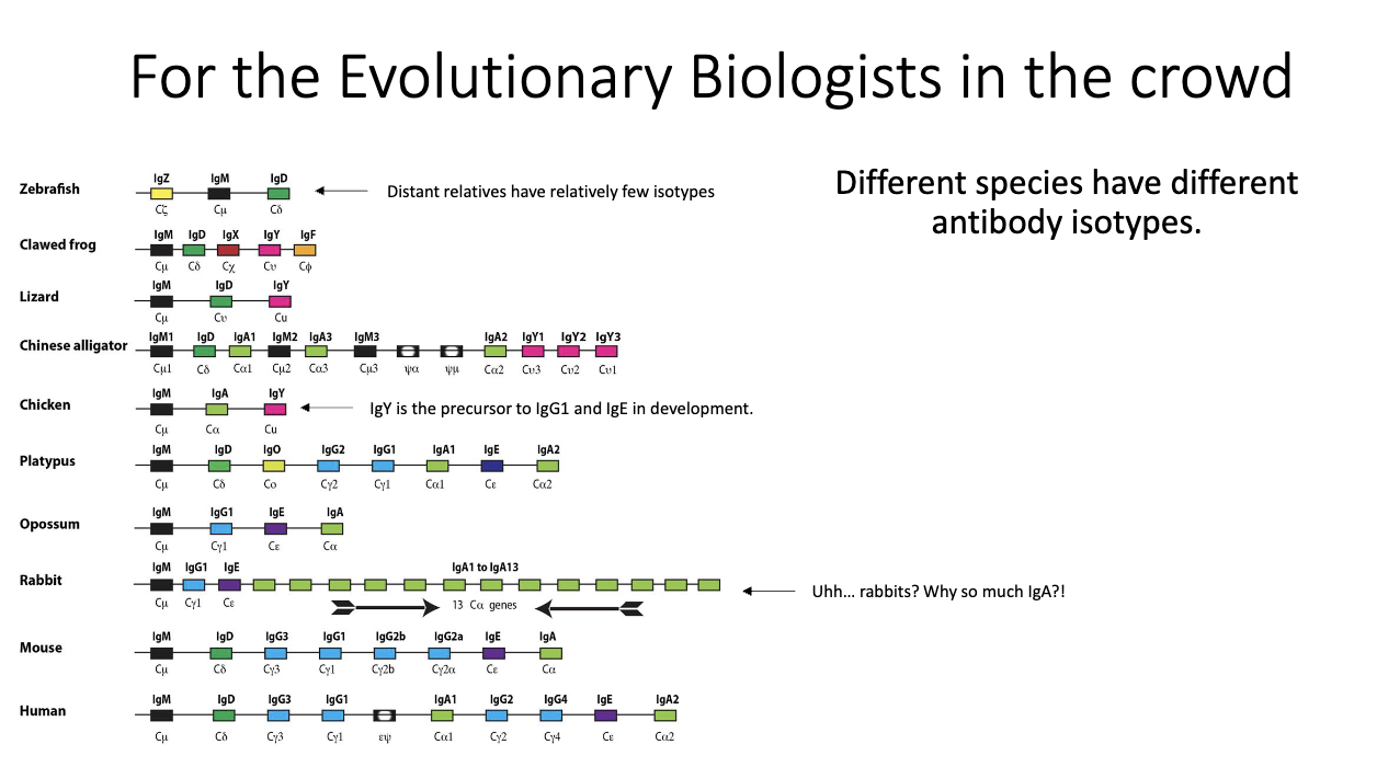
What does cross-species variation in antibody isotypes reveal?

  • Isotype repertoires vary widely between species (e.g., rabbits have 13 IgA genes).

  • IgY in birds is precursor to mammalian IgG1 + IgE.

  • All mammals have exactly one IgE, suggesting strong selection pressure.

  • Indicates rapid evolution driven by host–pathogen interactions.

<ul><li><p class="isSelectedEnd"><span>Isotype repertoires vary widely between species (e.g., rabbits have </span><strong><span>13 IgA</span></strong><span> genes).</span></p></li><li><p class="isSelectedEnd"><strong><span>IgY</span></strong><span> in birds is precursor to mammalian </span><strong><span>IgG1 + IgE</span></strong><span>.</span></p></li><li><p class="isSelectedEnd"><span>All mammals have exactly </span><strong><span>one IgE</span></strong><span>, suggesting strong </span><strong><span>selection pressure</span></strong><span>.</span></p></li><li><p><span>Indicates rapid evolution driven by </span><strong><span>host–pathogen interactions</span></strong><span>.</span></p></li></ul><p></p>
8
New cards

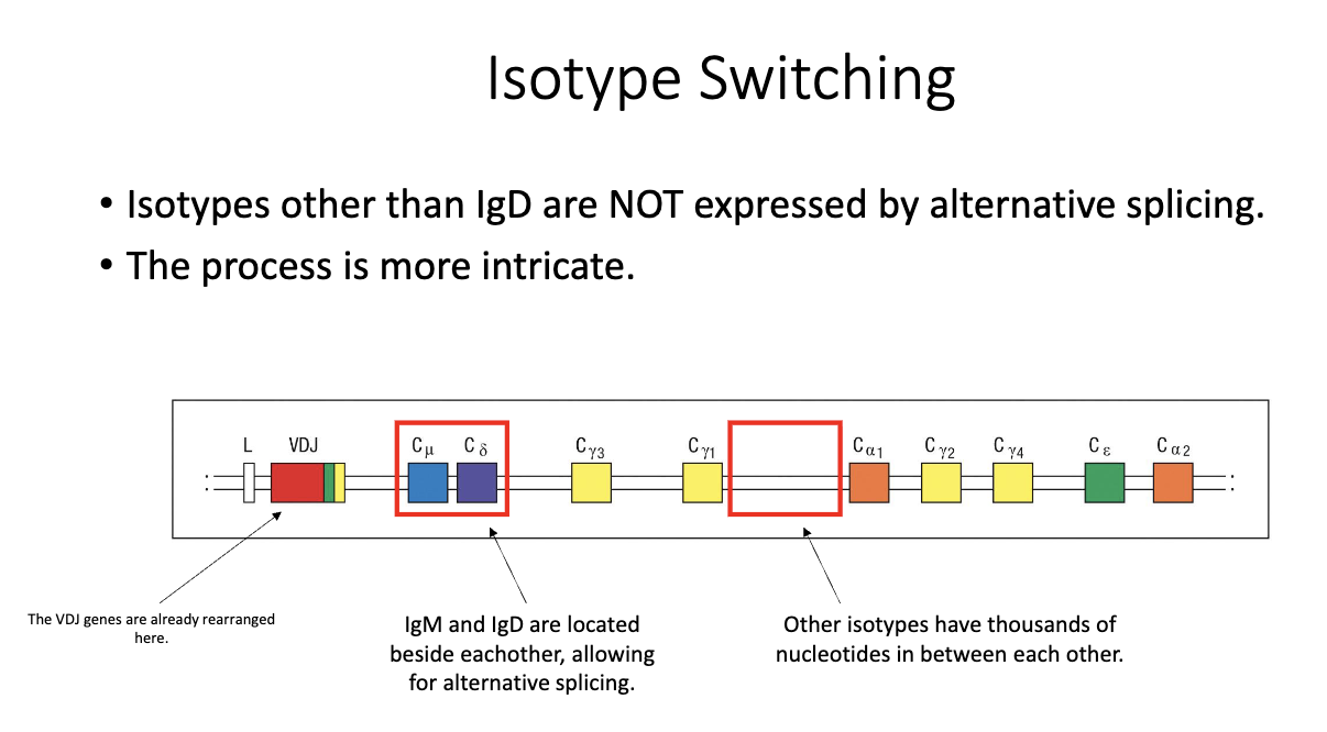
Why is IgD produced by alternative splicing but other isotypes require switching?

  • IgM and IgD lie adjacent in the genome → allow alternative splicing into two transcripts.

  • Other constant regions are separated by thousands of base pairs, preventing this mechanism.

  • All switching beyond IgM/IgD is after VDJ recombination and B-cell activation.

<ul><li><p class="isSelectedEnd"><strong><span>IgM and IgD</span></strong><span> lie adjacent in the genome → allow </span><strong><span>alternative splicing</span></strong><span> into two transcripts.</span></p></li><li><p class="isSelectedEnd"><span>Other constant regions are separated by </span><strong><span>thousands of base pairs</span></strong><span>, preventing this mechanism.</span></p></li><li><p><span>All switching beyond IgM/IgD is after </span><strong><span>VDJ recombination</span></strong><span> and B-cell activation.</span></p></li></ul><p></p>
9
New cards

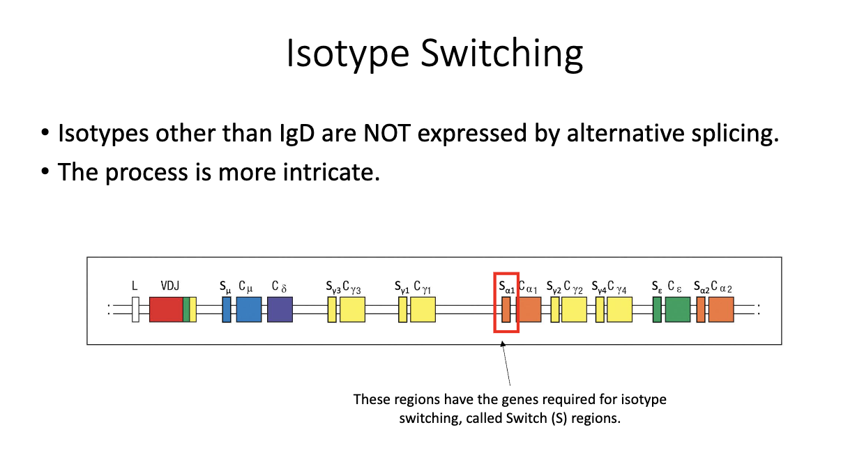
What are switch (S) regions, and why are they important for isotype switching?

  • Located upstream of each constant region (except δ/IgD).

  • Not translated; function is purely regulatory.

  • Required for the recombination process that enables class-switch recombination (CSR).

<ul><li><p class="isSelectedEnd"><span>Located </span><strong><span>upstream of each constant region</span></strong><span> (except δ/IgD).</span></p></li><li><p class="isSelectedEnd"><span>Not translated; function is purely regulatory.</span></p></li><li><p><span>Required for the recombination process that enables </span><strong><span>class-switch recombination (CSR)</span></strong><span>.</span></p></li></ul><p></p>
10
New cards

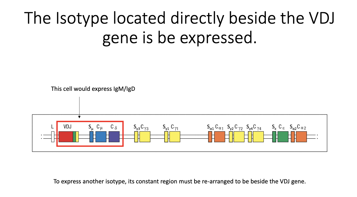
How does a B cell begin producing a new isotype at the DNA level?

  • Only the constant region directly adjacent to VDJ can be transcribed.

  • Naive state: IgM and IgD flank the VDJ → both expressed.

  • To express a new isotype, the relevant constant region must be moved next to VDJ.

  • Achieved by cutting out intervening DNA between the switch regions → irreversible CSR event.

<ul><li><p class="isSelectedEnd"><span>Only the constant region </span><strong><span>directly adjacent to VDJ</span></strong><span> can be transcribed.</span></p></li><li><p class="isSelectedEnd"><span>Naive state: </span><strong><span>IgM and IgD</span></strong><span> flank the VDJ → both expressed.</span></p></li><li><p class="isSelectedEnd"><span>To express a new isotype, the relevant constant region must be </span><strong><span>moved next to VDJ</span></strong><span>.</span></p></li><li><p><span>Achieved by </span><strong><span>cutting out</span></strong><span> intervening DNA between the switch regions → irreversible CSR event.</span></p></li></ul><p></p>
11
New cards

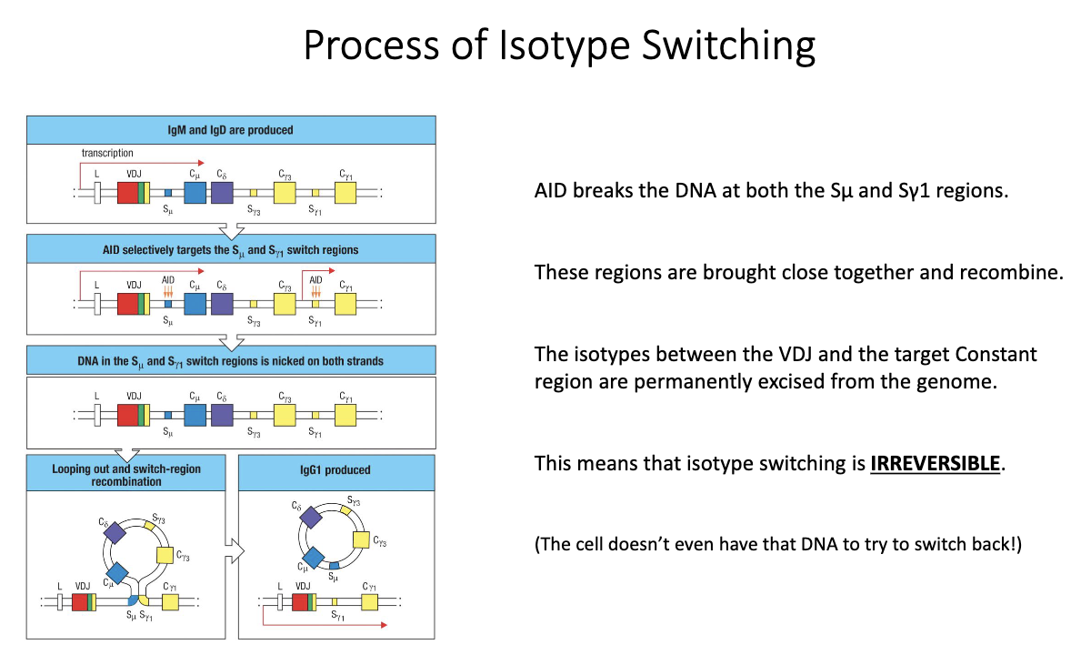
How does AID mediate class-switch recombination (CSR) to generate new isotypes?

  • Transcription begins upstream of VDJ, passing through IgM/IgD.

  • AID (upregulated after TFH help) introduces mutations/breaks in switch (S) regions.

  • Cytokines determine which S region AID targets (e.g., IL-4 → Sγ1, IFN-γ → Sγ3).

  • AID creates double-stranded breaks → DNA ends are joined; intervening DNA loops out as a switch circle.

  • New constant region is placed beside VDJ → irreversible isotype switch.

<ul><li><p class="isSelectedEnd"><span>Transcription begins upstream of </span><strong><span>VDJ</span></strong><span>, passing through </span><strong><span>IgM/IgD</span></strong><span>.</span></p></li><li><p class="isSelectedEnd"><strong><span>AID</span></strong><span> (upregulated after TFH help) introduces mutations/breaks in </span><strong><span>switch (S) regions</span></strong><span>.</span></p></li><li><p class="isSelectedEnd"><span>Cytokines determine </span><strong><span>which S region</span></strong><span> AID targets (e.g., IL-4 → Sγ1, IFN-γ → Sγ3).</span></p></li><li><p class="isSelectedEnd"><span>AID creates </span><strong><span>double-stranded breaks</span></strong><span> → DNA ends are joined; intervening DNA loops out as a </span><strong><span>switch circle</span></strong><span>.</span></p></li><li><p><span>New constant region is placed beside VDJ → </span><strong><span>irreversible isotype switch</span></strong><span>.</span></p></li></ul><p></p>
12
New cards

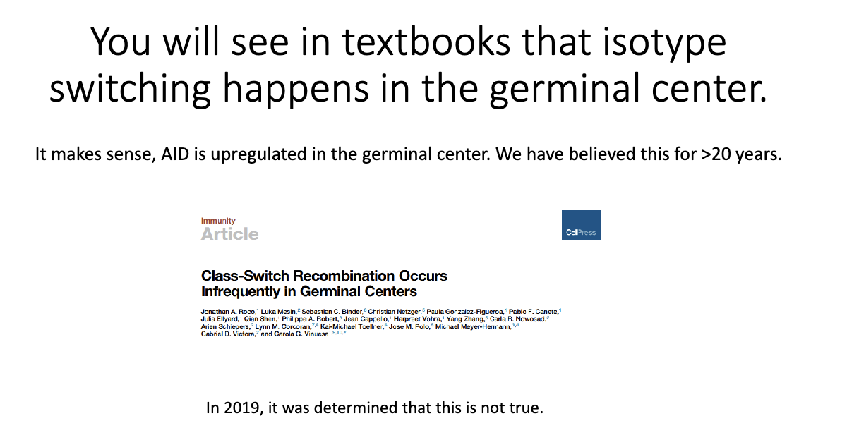
Does isotype switching occur inside germinal centers?

  • Textbooks say yes, but this is outdated.

  • Coróla Vinuesa (2019) showed CSR occurs before B cells enter germinal centers.

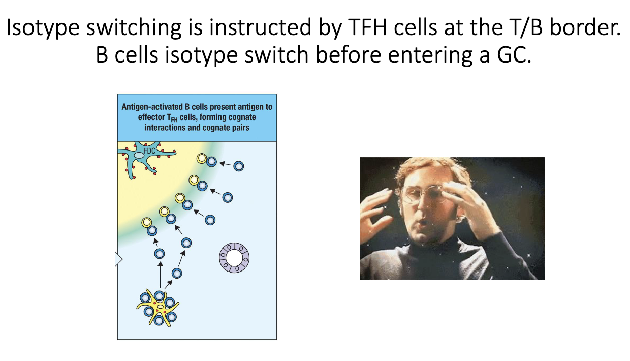
  • Switching happens at the T–B border, immediately after TFH–B cell interaction.

<ul><li><p class="isSelectedEnd"><strong><span>Textbooks say yes</span></strong><span>, but this is outdated.</span></p></li><li><p class="isSelectedEnd"><span>Coróla Vinuesa (2019) showed CSR occurs </span><strong><span>before</span></strong><span> B cells enter germinal centers.</span></p></li><li><p><span>Switching happens at the </span><strong><span>T–B border</span></strong><span>, immediately after TFH–B cell interaction.</span></p></li></ul><p></p>
13
New cards

When do B cells isotype switch relative to germinal center entry?

  • CSR is triggered during the T cell–B cell “hug” at the T–B border.

  • By the time a B cell migrates into the germinal center, it has already switched.

<ul><li><p class="isSelectedEnd"><span>CSR is triggered during the </span><strong><span>T cell–B cell “hug”</span></strong><span> at the T–B border.</span></p></li><li><p class="isSelectedEnd"><span>By the time a B cell migrates into the germinal center, it has </span><strong><span>already switched</span></strong><span>.</span></p></li></ul><p></p>
14
New cards

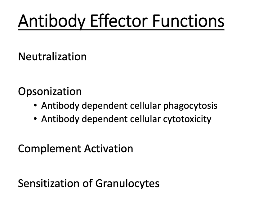
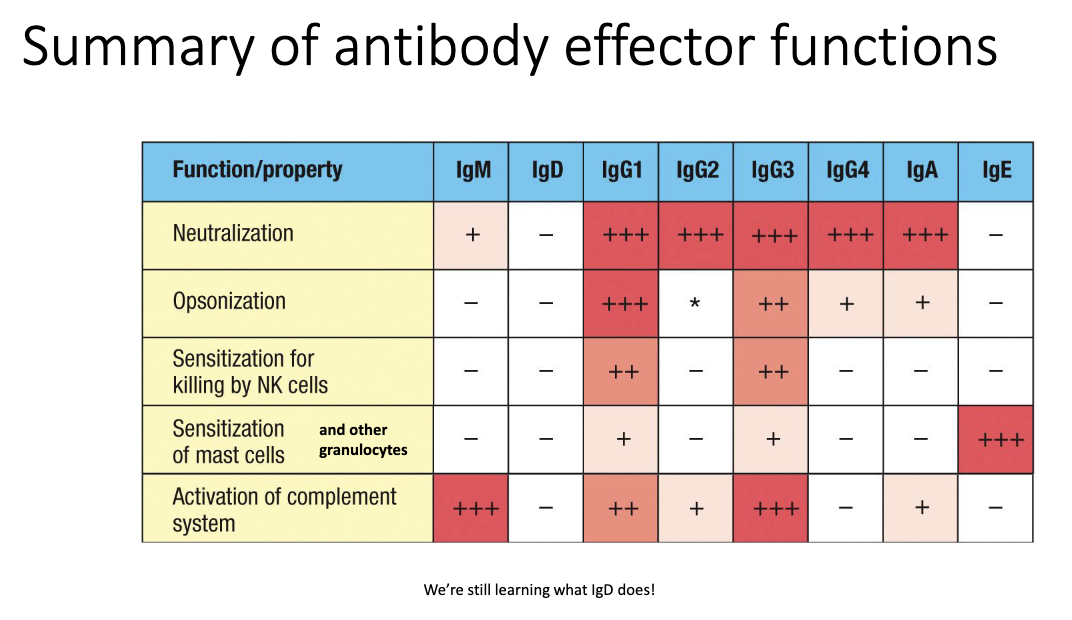
What are the four major pathogen-eliminating functions of antibodies?

  1. Neutralization

  2. Opsonization

  3. Complement activation

  4. Granulocyte sensitization

<ol><li><p class="isSelectedEnd"><strong><span>Neutralization</span></strong></p></li><li><p class="isSelectedEnd"><strong><span>Opsonization</span></strong></p></li><li><p class="isSelectedEnd"><strong><span>Complement activation</span></strong></p></li><li><p><strong><span>Granulocyte sensitization</span></strong></p></li></ol><p></p>
15
New cards

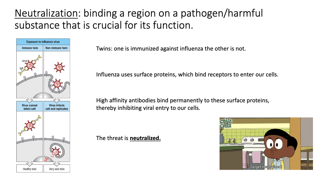
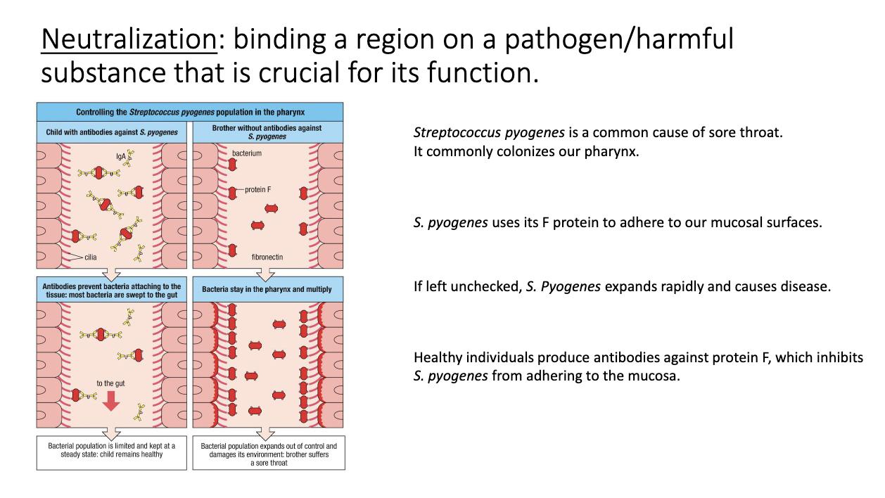
How does antibody-mediated neutralization work?

  • Antibody binds a pathogen’s entry/attachment site → blocks access to host cells.

  • No additional immune machinery needed.

  • High-affinity antibodies can permanently neutralize targets.

  • Classic example: mucosal IgA blocking influenza or bacterial adhesins.

<ul><li><p class="isSelectedEnd"><span>Antibody binds a pathogen’s </span><strong><span>entry/attachment site</span></strong><span> → blocks access to host cells.</span></p></li><li><p class="isSelectedEnd"><span>No additional immune machinery needed.</span></p></li><li><p class="isSelectedEnd"><span>High-affinity antibodies can </span><strong><span>permanently</span></strong><span> neutralize targets.</span></p></li><li><p><span>Classic example: mucosal </span><strong><span>IgA</span></strong><span> blocking influenza or bacterial adhesins.</span></p></li></ul><p></p>
16
New cards

How do antibodies neutralize bacterial pathogens like Strep pyogen?

  • Bacteria use adhesins (e.g., F protein) to bind mucosal surfaces.

  • IgA binds these F-protein → prevents attachment/colonization.

  • Pathogen is cleared before causing symptomatic infection.

<ul><li><p class="isSelectedEnd"><span>Bacteria use adhesins (e.g., </span><strong><span>F protein</span></strong><span>) to bind mucosal surfaces.</span></p></li><li><p class="isSelectedEnd"><strong><span>IgA</span></strong><span> binds these F-protein → prevents attachment/colonization.</span></p></li><li><p><span>Pathogen is cleared before causing symptomatic infection.</span></p></li></ul><p></p>
17
New cards

Why do IgM pentamers rely on avidity for neutralization?

  • Early IgM is low affinity (pre–germinal center).

  • Pentamer structure gives 10 binding sites → strong avidity even with weak individual interactions.

  • Compensates for poor affinity to still block pathogens effectively.

<ul><li><p class="isSelectedEnd"><span>Early IgM is </span><strong><span>low affinity</span></strong><span> (pre–germinal center).</span></p></li><li><p class="isSelectedEnd"><span>Pentamer structure gives </span><strong><span>10 binding sites</span></strong><span> → strong </span><strong><span>avidity</span></strong><span> even with weak individual interactions.</span></p></li><li><p><span>Compensates for poor affinity to still block pathogens effectively.</span></p></li></ul><p></p>
18
New cards

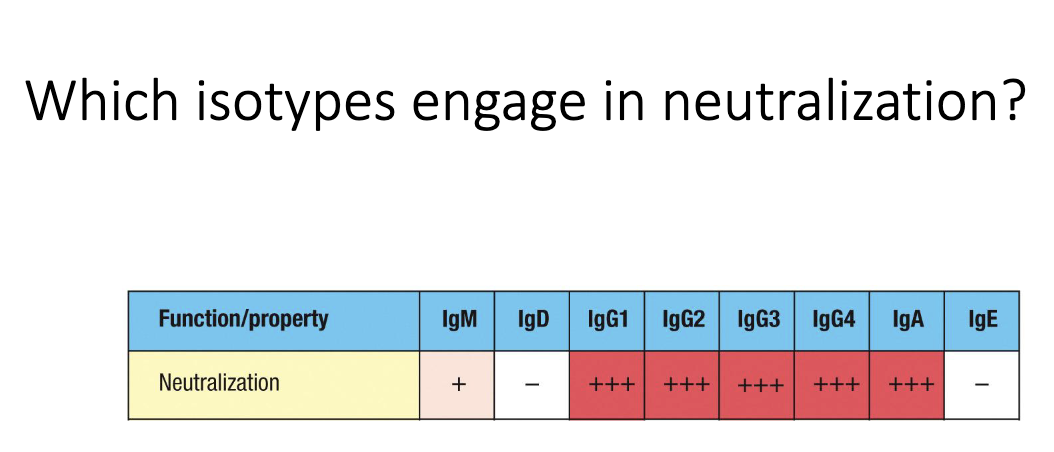
Which antibody isotypes are most effective at neutralization?

  • IgG (systemic) and IgA (mucosal) are strongest neutralizers.

  • IgM provides weaker but still meaningful neutralization via high avidity.

<ul><li><p class="isSelectedEnd"><strong><span>IgG</span></strong><span> (systemic) and </span><strong><span>IgA</span></strong><span> (mucosal) are strongest neutralizers.</span></p></li><li><p><strong><span>IgM</span></strong><span> provides weaker but still meaningful neutralization via high avidity.</span></p></li></ul><p></p>
19
New cards

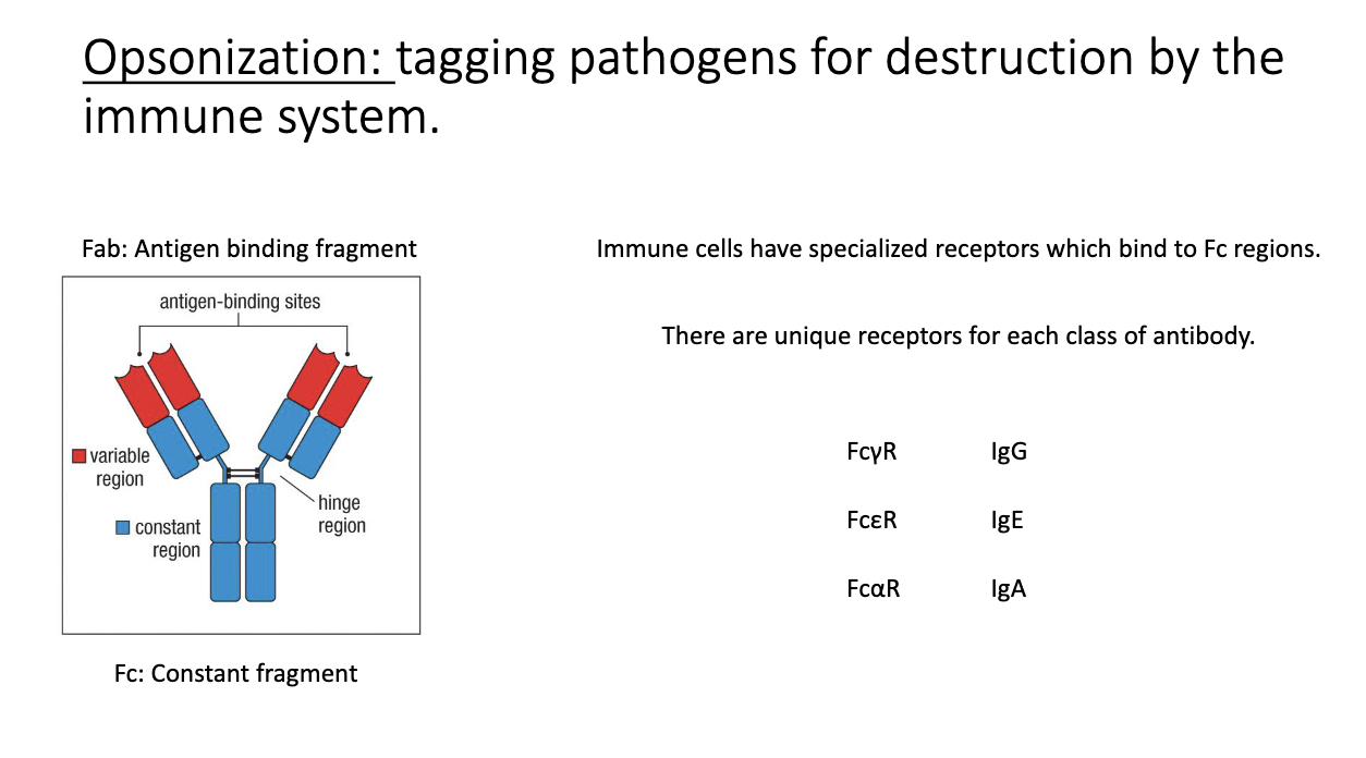
What is opsonization, and how do FC receptors participate?

  • Antibody binds a pathogen, and immune cells detect the Fc region via Fc receptors.

  • Fc receptor naming: Fc + isotype Greek letter + R (e.g., FcγR for IgG).

  • Leads to phagocytosis and destruction of the antibody-coated microbe.

<ul><li><p class="isSelectedEnd"><span>Antibody binds a pathogen, and immune cells detect the </span><strong><span>Fc region</span></strong><span> via </span><strong><span>Fc receptors</span></strong><span>.</span></p></li><li><p class="isSelectedEnd"><span>Fc receptor naming: </span><strong><span>Fc + isotype Greek letter + R</span></strong><span> (e.g., FcγR for IgG).</span></p></li><li><p><span>Leads to phagocytosis and destruction of the antibody-coated microbe.</span></p></li></ul><p></p>
20
New cards

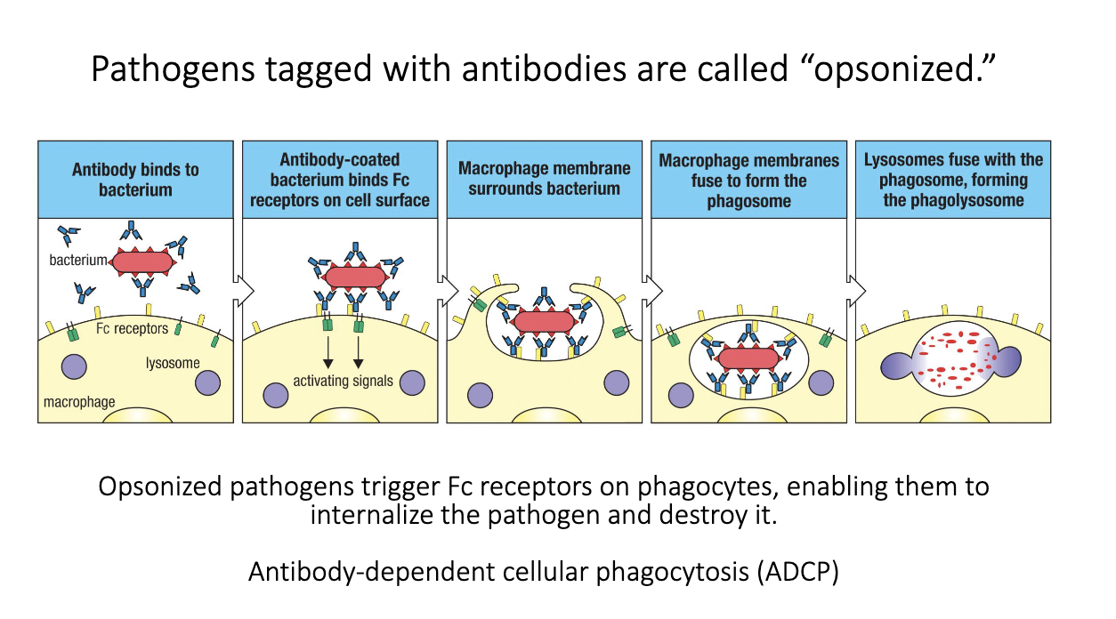
What is antibody-dependent cellular phagocytosis (ADCP)?

  • Pathogen coated with antibodies = opsonized.

  • Fc receptors on phagocytes bind Fc regions of those antibodies.

  • FcγR signaling tells the cell to engulf and destroy the target.

  • Leads to phagolysosome fusion and killing of the pathogen.

  • Antibodies augment innate immunity by making microbes easier to phagocytose.

<ul><li><p class="isSelectedEnd"><span>Pathogen coated with antibodies = </span><strong><span>opsonized</span></strong><span>.</span></p></li><li><p class="isSelectedEnd"><span>Fc receptors on phagocytes bind Fc regions of those antibodies.</span></p></li><li><p class="isSelectedEnd"><span>FcγR signaling tells the cell to </span><strong><span>engulf and destroy</span></strong><span> the target.</span></p></li><li><p class="isSelectedEnd"><span>Leads to phagolysosome fusion and killing of the pathogen.</span></p></li><li><p><span>Antibodies augment innate immunity by making microbes easier to phagocytose.</span></p></li></ul><p></p>
21
New cards

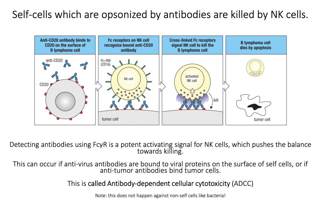
How does antibody-dependent cellular cytotoxicity (ADCC) work?

  • Antibodies bind antigens on infected or cancerous host cells.

  • NK cells use FcγRIII to recognize bound IgG.

  • Fc engagement → strong activating signal → target cell killing.

  • Used therapeutically (e.g., anti-CD20 antibodies killing B-cell cancers).

  • Does not work on bacteria because NK killing uses perforin/granzymes → apoptosis, which bacteria can’t undergo.

<ul><li><p class="isSelectedEnd"><span>Antibodies bind antigens on infected or cancerous host cells.</span></p></li><li><p class="isSelectedEnd"><span>NK cells use </span><strong><span>FcγRIII</span></strong><span> to recognize bound IgG.</span></p></li><li><p class="isSelectedEnd"><span>Fc engagement → strong activating signal → </span><strong><span>target cell killing</span></strong><span>.</span></p></li><li><p class="isSelectedEnd"><span>Used therapeutically (e.g., anti-CD20 antibodies killing B-cell cancers).</span></p></li><li><p><span>Does </span><em><span>not</span></em><span> work on bacteria because NK killing uses perforin/granzymes → apoptosis, which bacteria can’t undergo.</span></p></li></ul><p></p>
22
New cards

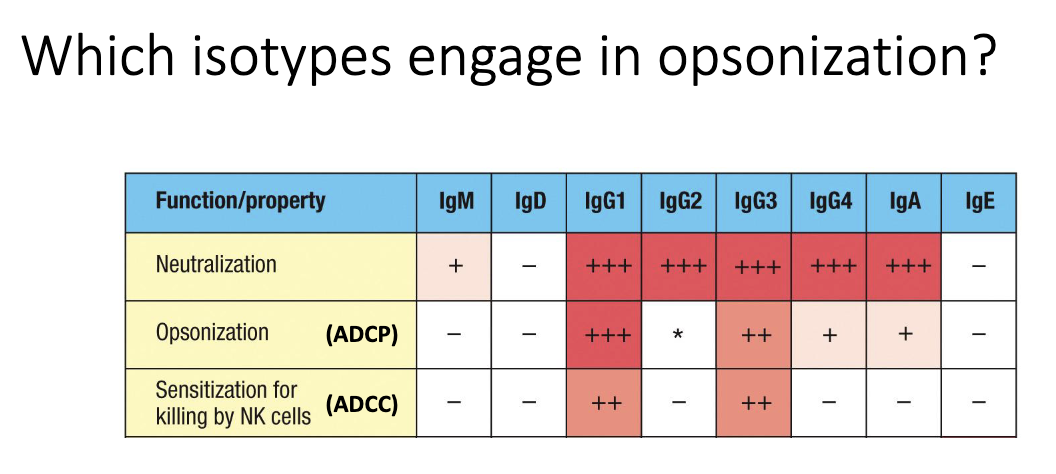
Which isotypes are effective at opsonization?

  • Primarily IgG1 and IgG3.

  • These bind Fcγ receptors strongly, enabling ADCP and ADCC.

  • IgG2 and IgG4 bind FcγRs poorly → weak opsonizing ability.

<ul><li><p class="isSelectedEnd"><span>Primarily </span><strong><span>IgG1</span></strong><span> and </span><strong><span>IgG3</span></strong><span>.</span></p></li><li><p class="isSelectedEnd"><span>These bind Fcγ receptors strongly, enabling ADCP and ADCC.</span></p></li><li><p><strong><span>IgG2</span></strong><span> and </span><strong><span>IgG4</span></strong><span> bind FcγRs poorly → weak opsonizing ability.</span></p></li></ul><p></p>
23
New cards

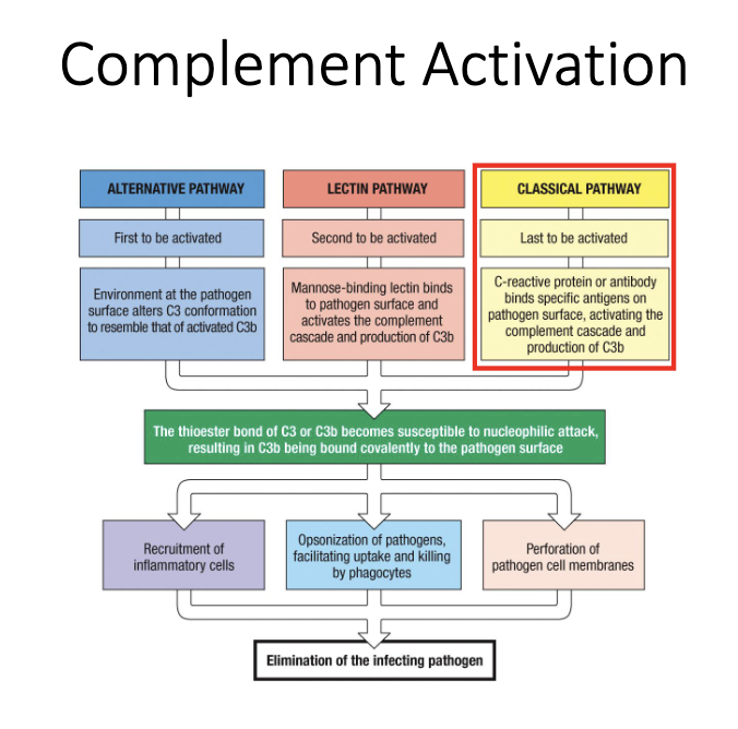
Which antibodies activate complement, and how?

  • IgM is the strongest complement activator (ideal structure for C1 binding).

  • IgG1 and IgG3 also activate complement effectively.

  • Complement binding to antibody-coated surfaces → classical pathway → pathogen lysis.

<ul><li><p class="isSelectedEnd"><strong><span>IgM</span></strong><span> is the strongest complement activator (ideal structure for C1 binding).</span></p></li><li><p class="isSelectedEnd"><strong><span>IgG1</span></strong><span> and </span><strong><span>IgG3</span></strong><span> also activate complement effectively.</span></p></li><li><p><span>Complement binding to antibody-coated surfaces → classical pathway → pathogen lysis.</span></p></li></ul><p></p>
24
New cards

Why is IgM exceptionally good at fixing complement?

  • Pentameric “planar” structure matches C1q binding sites perfectly.

  • C1q engagement rapidly triggers the classical complement cascade.

<ul><li><p class="isSelectedEnd"><span>Pentameric “planar” structure matches </span><strong><span>C1q binding sites</span></strong><span> perfectly.</span></p></li><li><p><span>C1q engagement rapidly triggers the </span><strong><span>classical complement cascade</span></strong><span>.</span></p></li></ul><p></p>
25
New cards

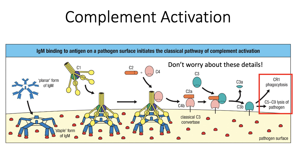
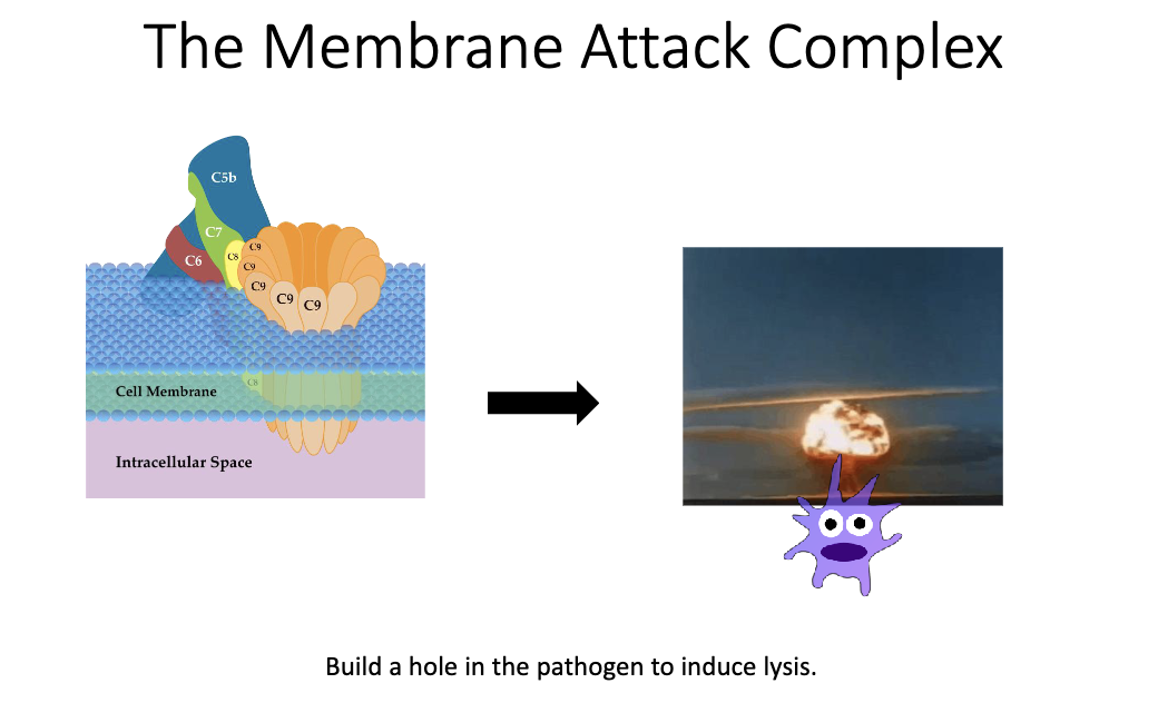
What is the end result of antibody-driven complement activation?

  • Complement cascade forms the MAC pore in microbial membranes.

  • Leads to osmotic lysis and death of the pathogen.

<ul><li><p class="isSelectedEnd"><span>Complement cascade forms the </span><strong><span>MAC pore</span></strong><span> in microbial membranes.</span></p></li><li><p><span>Leads to </span><strong><span>osmotic lysis</span></strong><span> and death of the pathogen.</span></p></li></ul><p></p>
26
New cards

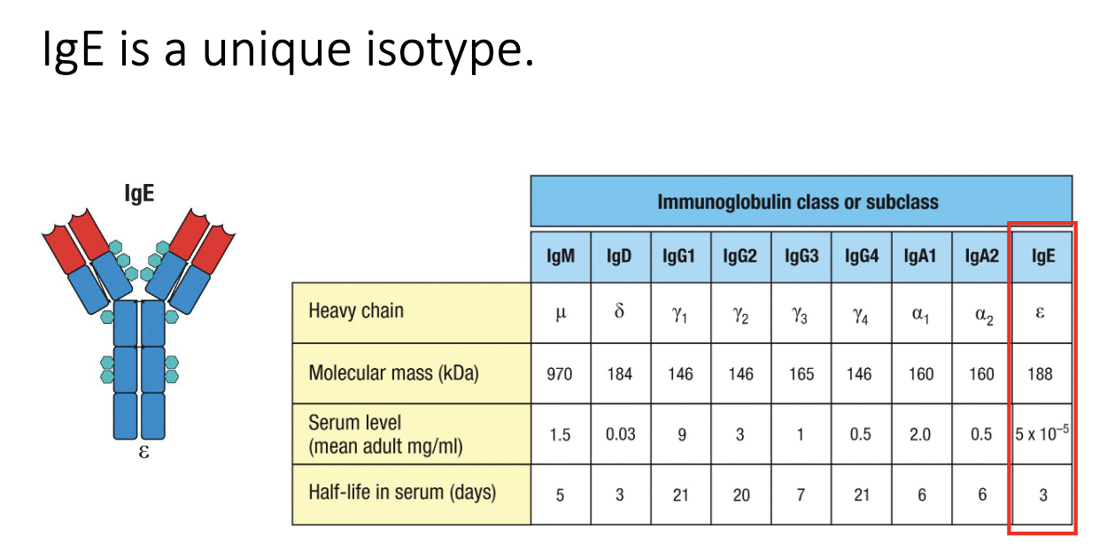
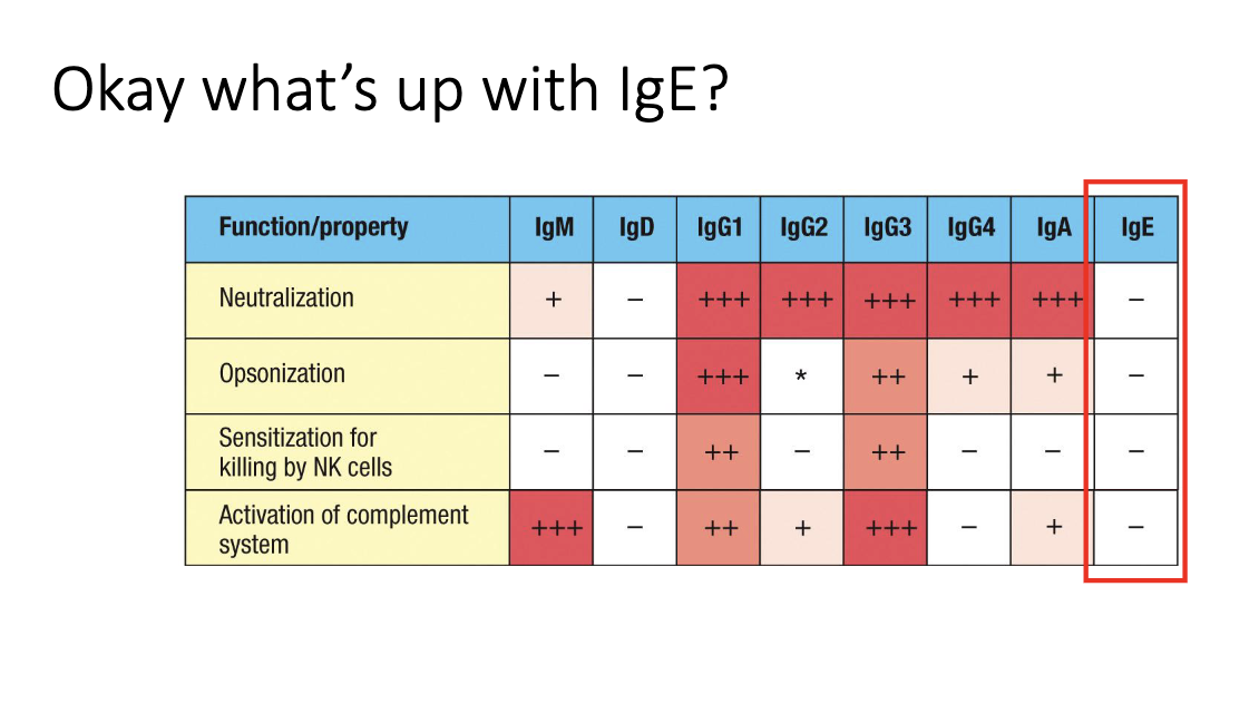
Why doesn’t IgE carry out neutralization or opsonization in circulation?

  • Serum IgE levels are extremely low (ng/mL range).

  • Too scarce to meaningfully neutralize or opsonize microbes.

  • Its role is instead mediated through FcεRI-bound IgE on granulocytes.

<ul><li><p class="isSelectedEnd"><span>Serum IgE levels are </span><strong><span>extremely low</span></strong><span> (ng/mL range).</span></p></li><li><p class="isSelectedEnd"><span>Too scarce to meaningfully neutralize or opsonize microbes.</span></p></li><li><p><span>Its role is instead mediated through </span><strong><span>FcεRI-bound IgE on granulocytes</span></strong><span>.</span></p></li></ul><img src="https://knowt-user-attachments.s3.amazonaws.com/9c2f0f42-b65c-4b31-a54e-03ef81af4c6e.png" data-width="100%" data-align="center" alt=""><p></p>
27
New cards

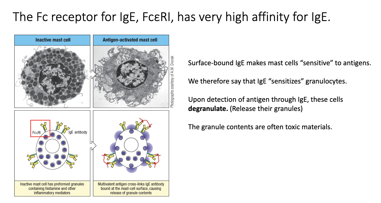
How does IgE sensitize mast cells and trigger degranulation?

  • IgE binds FcεRI on mast cells with very high affinity → remains for ~100+ days, compared to in serum IgE lasts for 7 days.

  • Antigen crosslinks IgE–FcεRI complexes → rapid degranulation (<1 hour).

  • Releases histamine, proteases, cytokines → drives allergy and anaphylaxis.

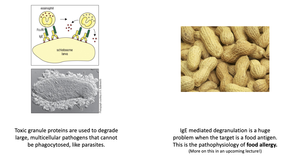
  • Same mechanism used to attack parasites via eosinophils/mast cells.

<ul><li><p class="isSelectedEnd"><span>IgE binds </span><strong><span>FcεRI</span></strong><span> on mast cells with very high affinity → remains for ~100+ days, compared to in serum IgE lasts for 7 days.</span></p></li><li><p class="isSelectedEnd"><span>Antigen crosslinks IgE–FcεRI complexes → </span><strong><span>rapid degranulation</span></strong><span> (&lt;1 hour).</span></p></li><li><p class="isSelectedEnd"><span>Releases histamine, proteases, cytokines → drives allergy and anaphylaxis.</span></p></li><li><p><span>Same mechanism used to attack </span><strong><span>parasites</span></strong><span> via eosinophils/mast cells.</span></p></li></ul><p></p>
28
New cards

How does IgE cause allergic reactions and anaphylaxis?

  • Pre-bound IgE on mast cells crosslinks upon re-exposure to allergen.

  • Triggers massive degranulation → vasodilation, itching, shock.

  • Same biology evolved for anti-parasite defense.

<ul><li><p class="isSelectedEnd"><span>Pre-bound IgE on mast cells crosslinks upon re-exposure to allergen.</span></p></li><li><p class="isSelectedEnd"><span>Triggers massive degranulation → vasodilation, itching, shock.</span></p></li><li><p><span>Same biology evolved for </span><strong><span>anti-parasite defense</span></strong><span>.</span></p></li></ul><p></p>
29
New cards

Which antibody isotype sensitizes mast cells and granulocytes?

  • IgE (via FcεRI).

  • IgD remains functionally unclear.

<ul><li><p class="isSelectedEnd"><strong><span>IgE</span></strong><span> (via FcεRI).</span></p></li><li><p><span>IgD remains functionally unclear.</span></p></li></ul><p></p>
30
New cards

How are antibody isotypes distributed throughout the body?

  • Dimeric IgA → mucosal surfaces (gut, lung, urogenital tract).

  • IgE → bound to mast cells/skin.

  • IgG & monomeric IgA → systemic circulation.

  • Brain/CNS normally antibody-free → antibodies here indicate pathology.

    • Ex: MS results from antibodies in brain/CNS.

<ul><li><p class="isSelectedEnd"><strong><span>Dimeric IgA</span></strong><span> → mucosal surfaces (gut, lung, urogenital tract).</span></p></li><li><p class="isSelectedEnd"><strong><span>IgE</span></strong><span> → bound to mast cells/skin.</span></p></li><li><p class="isSelectedEnd"><strong><span>IgG &amp; monomeric IgA</span></strong><span> → systemic circulation.</span></p></li><li><p><span>Brain/CNS normally </span><strong><span>antibody-free</span></strong><span> → antibodies here indicate pathology.</span></p><ul><li><p>Ex: MS results from antibodies in brain/CNS.</p></li></ul></li></ul><p></p>
31
New cards

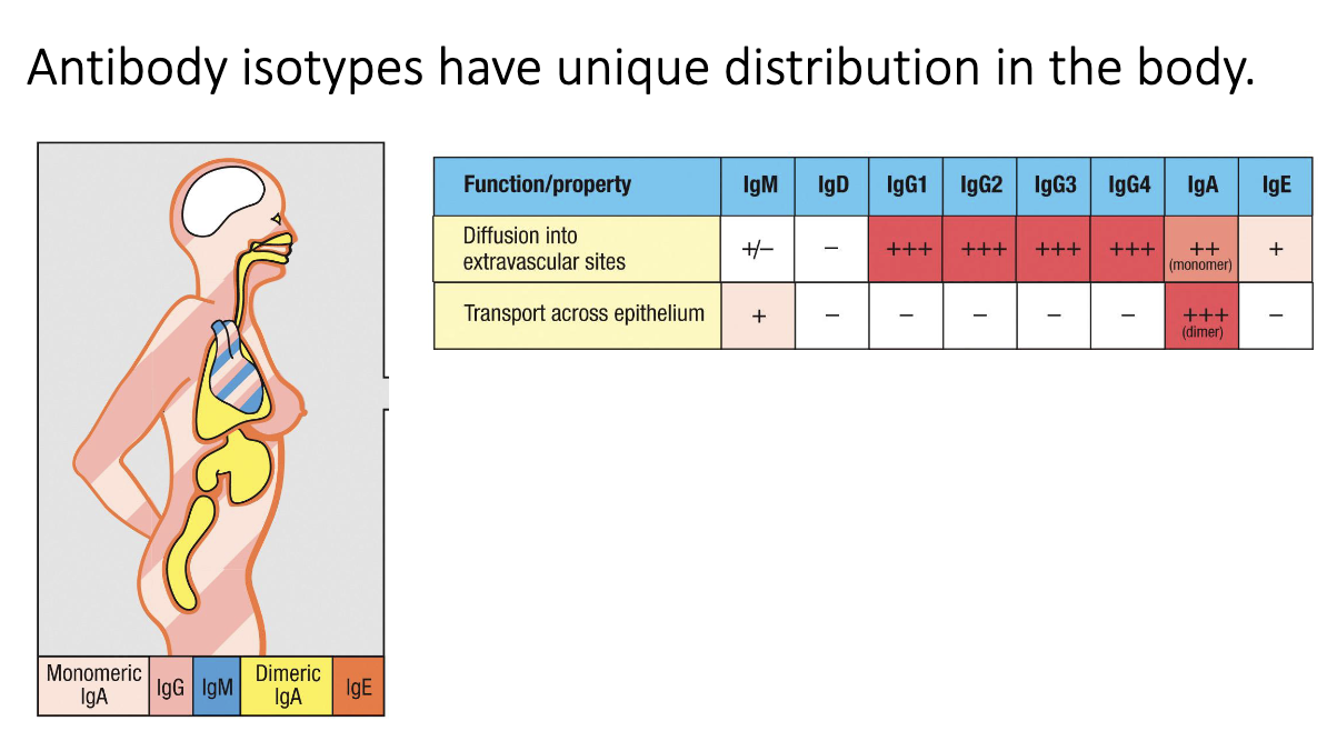
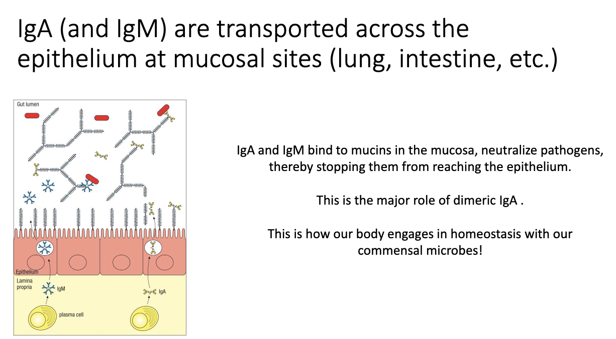
How are IgA and IgM transported into mucosal lumen?

  • Plasma cells secrete IgA/IgM.

  • Epithelial cells use the poly-Ig receptor to capture them.

  • Antibodies are transported and released into the lumen to bind microbes.

  • Maintains gut homeostasis by keeping bacteria out of host tissues.

<ul><li><p class="isSelectedEnd"><span>Plasma cells secrete IgA/IgM.</span></p></li><li><p class="isSelectedEnd"><span>Epithelial cells use the </span><strong><span>poly-Ig receptor</span></strong><span> to capture them.</span></p></li><li><p class="isSelectedEnd"><span>Antibodies are transported and released into the lumen to bind microbes.</span></p></li><li><p><span>Maintains gut homeostasis by keeping bacteria out of host tissues.</span></p></li></ul><p></p>
32
New cards

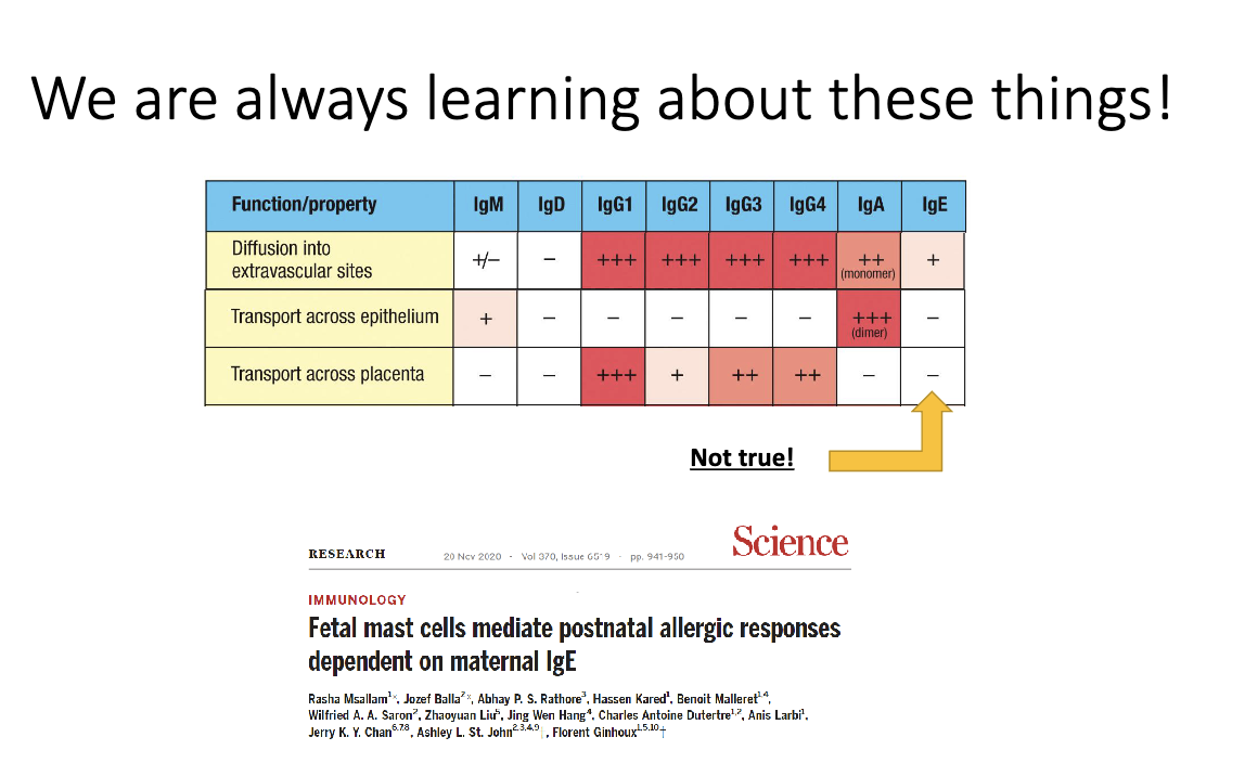
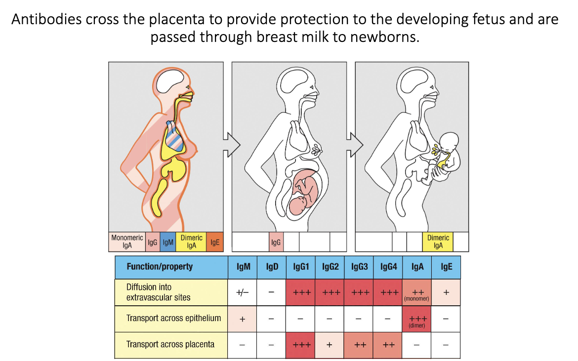
How do mothers pass antibodies to their babies?

  • IgG crosses the placenta → fetal protection in utero.

  • IgA from breast milk coats infant gut after birth.

  • “Passive transfer” of antibodies from parent to fetus/child allows for protection before fetus/infant antibody production matures.

<ul><li><p class="isSelectedEnd"><strong><span>IgG</span></strong><span> crosses the placenta → fetal protection in utero.</span></p></li><li><p class="isSelectedEnd"><strong><span>IgA</span></strong><span> from breast milk coats infant gut after birth.</span></p></li><li><p><span>“Passive transfer” of antibodies from parent to fetus/child allows for protection before fetus/infant antibody production matures.</span></p></li></ul><p></p>
33
New cards

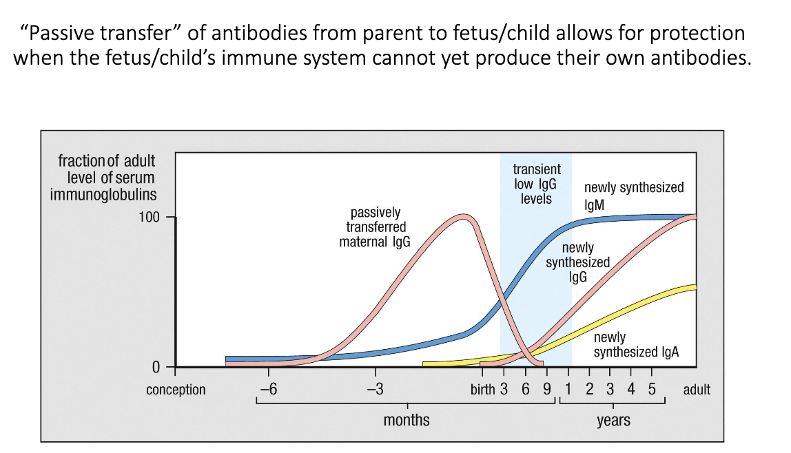
What is passive transfer of maternal antibodies, and how does it protect newborns?

  • During pregnancy, maternal IgG crosses the placenta to the fetus.

  • After birth, the newborn briefly has low antibody levels until its own IgG/IgA production ramps up.

  • During this window, maternal IgA from breast milk protects the infant’s gut.

  • Combined, these mechanisms provide early-life immunity before the baby’s immune system matures.

<ul><li><p class="isSelectedEnd"><span>During pregnancy, </span><strong><span>maternal IgG crosses the placenta</span></strong><span> to the fetus.</span></p></li><li><p class="isSelectedEnd"><span>After birth, the newborn briefly has </span><strong><span>low antibody levels</span></strong><span> until its own IgG/IgA production ramps up.</span></p></li><li><p class="isSelectedEnd"><span>During this window, </span><strong><span>maternal IgA from breast milk</span></strong><span> protects the infant’s gut.</span></p></li><li><p><span>Combined, these mechanisms provide </span><strong><span>early-life immunity</span></strong><span> before the baby’s immune system matures.</span></p></li></ul><p></p>
34
New cards

Can maternal IgE cross the placenta?

  • Historically thought no, but 2020 data show yes.

  • Fetal mast cells can acquire maternal IgE.

  • Provides a potential mechanism for inheritance of allergy risk.

<ul><li><p class="isSelectedEnd"><span>Historically thought </span><strong><span>no</span></strong><span>, but 2020 data show </span><strong><span>yes</span></strong><span>.</span></p></li><li><p class="isSelectedEnd"><span>Fetal mast cells can acquire maternal IgE.</span></p></li><li><p><span>Provides a potential mechanism for </span><strong><span>inheritance of allergy risk</span></strong><span>.</span></p></li></ul><p></p>