Invertebrate Zoology Exam 5
1/122
There's no tags or description
Looks like no tags are added yet.
Name | Mastery | Learn | Test | Matching | Spaced | Call with Kai |
|---|
No analytics yet
Send a link to your students to track their progress
123 Terms
Tagmosis
The fusion of segments to form functional units
arthropods have many segments, but they don’t operate independently, as the tagmata form distinct regions of the body
What are the three groups of Arthropoda?
Chelicerata
Crustacea
Mandibulata
What tagma make up the body of a chelicerate?
Prosoma
Opisthosoma
Prosoma
“Head” tagma of chelicerates; composed of 6 segments and bears the mouthparts and walking legs
Opisthosoma
“Body” tagma of chelicerates; contains the reproductive organs and gut
Differences in Hox genes of chelicerates
no antennae; chelicerae are the first pair of appendages
chelicerae are homologous to antennae, not mandibles
mouth is positioned between the first two segments
all 6 segments and the ocular fuse to form the prosoma during embryonic development
Crustacean body structure
body is divided into head, thorax, and abdomen, although the first two segments are often fused to form the cephalothorax
head: eyes, antennae, mouthparts
thorax: maxillipeds, walking limbs
abdomen: gut, abdominal appendages
What are the key features of Phylum Arthropoda?
Tagmosis
Each segment has jointed appendages with intrinsic musculature
Chitinous cuticle; forms sclerotized plates
Arthropodization
basically, “a worm-in-a-box” structure
the “box” is formed by a rigid cuticle that acts as an exoskeleton → this feature affects multiple biological functions
What are the divisions of an arthropod cuticle?
The cuticle is an exoskeleton that is divided into:
Tergites = dorsal plates
Sternites = ventral plates
Pleurites = side walls
Arthropod cardiovascular system
The body cavity is a hemocoel; the blood is pumped through an open circulatory system by a dorsal “heart”
What kind of respiratory pigment do arthropods use?
Hemocyanin
Arthropod musculature
Muscles attach to the body plates and extend into the limbs, but there are also intrinsic muscles in the limbs to manipulate the segments
Arthropod cuticle
A non-cellular, extracellular matrix that is secreted by the epidermis.
Key components of the cuticle
Epicuticle - continuous outer layer; made up of a waxy, waterproof substance secreted by gland cells and allows for flexibility between sclerotized plates
Exocuticle - middle layer; undergoes sclerotization
Endocuticle - lower layer; borders the epidermis
Why does the cuticle contain spines/setae?
some of these are solid, while others contain cells and/or sensory neurons
no cilia = not a scalid
extensions of trichogen cells form the innervated spines, while solid spines are cuticular processes
Sclerotization of the cuticle
also called “calcification” in crustaceans
this is a process used to harden the cuticle, as it is generally a flimsy material; this involves changes in cuticular proteins and usually results in a darkening of the cuticle (tanning)
the extend of sclerotization varies over the body and across life stages
only the exocuticle undergoes sclerotization
Arthropod molting process
Begins with a hormonal signal that causes the epidermis to secrete enzymes to dissolve the endocuticle (the dissolved materials are absorbed for later use)
this loosens the cuticle and provides a space in which new cuticle can be secreted
as unsclerotized cuticle is secreted, the old cuticle is cast off
after a period of time, the new cuticle is sclerotized and the animal can resume normal activity
Hormonal control of molting in insects
Key elements: neurosecretory cells, corpora cardiaca, prothoracic gland (suspended in the hemocoel)
Presence of appropriate stimulus → cuticle becomes too tight
Signal travels to the central nervous system → neurosecretory cells of the pars intercerebralis produce ecdysiotropin
Ecdysiotropin flows along the neurosecretory cells until it reaches the corpora cardiaca → stimulated to release PTTH (thoracotropic hormone)
PTTH travels through the blood and comes into contact with the prothoracic glands in the thorax → release ecdysone
Ecdysone travels through the blood to reach the target cells in the epidermis
The epidermal cells release the enzymes that begin to digest the endocuticle, leading to shedding of the exocuticle
How is crustacean molting different from insect molting?
While the brain still controls the process, the neurosecretory fibers extend into glands in the eyestalks to release a substance that inhibits molting
Effect of molting-inhibiting hormone (MIH) on crustacean molting
The X-organ produces MIH, which accumulates in the sinus gland and is released into the blood
MIH travels into the head and inhibits the Y-organ, which ordinarily secretes ecdysone
As long as MIH is produced, no ecdysone is released and molting does not occur
How does a crustacean molt if it has MIH?
An appropriate stimulus (cuticle tightening) will cause the neurosecretory cells to no longer stimulate the X-organ
this depletes the supply of MIH → unable to inhibit the Y-organ
Y-organ is able to produce ecdysone and initiate the molting process
Insects have wings on the dorsal surface of their bodies. What is the technical term for the cuticular plate that bears insect wings?
Tergite
Which of the following is NOT a feature of the Phylum Arthropoda?
Lobopods
Which of the following undergoes sclerotization?
Exocuticle
Which of the following secretes ecdysone in an insect?
Prothoracic gland
What is the principle secretion of the X-organ in crustaceans?
Molting-inhibiting hormone (MIH)
Modern chelicerates are almost entirely terrestrial. What is the one (modern) exception?
Horseshoe crab
What is the key feature of chelicerates?
Chelicerae (specialized mouthparts)
Chelicerae
Mouthparts on the prosoma; usually accompanied by poison glands to subdue prey
How do ticks feed?
Ectoparasites; use their chelicerae to slice open a wound in the skin
then, inserts a structure called a hypostome into the wound to anchor the tick while it sucks up blood
How do spiders feed?
They use their chelicerae to subdue prey
Once captured, digestive enzymes are secreted onto the prey and digestion take place outside the predator’s body
Then, they suck it up using a “sucking stomach” → brings the food into the gut
Spider digestive system
Sucking stomach
Midgut (lacks a cuticular lining) - expands in the opisthosoma and connects to a stercoral pocket
Digestive ceca in the prosoma - extends into the legs
What are the two ways that spiders take up oxygen?
Tracheae that penetrate into the body wall
Book lungs = plate-like extensions of the body wall housed in the opisthosoma
How do “book lungs” work?
Air enters the chamber and passes over individual lung plates while blood moves through the plates
Oxygenated blood is returned to the heart in special veins
Spider circulatory system
The dorsal heart is a contractile vessel surrounded by a pericardium
Openings in the heart (ostia) draw in the blood when the heart contracts
Contraction forces the blood out of the heart in both directions where it travels briefly in vessels until entering the hemocoel
How do spiders get rid of nitrogenous wastes?
Malpighian tubules attach to the gut and extend into the hemocoel, where they apparently take up nitrogenous wastes
their simple structure suggests that they are not involved in water regulation

Coxal glands (chelicerates)
Excretion and osmoregulatory glands
these glands are bathed in blood that eliminates wastes
extremely long tubule ensures that water is retained before wastes are eliminated at the base of the walking limbs on the prosoma
What are the three groups of crustaceans?
Branchiopoda
Maxillopoda
Malacostraca
Branchiopoda
contains about 1,500 species of mostly freshwater zooplankton
examples: brine shrimp, water fleas
key feature: gill appendages
What are the “gill appendages” of branchiopods?
flattened, paddle-like limbs that serve as gills; greater surface area increases gas exchange
also used in feeding → when water passes over them, the movement of the appendages traps food particles in suspension and passes them forward to the mouth
tiny setae help trap and move food particles
Maxillopoda
contains roughly 27,000 species, most of which occur in the ocean
example: copepods
key features: vibrating setae, filter chamber
How do maxillopods feed?
The second antennae and mouthparts contain vibrating setae that create swirls to direct water into a midline filter chamber
the second maxillae trap items in the filter chamber → setae brush the particles forward to the mouth
copepods are able to discriminate between low- and high-nutrient food items, and their feeding rate is higher when given more nutritious food items
Malacostraca
examples: shrimp, lobster, crab
key features: maxillipeds on thoracic appendages, cheliped (large, claw-bearing appendage), walking limbs
What is the body structure of malacostracans?
divided into three tagma: head, thorax, abdomen
sometimes, the head and thorax are fused into a cephalothorax
head has 2 pairs of antennae, mandibles, and 2 pairs of maxillae
thoracic appendages include maxillipeds and a large claw-bearing appendage (cheliped), as well as walking limbs
in most cases, the abdomen does not have jointed appendages
usually <8 pairs of thoracic limbs
What are the two chambers of a malacostracan stomach?
Cardiac stomach → contains the gastric mill
Pyloric stomach → contains the gland filter and filter press (setae rows)
What is the gastric mill?
Found in the cardiac stomach; grinds up food items to optimize enzymatic action and to minimize damage from sharp objects
Gas exchange of crustaceans
smaller species are able to conduct gas exchange directly across the body wall, while larger species use gills
gills = branches of the walking limbs that are partially enclosed within the carapace; housed inside the gill chamber
biramous appendages → have two branches
Gill bailers
Located on the second maxillae; used to create water flow
this causes water to enter the chamber and pass over the gills
oxygen is carried in solution in the body by hemocyanin
Coxal glands (crustaceans)
also called “green glands”
special excretory/osmoregulatory organs in large crustaceans that process liquid wastes from the hemolymph
located beneath the mouthparts → wastes are pushed away by the flickering appendages
essential solutes are reabsorbed as the liquid moves down the tubule before elimination at the front of the animal body
Ommatidia
Individual units that make up the compound eyes of crustaceans.
each unit has its own cornea to focus incoming light
the entire unit forms mosaic images
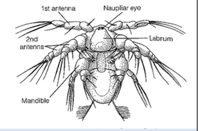
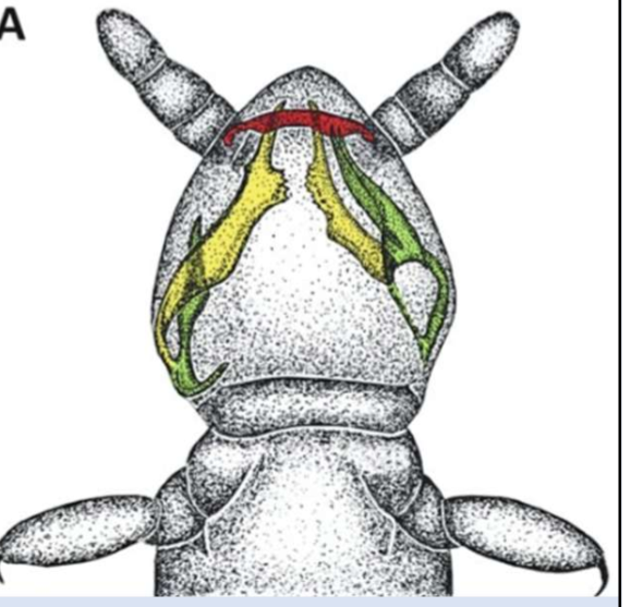
Barnacle larva
Called a nauplius.
appendages of the larva represent the first 3 segments of the head: 1st antennae, 2nd antennae, and mandibles → these appendages are used for swimming
Labrum
“Upper lip” found in nauplius larva; aids in holding food in the mouth (not an appendage)
Reproduction of barnacles
They are hermaphroditic; one extends a “groping penis” to fertilize the nearest individual
Brachiopod reproduction
Reproduce by parthenogenesis; have a resting egg stage
other freshwater and terrestrial crustaceans carry the young in brood chambers and release juveniles
Shrimp development
Indirect development, including “post-larval” stages.
shrimp cast their eggs on the seafloor
the nauplius molts to a zoea → molts to a post-larval mysis
mysis migrates into estuaries/shoreline habitats; these nurseries provide protection for the juvenile shrimp
Lobster development
lobsters carry their eggs through early development
post-larval stage = mysis; more fully-developed
the mysis has tons of setae for swimming through plankton
Crab development
the zoea molts into a post-larval stage, megalops, which has compound eyes and tiny claws
as segments and appendages are added, the abdomen folds under the body in a process called carcinization
Comparison between malacostracan body forms:
Shrimp:
cylindrical, flexible; build for swimming and crawling
~900 species
Lobster:
cylindrical; stout limbs are built for walking
~4,000 species
Crab:
carries the abdomen beneath the thorax → more efficient locomotion
~9,500 species
Features of carcinization in crabs
Carapace is wider than it is long
Sternites are fused into a single plate
Abdomen is bent beneath the thorax
What is the advantage of carcinization in crab evolution?
molds the body into a flattened and widened form that is able to move laterally, thereby avoiding front-facing predators
it also allows the animal to exploit habitats that provide hiding places from other predators
What prevents sharp food items from entering the crustacean mid-gut, which is not lined with cuticle?
The gastric mill
Which of the following appendages is found in a nauplius larva?
All of these are found in the nauplius
Which of the following is NOT a post-larval stage in crustaceans?
Nauplius
Mandibulata
almost entirely terrestrial
includes millipedes, centipedes, a group of wingless hexapods called Entognatha, and the insects
key features: mandibles, maxilla, 1 pair of antennae
What are the two clades of Mandibulata?
Myriapoda → “many-footed”
Hexapoda → “six-footed”
What are the two groups of Myriapoda?
Chilopoda → centipedes; 3,000 species
Diplopoda → millipedes; 11,000 species
What are the two groups of Hexapoda?
Entognatha → interior mouthparts; 10,000 species
Insecta → exterior mouthparts; >1 million species (largest species diversity of all arthropods)
Chilopod body structure
Divided into two tagma: head and trunk
in addition to the standard mandibulate mouthparts, they have a prehensorial claw on the first trunk segment and walking limbs on all remaining segments
maxillae cover the mandible, which lies beneath the first maxillae
second maxillae function as sensory palps
Prehensorial claws
attach to the first trunk segment of a chilopod and extend underneath the head (they are not mouthparts)
contain poison glands used to subdue their prey
How do chilopods grind up food items?
they have a gizzard → rigid “teeth” tear and chew food items before it travels to the midgut
additionally, the midgut produces a perforated peritrophic membrane to contain the food items and protect the gut lining from sharp objects and microbes
Chilopod excretion/osmoregulation
Uses a pair of Malpighian tubules in the hemocoel.
moves nitrogenous waste out of the hemocoel, where it can be processed and exit the body
How do chilopods breathe?
rows of spiracles along their back draw air into tubules called tracheae that deliver oxygen directly to tissues and organs
a dorsal blood vessel acts as a pump to circulate blood through the body
hemocyanin is present in solution to aid in transport of oxygen
Chilopod reproduction and development:
Male centipedes produce spermatophores → deposited on the soil; female passes over it and reproductive structures draw it up inside
centipedes exhibit direct development, and their young hatch with most of the segments and legs of the adult
there are some species that add segments and legs with additional molts
some centipedes brood the young after hatching
Diplopod body structure
mandibulate arthropods; 2 tagma: head and trunk (similar to chilopods)
trunk segments are fused to form diplosegments, each of which bears 2 pairs of legs
cuticle is typically well-sclerotized and forms a hard body covering
some species produce repellent fluids to deter predation
Gnathochilarium
The first pair of maxillae in diplopods are fused to form this structure; creates a “shelf” beneath the mandibles
millipedes lack the second pair of maxillae
Collum
A leg-less segment behind the head of a diplopod; acts as a blade as the millipede “bulldozes” through the detritus
Gut bacteria of millipedes
Millipedes are detritivores and herbivores → the gut is a lengthy tube, as is typical for animals that eat plant material
some have suggested that gut bacteria contributed to millipede nutrition by aiding in digestion of cellulose, but experimental studies show no differences in growth rates when bacteria are not present in the gut
this suggests that the bacteria may be providing other services, like de-toxification of secondary compounds
Millipede circulatory system
The entire circulatory system is a dorsal vessel.
ostia in this vessel draw in blood from the hemolymph and pump it forward into the head
millipedes also have hemocyanin to transport oxygen throughout the body
Movement of millipedes
Millipedes have short, stout legs that attach to the ventral surface of the body
Not all of the legs are in motion when a millipede moves → most remain in contact with the soil
This concentrates the energy of movement in power to move the millipede through the dense leaf litter on the forest floor
Movement style is appropriate for the detritivorous habit of millipedes

Movement of centipedes
Centipede legs are much longer than millipede legs; attached to the sides of the body
This arrangement suspends the body above the surface to make movement easier
When a centipede is running, only a few of the legs are in contact with the soil
As with annelids, a significant amount of energy is wasted in lateral movement when moving at high speeds
Entognatha
Non-insectan, soil-dwelling (edaphic) hexapods.
mouthparts are internal and confined within the head capsule
typically small and wingless
lack a waxy covering of the epicuticle, so they have to live in moist, underground environments
examples: collembola (“springtails”), diplura
Entognatha mouthparts
The mouthparts have limited mobility, as they are internal
structures: mandible, superlingua, maxilla, hypopharynx
cavity enclosed by oral folds
Entognathans include some carnivores, but detritivory is the dominant feeding strategy
Features of collembola
Furcula = abdominal appendage; used as a catapult to launch themselves away from predators
Collophore = a structure used to take up water
What are the two subclasses of Insecta?
Apterygota → wingless insects; ~4,500 species
Pterygota → winged insects; ~995,500 species
What are the two infraclasses of Pterygota?
Paleoptera → primitive, non-folding wings; ~7,500 species
Neoptera → folding wings; ~988,000 species
What are the three superorders of Neoptera?
Orthopterodea → no metamorphosis; ~33,000 species
Hemipterodea → partial metamorphosis; ~90,000 species
Holometabola → complete/complex metamorphosis; ~865,000 species
Apterygota
Wingless insects; include silverfish and bristletails
they are not part of Entognatha because they have external mouthparts
At what time period did insects start to evolve wings?
Around 350 million years ago
When did the earliest entognath evolve?
Around 474 million years ago
When did the earliest insect evolve?
Around 420-400 million years ago
Insect digestive system
Foregut and hindgut are lined with cuticle, while a peritrophic membrane is formed in the midgut
crop = food storage organ; leads into proventriculus
proventriculus = acts as a gizzard and regulates food entry into the midgut; contains spines to grind up food
What are “bio-reactors”?
Symbiotic microorganisms in the hindgut of termites → break down cellulose in a fermentation process
when they molt, they lose the symbionts and have to lick each other to re-obtain them
How do insects deal with wastes?
Malpighian tubules → extend into the hemocoel and are bathed by hemolymph
blind sacs; attach to the hindgut
remove solutes, but as the material passes into the hindgut, water is removed so only solid waste remains
converted into uric acid; passed out in feces
Respiratory system of insects
take in oxygen through spiracles → lead to tracheae → branch into finer tubules (tracheoles) to deliver gas to tissues
concentration of tracheae in the thorax is associated with the wing muscles
How do aquatic insect larvae breathe?
They utilize gills or “anal respiration” → takes up oxygen and passes it through a tracheal system
rings in the tracheae prevent collapse and the presence of fluid in the tracheoles regulates gas movement
tracheoles fill with fluid → blocks access to muscle → brings it closer to greater mitochondrial concentration

Insect circulation
Elongate dorsal vessel pumps blood forward into the head
additional pumps are associated with the wings
After it reaches the head, blood enters the hemocoel and flows to the rear, with side trips into the legs
septa within the leg segments allows blood to be delivered efficiently
Did insects co-evolve with flowering plants?
Probably, not as most trophic diversity in insects occurred before angiosperms even developed
mouthpart diversity pre-dates taxonomic diversity
the herbivorous group Paraneoptera displayed rapid diversification in late Paleozoic and early Mesozoic → made the greatest contribution to insect diversity during this time period
If not angiosperms, what caused the increase in insect diversity during the Cenozoic era?
Development of a constriction region (CR) between two sections of the midgut
this CR blocks passage into the next section for all materials except specific bacterial species
these bacteria take up residence in this section and their activity produces essential amino acids and nutrients
Elytra
In beetles, the forewings are modified to form this hardened covering to protect the hindwings
additionally, beetle larvae do not have external wing buds and are wormlike for burrowing/mining