chp. 7 the appendicular skeleton
1/100
There's no tags or description
Looks like no tags are added yet.
Name | Mastery | Learn | Test | Matching | Spaced |
|---|
No study sessions yet.
101 Terms
pectoral girdle
a skeletal structure that supports the anterior (front) limbs and lies posterior to the head.
It first developed in Devonian fishes (pre-historic) and has been modified in later vertebrates.
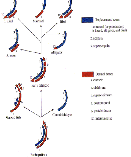
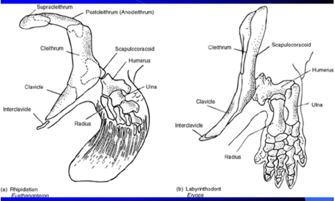
Primitive Pectoral Girdle (Devonian Fishes) elements
Composed of 7 paired bones:
Endochondral bones (endoskeleton):
Suprascapula (dorsal)
Scapula (middle)
Coracoid (ventral)
Dermal bones (ancestral dermal armor):
Postemporal (dorsal)
Supracleithrum
Cleithrum
Clavicle (ventral)
which were devonian fish endochondral pectoral girdle elements?
Suprascapula (dorsal)
Scapula (middle)
Coracoid (ventral)
which were devonian fish membranous bones pectoral girdle elements? (dermal bones)
Postemporal (dorsal)
Supracleithrum
Cleithrum
Clavicle (ventral)

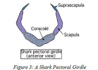
shark pectoral girdle
Cartilaginous fishes → cartilaginous pectoral girdle (no dermal bones)
Made of: suprascapula, scapula, coracoid.
Similar to the endochondral part of primitive fishes.
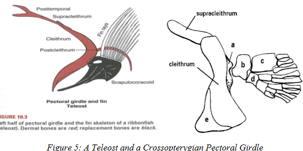
modern bony fish pectoral girdle (teleosts)
Cleithrum = major bone.
Clavicle and all other dermal bones are lost.
Coracoid fused with scapula → forms scapulocoracoid.
early tetrapods pectoral girdle
Pectoral girdle similar to primitive fishes but with key changes:
Lost: postemporal.
Gained: interclavicle (a singular midventral bone). MEMBRANE BONE
Early amphibians:
Dermal bones: Cleithrum, Clavicle, Interclavicle.
Endochondral bones: Scapula, Coracoid.
Later tetrapods that went on land: supracleithrum also lost.
interclavicle of amphibians
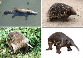
Present in amphibians, reptiles, birds, and monotreme mammals.
Examples:
Crocodilians → unpaired interclavicle with procoracoids.
Birds → interclavicle forms part of the furculum (“wishbone”).
Reptiles + monotremes → retain interclavicles.
fate of clavicle/coracoid
Clavicle + Coracoid roles are linked (both brace scapula against sternum, but usually one is reduced/lost). VENTRAL
Birds → strong clavicles (furculum), and retain coracoids.
Most reptiles → only procoracoids, no clavicles.
Coracoid formation (tetrapods)
Forms from the cartilaginous coracoid plate in lateral body wall.
Procoracoid from anterior centers, coracoid from posterior.
In eutherian mammals → both lost.
the coracoid bone has become reduced to a projection on the scapula
located over the Glenoid (the shoulder joint) called the Coracoid Process.

coracoid process
small hook-like projection on the scapula (shoulder blade) that sticks out near the shoulder joint.
It serves as an attachment point for muscles and ligaments of the arm and chest.
👉 In mammals, it’s all that remains of the original coracoid bone.
glenoid
shoulder joint, connects shoulder to humerus (in scapula)

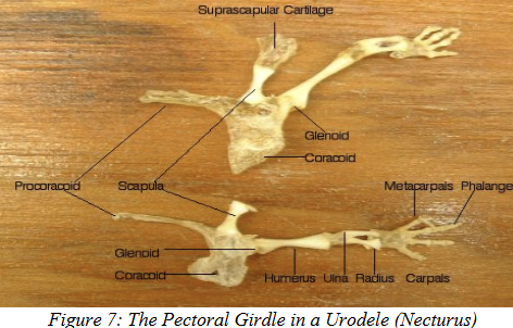
scapula in tetrapods
Present in all tetrapods with forelimbs.
Bears part/all of the glenoid.
Suprascapula usually fused to scapula (except in salamanders and frogs).
pectoral girdle in mammals
Derived from therapsid reptiles (mammal-like reptiles)
- Monotremes still have the same pectoral girdle as did the therapsids.
- the pectoral girdle has been reduced to a scapula (with an acromion and coracoid process) and (typically) a clavicle.

monotremes
mammals that lay eggs
eutherian mammals
every mammal except marsupials and monotremes. have a placenta for babies
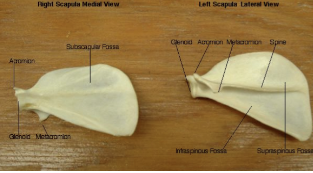
scapula in mammals
Lies posterolateral to ribs.
Underside: subscapular fossa (smooth to slide against ribs)
Dorsal side: spine dividing supraspinous & infraspinous fossae. (serves as origin for some shoulder muscles)
Projections near glenoid: acromion & coracoid process (for muscle/ligament attachment).
therapsid reptile pectoral girdle
"mammal-like reptiles," that lived during the Paleozoic eras and are significant because they are the direct ancestors of modern mammals.
Endochondral: scapula (with acromion), coracoid, procoracoid.
Dermal: clavicle, interclavicle.
Monotremes still have the same pectoral girdle as did the therapsids.
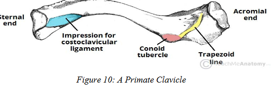
mammalian clavicle
Present/absent based on how a mammal uses its anterior limbs.
Large clavicles → moles (digging), bats (flying).
Reduced clavicle → cats (pectoral muscles form a muscular sling instead).
Lost clavicle → ungulates (horses, cows) & cetaceans (whales, dolphins).
pelvic girdle
supports hind limbs
Pelvic girdle in fish
is poorly developed.
It usually consists of a pair of Pelvic Plates (cartilaginous or bony) that meet at the Pelvic Symphysis, forming a base for the pelvic fins.
In cartilaginous fishes and lungfishes, the two pelvic plates fuse into one plate.
The position of the pelvic girdle varies among teleosts:
Some have it just behind and attached to the pectoral girdle.
Others have it near the tail base.
Fish and tetrapod pelvic girdles lack dermal bone!

pelvic girdle in tetrapod embryos
Cartilaginous pelvic plates develop.
Each plate has two ossification centers:
One forms the Pubis. (cranial)
One forms the Ischium. (caudal)
Another cartilage mass (blastema) forms dorsally, giving rise to the Ilium.
The ilium, ischium, and pubis grow together and meet at a joint cavity called the Acetabulum, which holds the head of the femur.
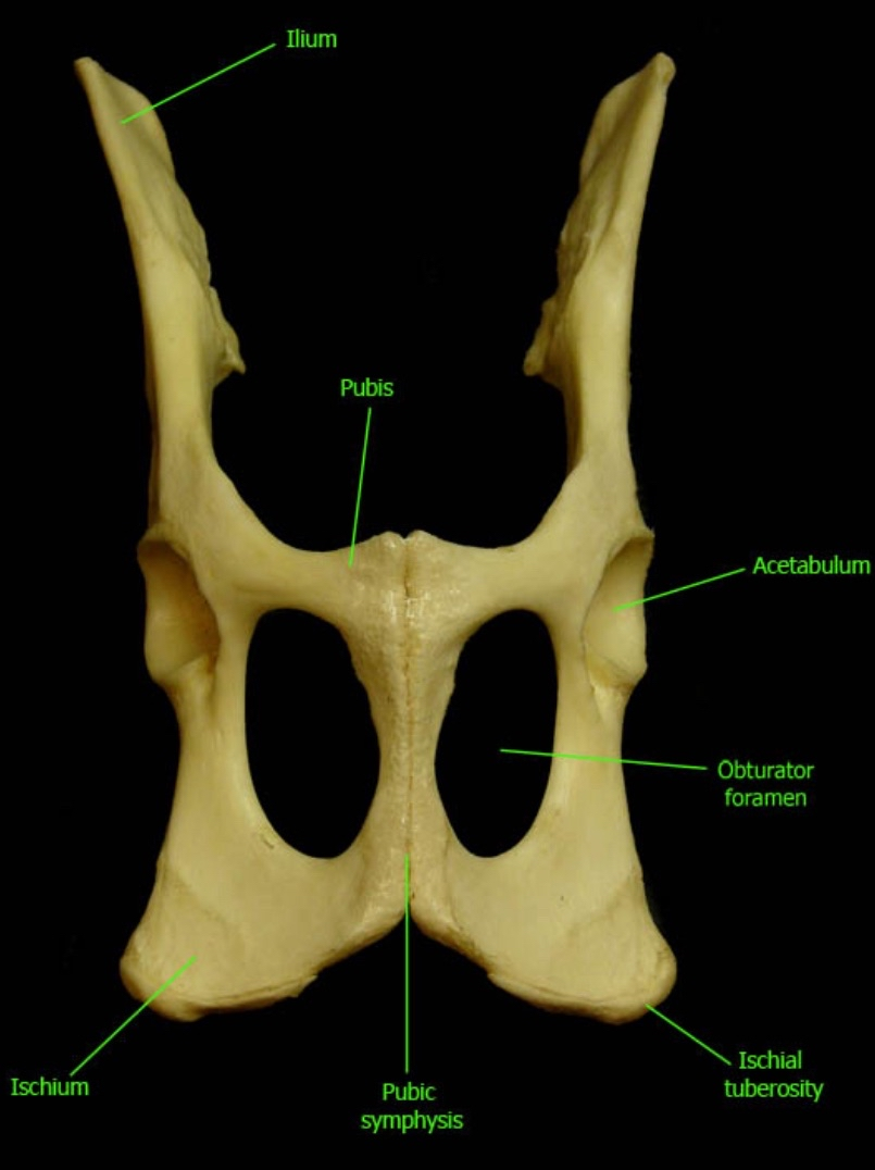
acetabulum
holds head of femur and articulates w pubis
joint cavity where illium, ischium, and pubis grow together
Ilium–sacrum connection
The ilium braces against the sacrum.
The sacrum has stout transverse processes (remember sacral vertrebra is fused):
1 pair in amphibians
2 pairs in reptiles
3+ pairs in birds and mammals
Some tetrapods also have Sacral Ribs, short ribs fused (ankylosed) to sacral transverse processes.
symphysis (in tetrapods)
created btwn 2 pubis bones or 2 ischium bones
Birds lack a symphysis (so they can lay eggs)
if the symphysis is between the two pubic bones it is the Pubic Symphysis. If the
symphysis is between the two ischia it is the Ischial Symphysis. (ex: ischipoubic bar in sharks)

force distribution in pelvic girdle
From the acetabulum, forces are directed:
Dorsally against the sacrum/vertebral column.
Ventrally against the symphysis.
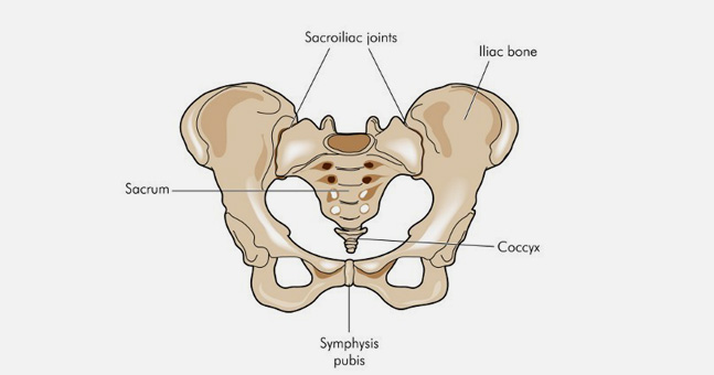
Pelvis (amniotes)
The sacrum and pelvic girdle join to form a bony enclosure called the Pelvis.
Pelvic bones are usually fused, but this depends on locomotion.
Humans: rigid sacroiliac joint. (synarthrotic)
Frogs (anurans): flexible (diarthritic) sacroiliac joint for hopping.
The pelvis surrounds the caudal end of the body cavity, forming the Pelvic Cavity, which contains urogenital organs and the end of the digestive tract.
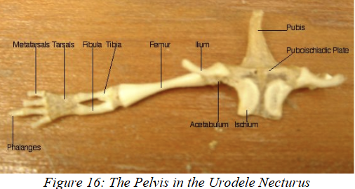
pelvic girdle of urodeles (salamanders)
Weak pelvic girdles, resembling fish.
Braced against one sacral vertebra by a weak ilium.
Terrestrial species develop a slender cartilage extending from the pelvic girdle into abdominal muscles, called Prepubic (Ypsiloid) Cartilage.
pelvic girdle of reptiles
Some have Epipubic and Hypoischial Bones.
These also appear in monotremes and marsupials.
Marsupials use the Epipubic Bone to support the pouch.
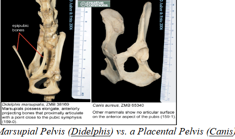
epipubic bones
slender bones that extend cranially from the pubic bones. They are found in monotremes, reptiles, and marsupial mammals.
Marsupials have an epipubic bone to support the pouch.

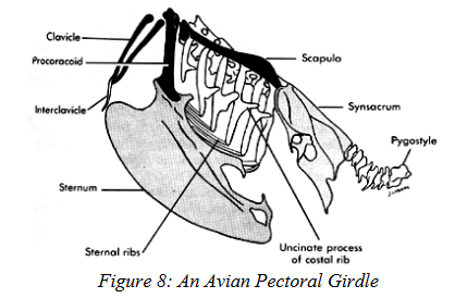
pelvic girdle of birds
Expanded ilium and ischium fuse into the synsacrum.
Pubic bones (CAUDAL) are reduced to long splinters running parallel to the ischium.
Birds lack a pubic symphysis, allowing space for laying large eggs.
Ornithischian dinosaurs independently evolved a similar pelvic structure.
However, birds evolved from Saurischian (“lizard hipped”) dinosaurs so this is an example of convergence.
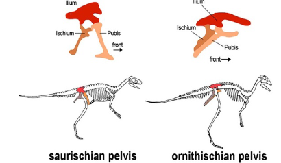
os coxa
Ilium, ischium, and pubis fuse completely into a single bone called the Os Coxa or Innominate.

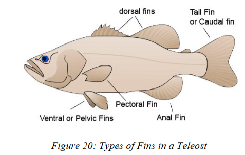
fins
Fins help fish:
prevent rolling,
control swimming angle,
steer,
brake/slow down,
and (in the caudal fin) provide thrust.
Fins can be paired (pectoral, pelvic) or singular (dorsal, anal, caudal).
Structure: two layers of skin supported by flexible rays radiating from a skeletal base.
fin rays
Found in the dermis.
2 types:
Bony fishes: rays = Lepidotrichia (jointed bony scales in series).
Distal end may have extra supports called Actinotrichia.
Cartilaginous fishes: rays = Ceratotrichia (long, hollow rays, like shark spines).
May also contain actinotrichia.
In elasmobranchs, scales grow into fins for stiffness.
lepidotrichia
type of fin ray found in bony fishes
-consists of jointed bony scales from end to end
May have actinotrichia to further reinforce fin
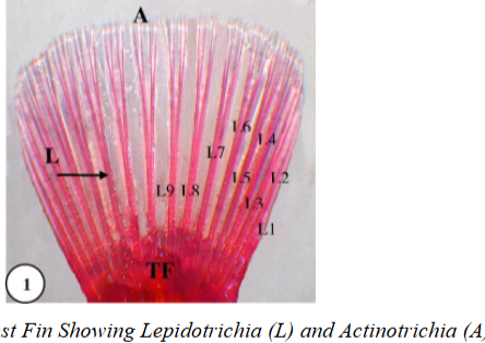
actinotrichia
part of fin rays: can be in lepidotrichia or ceratotrichia fin rays to strengthen fin
made of actinodins

ceratotrichia
fin rays in cartilagenous fishes (chondricythes)
They are long hollow rays similar to the dorsal spines found in some shark species
may contain actinotrichia in distal portion to support fin
In elasmobranchs, scales grow into fins for stiffness.
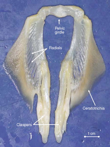
skeletal base of fins
Made of Basalia (cartilaginous/bony) and distal Radialia.
Together called Pterygiophores.
In some bony fishes, paired fins lack basalia and have reduced radialia.
Median and dorsal fin bases attach to the vertebral column.
skeletal base for paired or unpaired fins can be composed of either cartilage or bone
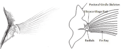
basalia
the pterygiophores that attach the fin to the body
in skeletal base of fin
pterygiophores
one of the cartilaginous or bony elements (as basalia and radialia) by which rays of the fin of a fish are supported
in skeletal base of fin

radialia
are distal to the basalia and attach to both the basalia and the fin rays
in skeletal base of fin

paired fins
Attachment:
Pectoral fins → articulate with the glenoid of the pectoral girdle.
Pelvic fins → braced against the pelvic plate.
Types of paired fins (in living fishes):
Lobed fins (Sarcopterygians):
Fleshy base with skeleton + muscles, plus distal rays.
Fin rays form a paddle.
Fin folds (Chondrichthyes):
Broad base with three basalia: Propterygium, Mesopterygium, Metapterygium.
In males, basalia form Claspers for reproduction.
Ray fins (Actinopterygians):
Teleosts have flexible ray fins with reduced basal skeleton.
Extinct (4th) type: Spiny fins (Acanthodians).
lobed fins
a type of paired fin found in Sarcopterygian (lobe-finned fish)
consists of a fleshy proximal lobe containing the fin skeleton and associated muscles and a membranous distal portion stiffened by fin rays.
The fin rays have a narrow base and form a paddle-like shape.
The lobe fin gave rise to the tetrapod limb.
fin folds
a type of paired fin found in Chondrichthyes (sharks)
Broad base
3 basalia in pectoral fin: Propterygium, Mesopterygium, Metapterygium.
2 basalia in the pelvic fin: propterygium and metapterygium
In males, basalia form Claspers for reproduction.
ray fins
a type of paired fin found in actinopterygians (bony fish)
in some teleosts the ray fins have lost skeletal elements to give more mobility.
lost components of basal skeleton and show great flexibility
median fins
Types: Dorsal fins (1 or more) and Anal fins (if present).
Structure similar to paired fins: proximal basalia, distal radialia, and rays.
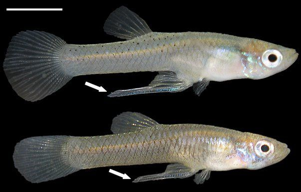
In some male teleosts, the anal fin is modified into a Gonopodium (intromittent organ, e.g., poecilids).
gonopodium
(from median fins) in male teleosts, the anal fin is modified into this
anal fin in male live-bearing fish, such as guppies, mollies, and platies, used for internal fertilization by delivering sperm into the female's genital opening
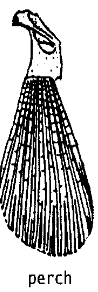
caudal fins
classified based on shape and the relationship between the tail, notochord, and vertebral column.
3 types:
- Heterocercal
- Diphycercal
- Homocercal
- Hypocercal (extinct 4th type)

Heterocercal tail
type of caudal fin
Vertebral column/notochord extends into dorsal lobe.
Dorsal lobe is GREATER than ventral lobe (ex: sharks)

Diphycercal tail
type of caudal fin
Vertebral column barely enters dorsal lobe.
Evolved from heterocercal tails
Dorsal and ventral lobes EQUAL in size. (Ex: lungfish, coelacanths)

homocercal tail
type of caudal fin
Notochord extends deeply into dorsal lobe, enclosed in caudal vertebrae forming a Urostyle.
Also evolved from heterocercal tails.
Dorsal and ventral lobes EQUAL in size! (most teleosts, mackerel sharks)
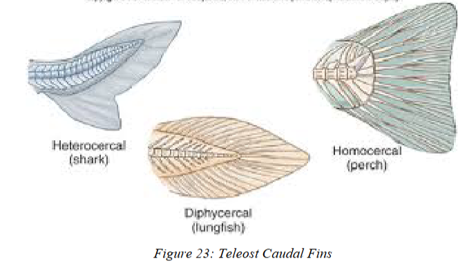
hypocercal tail
type of caudal fin (EXTINCT) Ex: Ichthyosaurs.
Vertebral column/notochord extends into ventral lobe.
Dorsal lobe is SMALLER than ventral lobe (reverse of heterocercal tail)
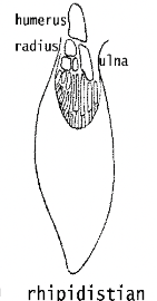
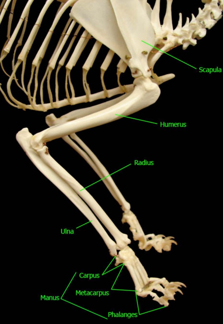
tetrapods limbs
A tetrapod limb has 3 main parts:
Propodium = upper arm (forelimb) or thigh (hindlimb).
Epipodium = lower arm (forelimb) or leg (hindlimb).
Autopodium = manus (hand) or pes (foot).
Evolutionary trend: increased mobility through more joints.
Propodium + epipodium are relatively consistent across tetrapods in skeletal lvl.
propodium
upper arm (forelimb) or thigh (hindlimb).
humerus or femur
epipodium
lower arm (forelimb) or leg (hindlimb).
radius/ulna or tibia/fibula
autopodium
manus (hand) or pes (foot).
forelimbs
Propodium = Humerus (similar across tetrapods and early fishes).
Epipodium =
Radius (preaxial, thumb side MEDIAL)
Ulna (postaxial, pinky side LATERAL).
radius and ulna r antiparallel
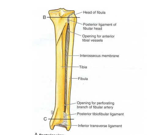
hindlimbs
Propodium = Femur.
Epipodium =
Tibia (preaxial, MEDIAL) weight-bearing
Fibula (postaxial, LATERAL).
Variations:Tibia + fibula may fuse = Tibiofibula (horses, frogs).
Fibula reduced (birds).
Fibula lost (deer).
Tibia + tarsus fused = Tibiotarsus (birds).
Additional bone: Patella (sesamoid, found in reptiles and mammals).
distal aspect of limbs are most variable among tetrapods.
tibiofibula
fibula unites partially/completely to tibia (ex: horses, frogs)
tibiotarsus
tibia will fuse to tarses (in birds) why birds can’t drive
fibula is v reduced in size in birds
manus (hand)
manus = autopodium
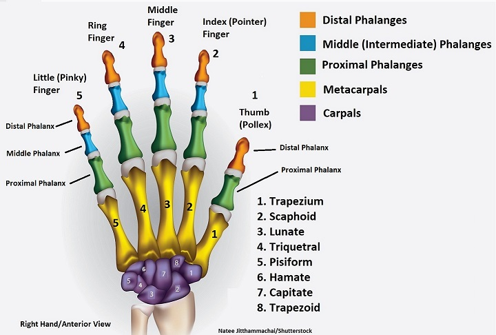
3 Components: Carpus (wrist), Metacarpus (palm), Phalanges (digits).
Carpus (wrist):
Proximal row: Radiale (articulates with radius), Intermedium, Ulnare (articulates with ulna).
Extra bone: Pisiform (sesamoid; articulates with ulnare).
Human equivalents: Scaphoid (radiale), Lunate (intermedium), Triquetral (ulnare), Pisiform.
Middle row: 0–4 Centralia (early tetrapods had 3–4; reduced in reptiles; fused in humans).
Distal row: 5 carpals (digits 1–5); humans have 4: Trapezium, Trapezoid, Capitate, Hamate (4+5 fused).
Metacarpus: 5 metacarpals in generalized hand.
Phalanges: 2–3 phalanx bones per digit. Humans = 2-3-3-3-3 (pollex = 2).

pollex
2 phalanx bones (distal + proximal) instead of 3 in phalanges (thumb)
metacarpus
palm
Metacarpals = long bones between carpus and phalanges.
General tetrapod: 5 metacarpals, numbered 1–5 (thumb → pinky).
Function: support digits, provide leverage for grasping or locomotion.

phalanges
Each digit = a Phalange(fingers), made of smaller bones called Phalanx bones.
General tetrapod: usually 2–3 phalanges per digit. (pollex has 2)
Human phalangeal formula: 2-3-3-3-3 (thumb has 2 bones, fingers 2–5 have 3 each).
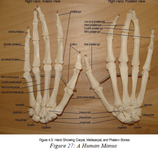
carpus
carpus = cluster of small bones forming the wrist joint.
In the generalized pentadactyl hand, there are three rows of carpal bones:
Proximal row (articulates with radius & ulna):
Radiale → bone that articulates with the radius.
Intermedium → middle proximal bone.
Ulnare → bone that articulates with the ulna.
Pisiform → sesamoid bone that articulates with the ulnare.
Middle row:
Contains Centralia (0–4 bones).
Early tetrapods: 3–4 centralia.
Reptiles: reduced to 2.
Humans: centralia fuses with the scaphoid during development.
Distal row (articulates with metacarpals):
Typically 5 carpal bones, one for each digit (numbered 1–5).
Humans have 4 bones here:
Trapezium (digit 1/thumb),
Trapezoid (digit 2),
Capitate (digit 3),
Hamate (digits 4 & 5 fused).

proximal row of carpus
(articulates with radius & ulna):
Radiale → bone that articulates with the radius.
Human equivalent = Scaphoid.
Intermedium → middle proximal bone.
Human equivalent = Lunate.
Ulnare → bone that articulates with the ulna.
Human equivalent = Triquetral.
Pisiform → sesamoid bone that articulates with the ulnare.
Present in many reptiles & mammals.
In humans, also called Pisiform.

medial row of carpus
Contains Centralia (0–4 bones).
Early tetrapods: 3–4 centralia.
Reptiles: reduced to 2.
Humans: centralia fuses with the scaphoid during development.

distal row of carpus
(articulates with metacarpals):
Typically 5 carpal bones, one for each digit (numbered 1–5).
Humans have 4 bones here:
Trapezium (digit 1) thumb
Trapezoid (digit 2) index finger
Capitate (digit 3) middle finger
Hamate (digits 4 & 5 fused) ring and pinky finger

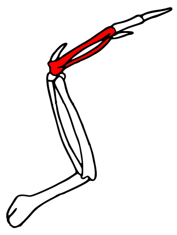
manus modifications for birds
has aerodynamic effect
Carpals: 2 proximal + 3 fused distal = Carpometacarpus.
Digits reduced to 3, most clawless.
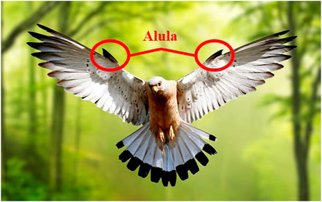
Special digit: Alula: the 1st digit (pollex), mobile and feather-bearing. Functions like a "thumb flap" to improve lift and prevent stalling at low speeds.
manus plays in a role in braking, hovering, and steering.
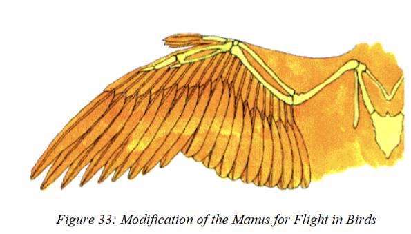
carpometacarpus
The 3 distal carpal bones will fuse with the metacarpals to form the Carpometacarpus in birds.
(rigid unit that strengthens the wing).
alula
the first finger of a bird that became elongated, slender, and independently mobile
allows for more precise flight and aids to prevent stalling in air.
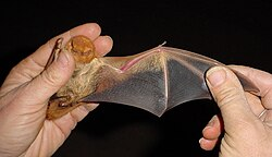
manus modifications for bats (mammals)
Digits: Retain 5 digits. Digits 2-5 are greatly elongated, forming the skeletal supports of the patagium (wing membrane).
1st digit (thumb): Free, clawed, used for climbing and grasping.
Carpals: Several fused into a compound wrist unit for wing strength
manus main component of wing
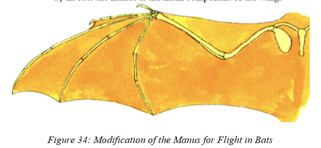
patagium
wing membrane that assists an animal in obtaining lift when gliding or flying.
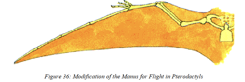
manus modifications for Pterosaurs
Digits: Only 4 digits and 4 metacarpals in manus
Digits 1-3 = small, clawed (used for climbing/feeding).
Digit 4 = extremely elongated → supports the main wing membrane (patagium).
Gave them a unique wing structure, different from birds and bats.
like bats, manus is main component of wing
It is composed of a very elongated phalange and metacarpal embedded in a patagium
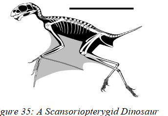
manus modifications for flight in scansoripterygid dinosaurs
Digits 3–5 elongated, especially digit 5, supporting a membranous wing surface (patagium)
more of bat-like wing
Modifications of the Terrestrial Manus for Aquatic Life
Flippers evolved in seals, whales, sirenians, penguins, ichthyosaurs, plesiosaurs.
flippers often flattened + broad.
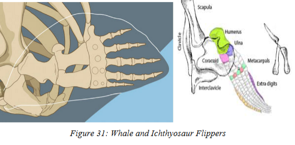
Some species = extreme increase in phalanges (e.g., ichthyosaurs up to 26 per digit).
manus modifications for speed on land
Plantigrade: whole palm/sole on ground. PRIMITIVE (ex: humans, bears).
Digitigrade: walk on digits 2–5, carpus/tarsus is raised off ground, first digit reduced (e.g., cats, dogs).
Unguligrade: walk on tips of toes, carpus/tarsus kept off ground → claws turn into hooves, digits/metacarpals reduced/fused.
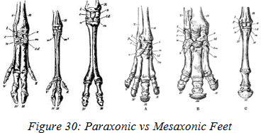
Perissodactyls (odd-toed ungulates): 1 or 3 hooves; mesaxonic (weight on middle digit).
Artiodactyls (even-toed ungulates): 2 or 4 hooves; paraxonic (weight shared equally on digits 3 & 4).
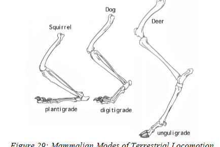
plantigrade
whole palm/sole on ground.(ex: humans, bears)
considered to be primitive stance

digitigrade
walk on digits 2–5, carpus/tarsus is raised off ground
first digit reduced and doesn’t play a role in walking (e.g., cats, dogs).

unguligrade
walk on tips of toes, carpus/tarsus kept off ground
claws turn into hooves
digits/metacarpals reduced/fused.
2 types:
Perissodactyls (odd-toed ungulates)
Artiodactyls (even-toed ungulates)

Perissodactyls
ODD TOED
1 or 3 hooves bears all the weight
mesaxonic (weight on middle digit).
if they have only one walking hoof, it will be 3rd
if they have 3 walking hooves, it’ll be 2,3, and 4
Mesaxonic foot- forces are directed along the middle of the foot and are primarily handled by one digit, the 3rd digit

Artiodactyls
EVEN TOED
2 or 4 hooves for walking
if it has 2 hooves, digits 3 and 4 used
if it has 4 hooves, the digits 2-5 used but 3 and 4 carry bulk of weight
paraxonic- weight shared equally on digits 3 & 4

paraxonic foot
forces are directed on the middle of the foot but shared by 2 digits, not primarily one as is seen in the mesaxonic foot.
These two digits are numbers 3 and 4.

mesaxonic foot
forces are directed along the middle of the foot and are primarily handled by one digit, the 3rd digit.
manus modifications for grasping
Some mammals flex metacarpal–phalanx joints to grasp (e.g., rodents).
True grasping (curling all digits): raccoons, primates.
Opposable thumb:
Opposition = touching tip of each digit with thumb.
Requires saddle joint ( btwn 1st metacarpal + proximal phalanx) + strong adductor pollicis.
Found in humans, apes, monkeys.
pes
The pes (hind foot) is similar to the manus (forefoot), especially in primitive forms.
Key difference: the pes lacks the pisiform bone.
Structure:
The ankle = Tarsus, arranged in three rows.
a) Proximal Row (3 bones):
Tibiale (articulates with tibia)
Fibulare (articulates with fibula)
Intermedium – present in some amphibians, lost in most vertebrates
b) Middle Row:
Bones called Centralia
c) Distal Row:
5 Distal Tarsals, numbered from the big toe (1–5)
Metatarsus (Foot Body)
Primitive form: 5 metatarsals (numbered 1–5, medial → lateral).
.
Digits (Phalanges)
Similar to manus phalanges.

tarsus
a) Proximal Row (3 bones):
Tibiale (articulates with tibia)
Also called Astragalus or Talus
Equivalent to Radiale
Fibulare (articulates with fibula)
Also called Calcaneus
Equivalent to Ulnare
Intermedium – present in some amphibians, lost in most vertebrates
In some reptiles, tibiale + fibulare fuse → Astragalocalcaneus
b) Middle Row:
Bones called Centralia
Originally 4, but reduced in most vertebrates
Sometimes completely lost (e.g., humans have only 1 = Navicular/Scaphoid)
c) Distal Row:
5 Distal Tarsals, numbered from the big toe (1–5)
Some fuse or are lost in evolution
In humans:
1st = Medial Cuneiform
2nd = Intermediate Cuneiform
3rd = Lateral Cuneiform
4th + 5th = Cuboid

proximal row of tarsus
3 bones:
Tibiale (articulates with tibia)
Also called Astragalus or Talus
Equivalent to Radiale
Fibulare (articulates with fibula)
Also called Calcaneus
Equivalent to Ulnare
Intermedium – present in some amphibians, lost in most vertebrates
In some reptiles, tibiale + fibulare fuse → Astragalocalcaneus

medial row of tarsus
Bones called Centralia
Originally 4, but reduced in most vertebrates
Sometimes completely lost (e.g., humans have only 1 = Navicular/Scaphoid)

distal row of tarsus
5 Distal Tarsals, numbered from the big toe (1–5)
Some fuse or are lost in evolution
In humans:
1st = Medial Cuneiform
2nd = Intermediate Cuneiform
3rd = Lateral Cuneiform
4th + 5th = Cuboid

metatarsus
body of foot
Very similar to the metacarpus.
Primitive form: 5 metatarsals (numbered 1–5, medial → lateral).
Support the digits (phalanges)
Reduction of digits = reduction of metatarsals.

Astragalocalcaneus
in some reptiles, the tibiale and fibulare fuse to form this in the proximal ankle.

phalanges of pes
Similar to manus phalanges.
Primitive condition = 5 digits with 2+ phalanx bones each.
Example (humans): 5 digits with formula 2-3-3-3-3.
Big toe = Hallux, only 2 phalanges.

hallux
big toe on tarsus with only 2 phalanges
modifications of pes in amphibians
Labyrinthodonts (extinct) & anurans (frogs) → Prehallux, a vestigial (remnant) tarsal/metatarsal from a lost digit.
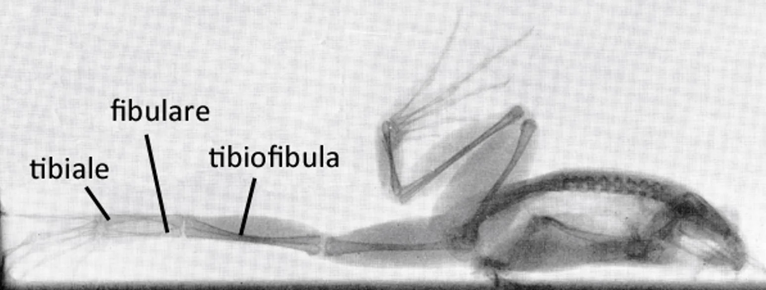
Anurans: elongated tibiale + fibulare (look like tibia + fibula).
These attach to a fused tibiofibula → increases hopping ability.
prehallux
the remains of a metatarsal or tarsal bone (6th toe?) in anurans and labyrinthodonts.
pes modifications in reptiles
Some (ex: sphenodon (iguana)) → tibiale + fibulare fuse into Astragalocalcaneus, intermedium lost.
pes modifications for birds
Proximal tarsals fuse with tibia → Tibiotarsus
Centralia all lost
Distal tarsals fuse with proximal metatarsals → Tarsometatarsus
Metatarsals also fused
Joints:
Between tibiotarsus & tarsometatarsus = Intratarsal Joint
Between tarsometatarsus & toes
Toes: usually 4 (numbered 1–4)
Typical: 3 forward, 1 backward (posterior)
Some birds (parrots, woodpeckers): 2 forward, 2 backward
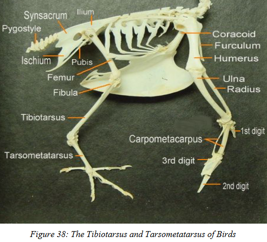
tarsometatarsus
fused distal tarsals and metatarsals in birds

intertarsal joint
joint btwn the tibiotarsus and tarsometatarsus in birds
