IB BIO - U1 L3 P2 - MICROSCOPES AND MICROSCOPE SKILLS (SCIENTIFIC DRAWINGS).
1/21
There's no tags or description
Looks like no tags are added yet.
Name | Mastery | Learn | Test | Matching | Spaced | Call with Kai |
|---|
No analytics yet
Send a link to your students to track their progress
22 Terms
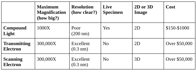
Microscope Types
Ocular lens
Lens in eye piece 10x

Arm
Supports tube and connects it to base

Coarse Adjustment Knob
Moves stage up/down to focus specimen

Fine Adjustment Knob
Precisely focuses specimen

Nose Piece
Holds objective lenses - rotate to change magnification

Stage
Platform where slide is placed

Light Source
Steady light source which illuminates specimen

Base
Supports microscope

Objective Lens
Magnifies specimen - 4X, 10X, 40X and 100X Multiply by 10X (ocular lens) to get total magnification

Diaphragm
Rotating disk under stage - varies amount of light
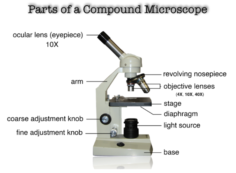
Stage Clips
Hold slide in place

How to Use Microscope
Uncover, plug in and turn on light source
Begin with lowest power objective lens - 4X or 10X
Place slide on stage - use stage clips to hold in place
Look through eye piece and focus specimen by turning course adjustment knob slowly
If too dark/bright, adjust diaphragm
Move to medium power objective. DON’T touch coarse adjustment knob anymore!
Sharply focus specimen using fine adjustment knob
Record observations - scientific drawing
Packing Up Microscope
Turn objective lens back to low power (4X or 10X)
Move stage all the way down and remove slide
Wrap cord around base
Put on cover
SCIENTIFIC DRAWINGS
Use a sharp pencil
Drawing should be neat and simple - don’t include unnecessary detail (like shading/colour)
Make large drawings (min 1/4 page)
Include descriptive title at top of page - underline it
Use a ruler to draw labelling lines - parallel, column, right of F.O.V
Include total magnification and scale line (1 cm) at bottom right
Draw only what you see. Not what you THINK you see!!
Determining SIZE of Imaging Using a Scale Bar
Estimate number of scale bars which fit into image, and estimate length
Measure length of diagram in millimeters (not cm - because it's easier to divide a larger number and avoid decimals)
Measure length of scale bar in millimeters
Divide length of diagram by length of scale bar and multiply by size written on scale bar
Determining MAGNIFICATION Using A Scale Bar
Measure length of scale bar in mm
Convert this to same units as written on scale bar - μm
Divide actual diagram size by measurement written on bar
Determining MAGNIFICATION If Given Actual Size
Measure image size with a ruler
Convert actual size and image size to same units
Divide image size by actual size of specimen
Magnification of a micrograph
magnification = image size/specimen size
1mm=1000μm (micrometres)
Animal Cells Size.
10 to 30 micrometres (µm) in diameter
Plant Cells Size.
10 to 100 micrometres (µm) in diameter
Plant Cells Size.
0.5 to 5 micrometres (µm) in diameter.