Looks like no one added any tags here yet for you.
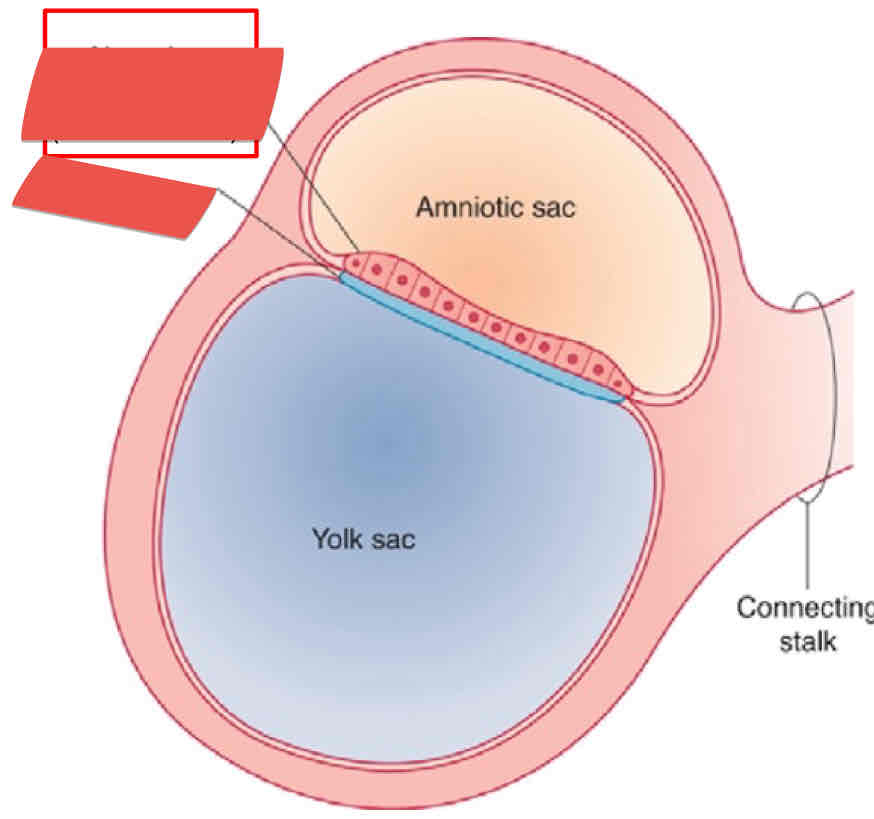
What is the red structure in the amniotic sac? Is it apart of the endoderm or ectoderm?
Neural Plate, ectoderm
What are the 3 layers of the trilaminar germ disc?
Ectoderm, mesoderm, endoderm
The notochord is located in what trilaminar germ disc?
Mesoderm
T/F The yolk sac is located in the Ectoderm
False (endoderm)
What trilaminar layer is the amniotic cavity located?
Ectoderm
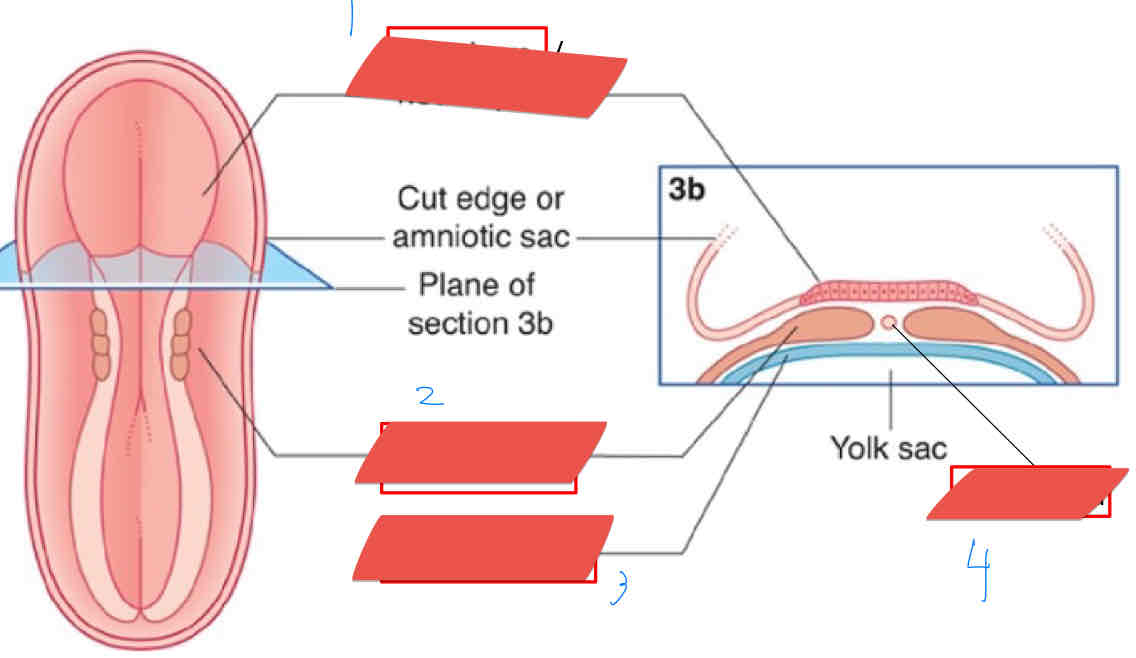
What structure is being shown at number 4?
Notochord
What structure is being show a number 1?
Ectoderm/neural plate
Number 3 is showing which structure?
Endoderm
What structure is shown in number 2?
Mesoderm
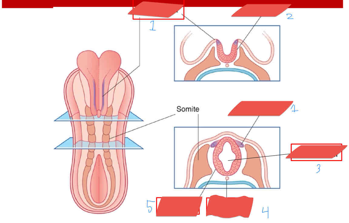
What is the structure being shown in number 1?
Neural crest
What structure is being shown in number 3?
Neural canal
What structure is being shown in number 5?
Neural tube
What structure is being shown in number 4?
Notochord
What structure is being shown in number 2?
Neural fold
T/F Schwann cells are myelin cells for the PNS
True
T/F the neural tube has neurons, supporting cells, lower motor neurons, and preganglionic autonomic neurons located in the PNS.
False (slide 7)
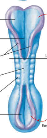
The cranial part of the embryo is _______ to the fused neural folds. What happens when the cranial part of embryo doesn’t close?
Rostral, the baby is stillborn
In the image, the end of the spinal cord is ______ to the fused neural folds. What happens if that region doesn’t close?
Caudal, spinal bifida occurs
What is the ventral tube called in a developing embryo? Is it sensory or motor?
Basal plate, motor
What is the dorsal tube called in the embryo? Is it sensory or motor?
Alar plate, sensory
T/F When you add the dorsal and ventral nerve roots, they create the spinal cord of the embryo
False (they create the spinal nerves, slide 10)
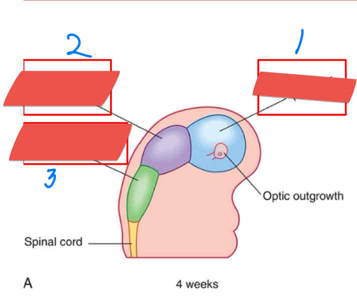
Identify the structures Labeled 1-3.
Prosencephalon
Mesencephalon
Rhombencephalon
This primary vesicle gives rise to secondary vesicles telencephalon and diencephlon
Prosencephalon
T/F the primary vesicle mesencephalon eventually gives rise to the metencephalon and then the midbrain
False (still mesencephalon)
What adult derivatives comes from the Prosencephalon?
Cerebral cortex, corpus striatum, thalamus, hypothalamus
What adult derivatives come from he mesencephalon?
Midbrain
What adult derivatives com from the Rhombencephalon?
Cerebellum, pons, medulla
The cerebral cortex and corpus striatum arise from what secondary vesicle?
Telencephalon
The cerebellum and pons arise from what secondary vesicle?
Mesencephalon
The myelencephalon gives rise to what adult derivative?
Medulla
The Rhombencephalon gives rise to what secondary vesicles?
Metencephalon and myelencephalon
The Prosencephalon gives rise to what secondary vesicles?
Telencephalon and diencephalon
This structure contains the lateral ventricles
Telencephalon (hemispheres)
Which structure contains the third ventricle in the ventricular system?
Diencephalon
What structure contains the cerebral aqueduct in the ventricular system?
Mesencephalon (midbrain)
At what point during fetal development does the frontal, parietal, temporal, and occipital lobes are identifiable?
week 14
At what point during fetal development do we see the lateral, central, and calcarine sulcus?
Week 28
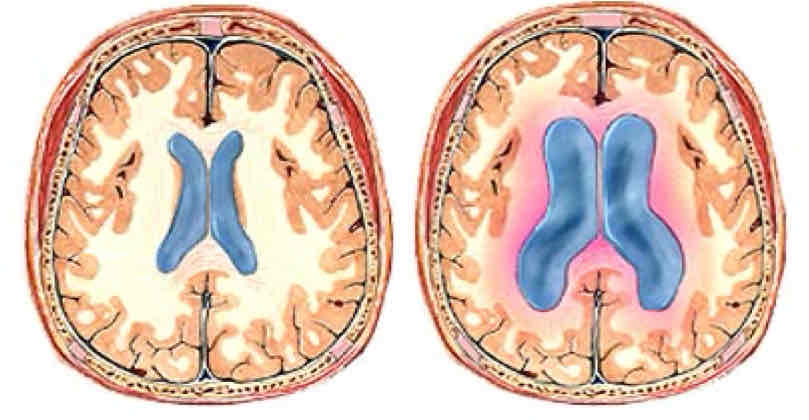
This condition is characterized by excess CSF in the ventricular system due to an imbalance between production and absorption of CSF.
Hydrocephalus
What are some causes for hydrocephalus
Arnold-Chiari malformation, congenital aqueductal stenosis, meningitis
This malformation describes the foramina being obstructed from the 4th ventricle to subarachnoid space. This is one of the causes for what condition?
Arnold-chairing malformation, hydrocephalus
This condition is characterized by inflammation of the meninges
Meningitis
Which brain is considered hydrocephalic? How can you tell?
Right brain, excess fluid in the lateral ventricles
This condition causes atrophy of cerebral cortex and white matter, thinning of bones in calvaria, prominent forehead, and compression of basal ganglia and diencephalon.
Hydrocephalus
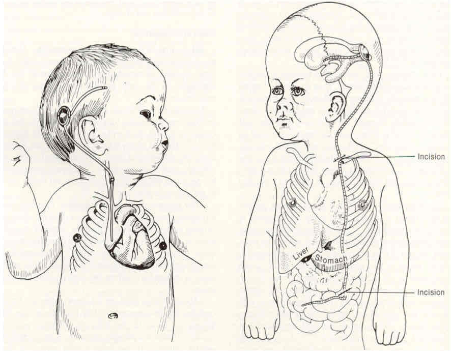
How would we treat hydrocephalus?
Early prevention, pressure sensitive catheter/shunt into internal jugular vein
What condition is being treated in the image below?
Hydrocephalus
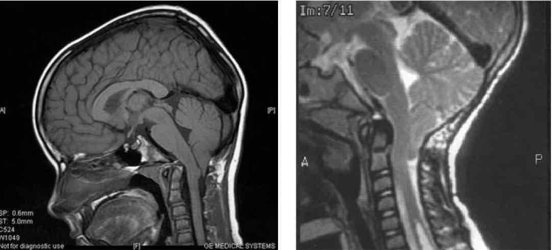
This condition is the most common congenital anomaly involving the lower brain stem and the cerebrum
Arnold-Chiari malformation
What is another name for Arnold-Chiari malformation
Chiari malformation
This condition is characterized by a structural defect of the cerebellum and herniation of the medulla causing part of cerebellum to go through foramen magnum.
Arnold-Chiari malformation
T/F Arnold-chiari malformation may cause hydrocephalus.
True
How often does Arnold-Chiari malformation occur and what conditions are associated with it?
Occurs 1/1000 births, spina bifida and lumbar meningomyelocele
What are some signs and symptoms of Arnold-Chiari Malformation?
Neck pain, balance problems, muscle weakness
T/F Hand coordination and fine motor skills may be affected with Arnold-Chiari malformation
True
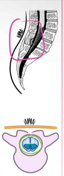
What is happening in this image? What condition is causing this?
Cerebellum is plugging the foramen magnum, Arnold-Chiari malformation
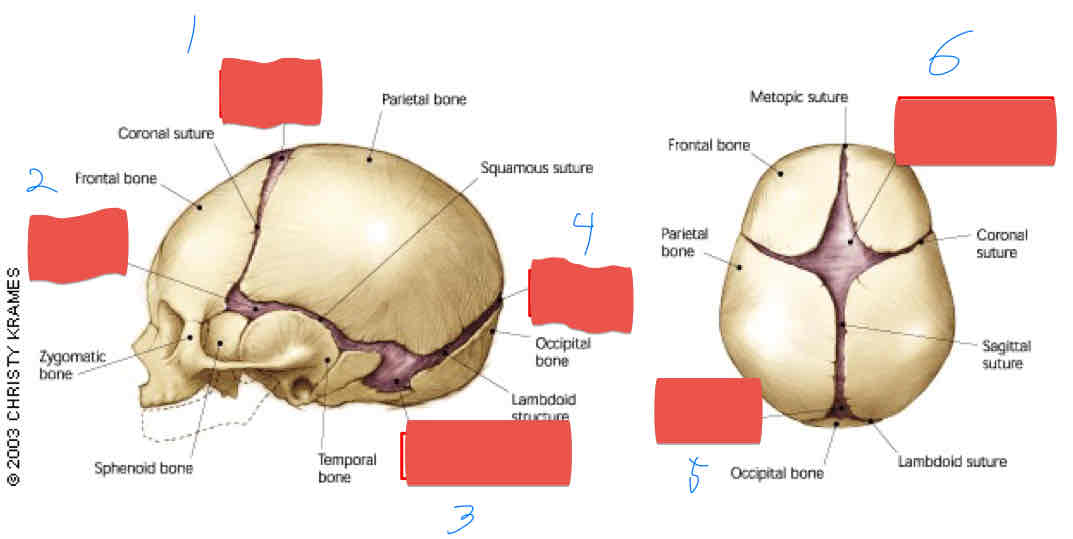
What structure is shown in number 1?
Anterior fontaneal
What structure is shown in number 2?
Sphenoid fontaneal
What structure is shown in image 3?
Mastoid fontanel
What structure is shown in number 4?
Posterior fonatnel
What structure is shown in number 5?
Posterior fontanel
What structure is shown in number 6?
Anterior fontanel
What is the cause of microencephaly
Premature closing of fontanelles and sutures
T/F Even though the sutures and fontanelles close prematurely in a patient with microcephaly, the brain is fully developed. (Slide 33)
False
What are some side effects of microcephaly?
Gross cognitive impairment, poor motor function, seizures, dwarfism
This neural tube defect is caused by a failure to close the entire neural tube.
Craniorachischisis
What is another name for craniorachischisis?
Total dysraphism
This defect is caused by a failure of the cranial neural tube to close during development?
Cranioschisis
Neural defect where the neural tube doesn’t fully close and leaves the entire lower spinal cord open
Rachischisis
This neural defect is caused by a failure of closure of the anterior neuropore causing Acrania
Anencephaly
T/F Anencephaly is a condition that happens in the late 2nd trimester, gives the fetus a “frog-like” appearance. (Slide 35)
False
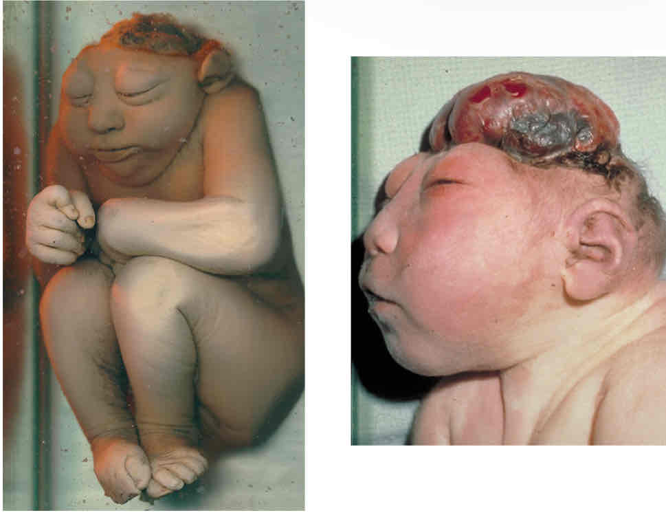
What condition is shown in the image below
Anencephaly
What is another term used to describe Anencephaly?
Cranioschisis
This is the most common severe anomaly seen in stillborn fetuses, making up ½ of the severe neural tube defects.
Anencephaly
Wha are some causes of anencephaly?
genetic factors, high exposure to nickel, chromium, lead, and mercury
This condition can be induced by rats by teratogenic agents
Anencephaly
What are some prevention methods we can use to treat anencephaly?
Folic acid= up to 0.4 mg/day before pregnancy
T/f You should take 0.4 mg/day of folic acid during your pregnancy to reduce the risk of Anencephaly (slide 38)
False
What are some ways we can diagnose Anencephaly
Ultrasound, alpha-fetoproteins in amniotic fluid
T/F When we look at Alpha-fetoproteins in amniotic fluid, we often see a decrease in neural tube defect and an increase in patients with Down syndrome (Slide 38)
false
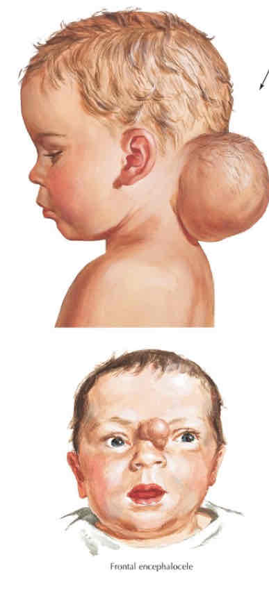
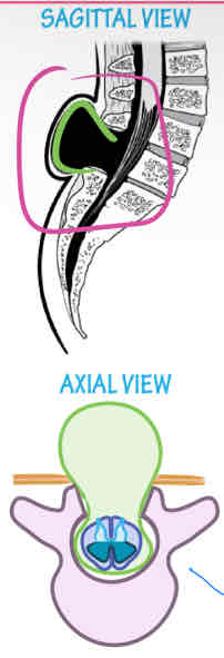
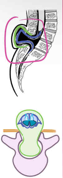
This condition is characterized by a sac-like protrusion of the cerebrum and meninges through a skull defect. Where does this condition commonly occur? (Slide 39) 2-part question
Encephalocele; in the Occiput, top of the head, Forehead-nose junction
What condition is being shown in the image below?
Encephalocele
This condition is caused by a nonfusion of the embryonic halves of the vertebral arches during the 4th week of development
Spina bifida
What are the types of spina bifida that are present with a cyst?
Spina Bifida Meningomyelocele, Spina Bifida Meningocele
What are the types of spina bifid that are considered closed?
Spina Bifida occulta
List the spina bifida that are considered open (aperta).
Spina bifida myelocele, spina bifida meningocele, spina bifida meningomyelocele
This neural tube defect is usually asymptomatic and occurs in L5 or S1 in about 10% of otherwise normal people.
spina bifida occulta
This neural tube defect is characterized by a small dimple with a tuff of hair, a small percentage of cases may have functional defects.
spina bifida occulta
What condition is being shown in the image below?
Spina bifida occulta
This is the most severe form of spina bifida
Spina bifida myelocele (rachischisis)
This neural tube defect is characterized by neural folds remaining open, causing CSF to leak out.
Spina bifida Myelocele (rachischisis)
T/F Patients with spina bifida myelocele have a strong clinical outlook (slide 43)
False
This neural tube defect is characterized by a cyst-like sac covered by skin or easily ruptured membrane, most commonly found in the lumbar region.
Spina bifida cystica
T/F Of the different types of spina Bifida cystica, 10% are menigoceles and 90% are considered meningomyeloceles. (Slide 44)
True
T/F Of the different types of spina Bifida cystica, 90% are menigoceles and 10% are considered meningomyeloceles. (Slide 44)
False
What are some possible causes of spina bifida cystica?
Obesity, Diabetic/anticonvulsant meds, fever during neural tube development
What condition is shown in the images below?
Spina bifida meningocele
This neural tub defect is characterized by a protrusion through a defect in the vertebral arch
Spina bifida meningocele
T/F in a spina bifida meningocele, the neural structures are intact, in normal position, and has low alpha-fetoproteins (slide 46)
False
This neural tube defect is characterized by a protrusion of meninges and spinal cord through a defect in the vertebral arch.
Spina Bifida meningomyelocele
This type of neural tube defect is more serious than a meningocele and causes paralysis in the lower limb, bladder, and bowel.
Spina bifida meningomyelocele
T/F The spina bifida meningomyelocele causes marked neurologic defects superior to the level of the cyst (slide 48)
False (inferior)
What neural tube defect is shown in the image below
Spina bifida meningomyelocele