SLT1204 Gastrointestinal Anatomy
5.0(1)
Studied by 2 peopleCard Sorting
1/53
Earn XP
Description and Tags
Last updated 1:02 PM on 1/12/23
Name | Mastery | Learn | Test | Matching | Spaced | Call with Kai |
|---|
No analytics yet
Send a link to your students to track their progress
54 Terms
1
New cards
What forms the roof of the oral cavity?
The hard and soft palate
2
New cards
What comprises of the floor of the oral cavity? (covering, muscles, etc.)
* Thin and Vascular layer of mucosa
* Supported by geniohyoid and mylohyoid muscles
* Tongue rests on the floor of the oral cavity.
* Supported by geniohyoid and mylohyoid muscles
* Tongue rests on the floor of the oral cavity.
3
New cards
What supports the lateral walls of the oral cavity?
The lateral walls of oral cavity are supported by pads of fat and the buccinator muscle.
4
New cards
What are the 4 functions of the oral cavity?
1. Sensation of taste
2. Grinding of food through the actions of the teeth, tongue, and palatal surfaces
3. Lubrication by mixing with mucus and saliva
4. Limited digestion of carbohydrates and lipids.
5
New cards
What are the major components of the oral cavity that allow it to achieve its functions?
The teeth, tongue and salivary glands.
6
New cards
What is the pharynx and how can it be further divided?
The pharynx is a connection between the oral cavity and the esophagus, which serves as a common pathway for solid food, liquid and air
Further divided into:
* Nasopharynx
* Oropharynx
* Laryngopharynx
Further divided into:
* Nasopharynx
* Oropharynx
* Laryngopharynx
7
New cards
What kind of cells line the regions in the pharynx?
Stratified squamous epithelium (similar to oral cavity)
8
New cards
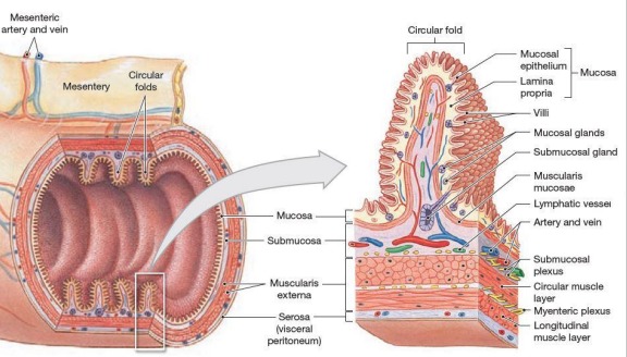
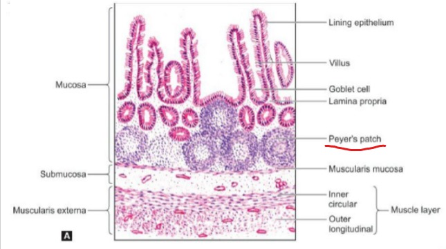
What are the 4 main histological layers of the gastrointestinal tract?
1. Mucosa
2. Submucosa
3. Muscularis Externa
4. Serosa
9
New cards
What are the components of the mucosal layer of the gastrointestinal tract?
1. Epithelium
2. Lamina Propria
3. Muscularis Mucosae
10
New cards
What are the components of the muscularis externa layer of the gastrointestinal tract?
1. Inner circular
2. Outer longitudinal
11
New cards
Describe what the Serosa is. (What is it, Purpose, Structures found, etc.)
What it is - the **outermost layer** of the gut, a **serous membrane** lined by **simple squamous epithelium**
Purpose - a visceral peritoneum that **covers most of the gastrointestinal tract** and extends over the abdominal wall to **form the parietal peritoneum**
Structures found - Large **blood vessels**, **lymphatics** and **nerve trunk** run through the serosa
Purpose - a visceral peritoneum that **covers most of the gastrointestinal tract** and extends over the abdominal wall to **form the parietal peritoneum**
Structures found - Large **blood vessels**, **lymphatics** and **nerve trunk** run through the serosa
12
New cards
Explain the difference between the Serosa and Adventitia.
The Serosa covers the gastrointestinal parts that are within the peritoneal cavity
The adventitia covers the gastrointestinal parts that are not within the peritoneal cavity
The adventitia covers the gastrointestinal parts that are not within the peritoneal cavity
13
New cards
Describe the Esophagus. (What it is, Where it is located within the body)
What it is - It is a **muscular tube** that starts from the pharynx and descends through the thoracic cavity to end in the stomach, **allowing for the passing of food** from the oral cavity to the stomach.
\
Where it is located within the body - **Posterior to the trachea** and **anterior to the vertebral column**
\
Where it is located within the body - **Posterior to the trachea** and **anterior to the vertebral column**
14
New cards
What is the opening between the esophagus and stomach called?
The opening between esophagus and stomach is controlled by **esophageal sphincter** (also known as **cardiac sphincter**).
15
New cards
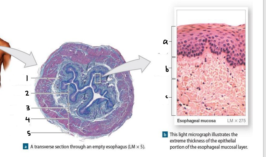
Identify the layers labelled 1 to 5 and a to c.
1. Muscularis mucosae
2. Mucosa
3. Submucosa
4. Muscularis externa
5. Adventitia
\
a. Epithelium (Stratified squamous epithelium)
b. Lamina Propria
c. Muscularis Mucosae
16
New cards
What are the 3 main sections of the stomach?
Fundus (dome-shaped)
Body (contains greater/lesser curvature)
Pylorus (contains antrum and pyloric canal)
Body (contains greater/lesser curvature)
Pylorus (contains antrum and pyloric canal)
17
New cards
What are the two openings of the stomach?
The esophageal sphincter (aka cardiac sphincter) and the pyloric sphincter.
18
New cards
What are the folds of the inner surface called?
Rugae
19
New cards
What are the main differences present in the wall of the stomach ?
1. Mucosa contains gastric pits
2. Muscularis Externa has 3 layers instead of 2
* Inner layer: Oblique muscle layer
* Middle layer: Circular muscle layer
* Outer layer: Longitudinal muscle layer
20
New cards
What are the main cells of the gastric glands and what are their functions?
Parietal Cells - Release hydrochloric acid (HCl)
G Cells - Releases gastrin that stimulates the release of HCl
Chief cells - Release pepsin (breaks down protein), an enzyme activated by HCl
G Cells - Releases gastrin that stimulates the release of HCl
Chief cells - Release pepsin (breaks down protein), an enzyme activated by HCl
21
New cards
What are the 5 functions of the stomach?
1. Mechanical Digestion - the presence of the 3 muscle layers allows the stomach to turn and mix food into chyme
2. Enzymatic Digestion - Proteins are digested by pepsin
3. Neutralization of bacteria - done by HCl (hydrochloric acid)
4. Absorption - of alcohol, sugar, salt, water and drugs
5. Container/Reservoir for food - due to the presence of rugae in the stomach, it is distensible and can store food for up to 4 hours.
22
New cards
What is the omental foramen?
The omental foramen (or foramen of winslow) is the only connection for the greater and lower sac.
23
New cards
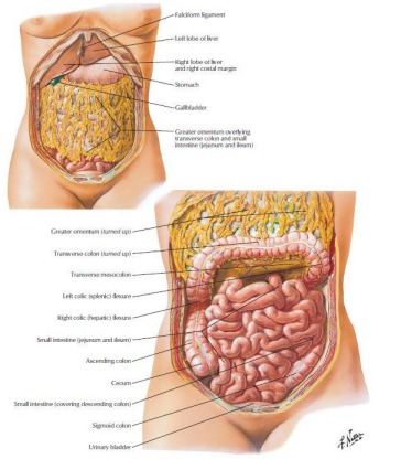
What is the omentum and what are its functions?
The omentum is a fold of the peritoneum that surrounds some of the abdominal organs.
Only the top border of the greater omentum is attached.
Aside from fat storage, it is involved in immune response - its mobility allows it to find and neutralize any potential infection.
Only the top border of the greater omentum is attached.
Aside from fat storage, it is involved in immune response - its mobility allows it to find and neutralize any potential infection.
24
New cards
What is heartburn and what causes it?
Heartburn is a burning feeling in the chest, due to the backing up of stomach acid into the eosophagus.
This is likely due to the relaxation of the esophageal sphincter (aka cardiac sphincter). It can be brought on by consumption of alcohol or spicy food.
This is likely due to the relaxation of the esophageal sphincter (aka cardiac sphincter). It can be brought on by consumption of alcohol or spicy food.
25
New cards
Explain the blood supply of the stomach.
The abdominal aorta gives rise to the celiac trunk, which is the source of blood supply for the stomach.
\
The right gastric artery (from the common hepatic artery) and left gastric artery (directly from the celiac trunk) are found on the lesser curvature of the stomach.
\
The right gastro-omental artery (from the gastroduodenal artery that arose from the common hepatic artery) and left gastro-omental artery (from the splenic artery) are found on the greater curvature of the stomach
\
The right gastric artery (from the common hepatic artery) and left gastric artery (directly from the celiac trunk) are found on the lesser curvature of the stomach.
\
The right gastro-omental artery (from the gastroduodenal artery that arose from the common hepatic artery) and left gastro-omental artery (from the splenic artery) are found on the greater curvature of the stomach
26
New cards
Describe the innervation and lymphatics of the stomach.
Innervation - Parasympathetic stimulation from the vagus nerve and sympathetic stimulation from the celiac plexus
\
Lymphatics - Drains into the gastric and gastro-omental lymph nodes found at the curvatures that drain in the celiac lymph nodes
\
Lymphatics - Drains into the gastric and gastro-omental lymph nodes found at the curvatures that drain in the celiac lymph nodes
27
New cards
Where is the main site of digestion and absorption of nutrients?
The small intestine.
28
New cards
How long does the absorption process take in the small intestine?
3 - 6 hours.
29
New cards
What are the 3 main parts of the small intestine?
1. Duodenum
2. Jejunum
3. Ileum
30
New cards
List the parts of the small intestine in order of length (shortest to longest)
Shortest: Duodenum
Jejunum (2.5m\~)
Longest: Ileum (3.5m\~)
Jejunum (2.5m\~)
Longest: Ileum (3.5m\~)
31
New cards
Describe further the process of digestion in the duodenum.
Duodenum can be further subdivided into 4 parts
Pancreatic enzymes reach small intestine through 2nd part of duodenum - then mixed with chyme
Duodenum receives bile from liver and gallbladder
Pancreatic enzymes reach small intestine through 2nd part of duodenum - then mixed with chyme
Duodenum receives bile from liver and gallbladder
32
New cards
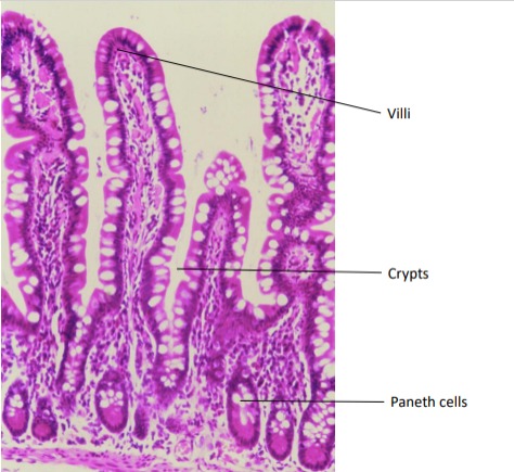
What are the anatomical modifications that allow for better absorption in the small intestine?
Plica Circularis - circular folds in inner surface of small intestine
Villi - finger-like projections covered with simple columnar epithelium
Microvilli
Villi - finger-like projections covered with simple columnar epithelium
Microvilli
33
New cards
What are the 4 main functions of the small intestine?
1. Complete process of digestion
2. Selective absorption
3. Hormone secretion
4. Deliver chyme from stomach to large intestine
34
New cards
What is the most distinctive histological feature of the duodenum?
Presence of Brunner’s gland in submucosa.
35
New cards
Where are the intestinal juices secreted from?
Secreted from the Crypts of Lieberkuhn
36
New cards
What is the most distinctive histological feature of the jejunum?
Absence of Brummer’s gland in submucosa
More tall and slender villi
More tall and slender villi
37
New cards
What is the most distinctive histological feature of the Ileum?
Presence of Peyer’s patches in mucosa.
38
New cards
What is the mesentery?
It is a membrane that attaches the intestine to the abdominal wall and holds it in place.
39
New cards
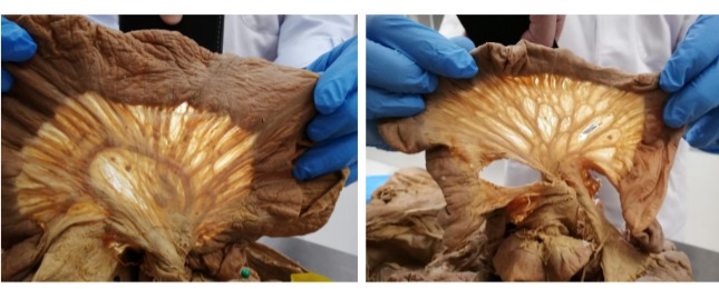
Visually (by looking at the mesentery), how can we differentiate the jejunum and ileum?
Jejunum mesentery has less arcades and longer vasa recta
\
Ileum mesentery has more arcades and shorter vasa recta
\
\
Ileum mesentery has more arcades and shorter vasa recta
\
40
New cards
What is the name of the meeting point of the small and large intestines?
Ileo-cecal junction
41
New cards
What are the 8 parts of the large intestine?
1. Caecum
2. Ascending Colon
3. Right Colic/ Hepatic flexure
4. Transverse Colon
5. Left Colic/ Splenic Flexure
6. Descending Colon
7. Sigmoid Colon
8. Rectum
42
New cards
What are the 3 characteristic features of large intestine?
1. Haustrations - mucosal folds within the colon
2. Appendices epiploicae (or omental appendices) - small pouches of the peritoneum filled with fat and situated along the colon
3. Taeniae Coli - three bands of longitudinal smooth muscle on the colon surface
43
New cards
What are the histological identification features for the large intestine?
* Simple columnar epithelial lining
* Numerous goblet cells - secrete mucus to allow for passing of faeces
* Presence of taenia coli
* Numerous goblet cells - secrete mucus to allow for passing of faeces
* Presence of taenia coli
44
New cards
What are the 3 main branches of the abdominal aorta?
1. Celiac trunk
2. Superior Mesenteric Artery
3. Inferior Mesenteric Artery
45
New cards
How can we divide the alimentary canal and what are the organs in each division?
Foregut - Esophagus to 1st and 2nd part of duodenum
Midgut - 3rd and 4th parts of the duodenum to proximal two thirds of the transverse colon
Hindgut - distal one-third of transverse colon to the urogenital sinus
Midgut - 3rd and 4th parts of the duodenum to proximal two thirds of the transverse colon
Hindgut - distal one-third of transverse colon to the urogenital sinus
46
New cards
Match the divisions of the alimentary canal to the blood supply.
Foregut - Celiac Trunk
Midgut - Superior Mesenteric Artery
Hindgut - Inferior Mesenteric Artery
Midgut - Superior Mesenteric Artery
Hindgut - Inferior Mesenteric Artery
47
New cards
What are 3 main differences in the rectum compared to the colon?
1. Continuous coat of longitudinal muscle present with no taenia coli
2. Peritoneum only covers front and sides of upper one third of rectum and only front of middle third - rest of the rectum is devoid of serous covering
3. No appendices epiploicae
48
New cards
What cells line the anal canal?
Stratified squamous epithelium - due to wear and tear
49
New cards
What is the appendix, what is its function and what is its blood supply?
The appendix is an extension of the cecum and has an unknown function. It receives blood from the appendicular artery and vein.
50
New cards
What is a common surgery performed with relation to the appendix and why is it done?
An appendicectomy - the removal of the appendix. It is done in case of inflammation.
51
New cards
What are the histological features of the appendix?
* Absence of villi
* Presence of lymphatic follicles
* Presence of lymphatic follicles
52
New cards
What is appendicitis?
It is the acute inflammation of the appendix, and the most common cause for acute, severe abdominal pain.
53
New cards
What are the clinical presentations, prevalence and treatment for of appendicitis?
Clinical presentation - tenderness over McBurney’s point, which corresponds to the base of the appendix
\
Prevalence - seen more commonly in older children and young adults, and is uncommon at the extremes of age. The disease is seen more frequently in the West and in affluent societies.
Treatment - Removal of appendix to avoid rupture (rupture can result in peritonitis)
\
Prevalence - seen more commonly in older children and young adults, and is uncommon at the extremes of age. The disease is seen more frequently in the West and in affluent societies.
Treatment - Removal of appendix to avoid rupture (rupture can result in peritonitis)
54
New cards
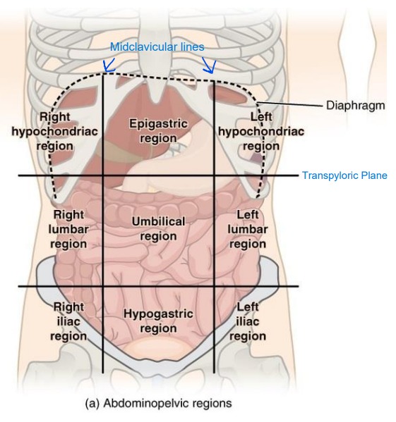
What are the names 9 regions of the abdomen?
1. Right hypochondrium
2. Epigastric
3. Left hypochondrium
4. Right lumbar
5. Umbilical
6. Left lumbar
7. Right Iliac or Inguinal
8. Pubic or hypogastric
9. Left Iliac or Inguinal