Bone Processes and Landmarks
1/184
There's no tags or description
Looks like no tags are added yet.
Name | Mastery | Learn | Test | Matching | Spaced | Call with Kai |
|---|
No analytics yet
Send a link to your students to track their progress
185 Terms
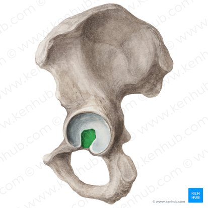
Name the highlighted portion.
Acetabular fossa.
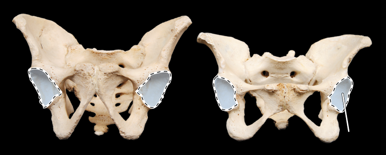
Name the highlighted portion.
(Pelvis, anterior view).
Acetabulum.
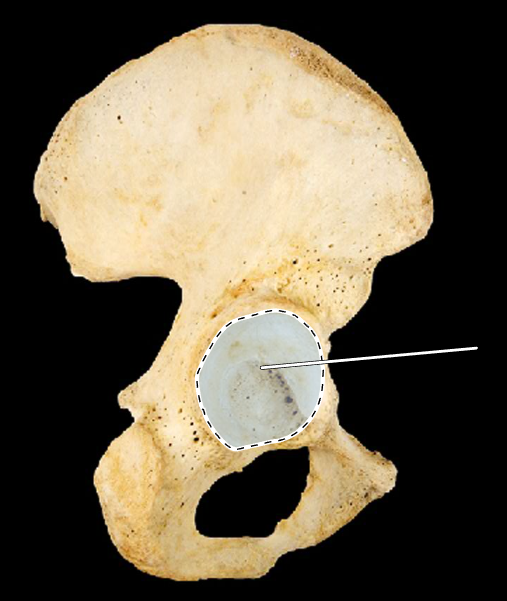
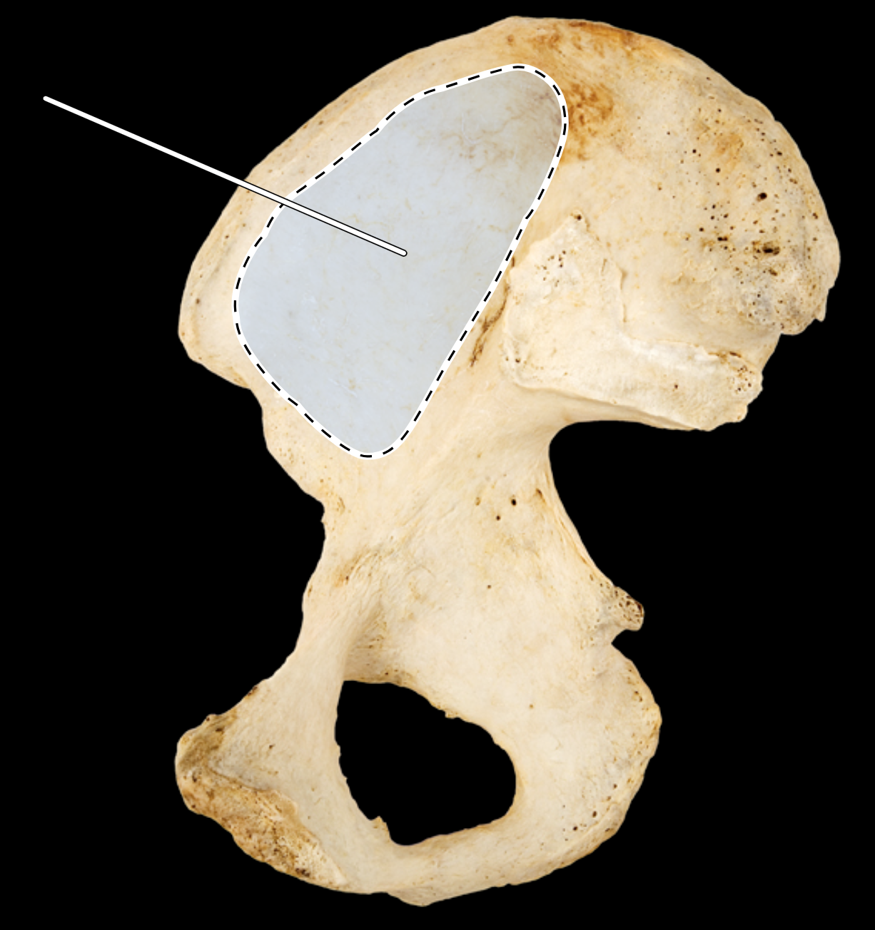
Name the highlighted portion.
(Hip bone, lateral view, right side).
Acetabulum.
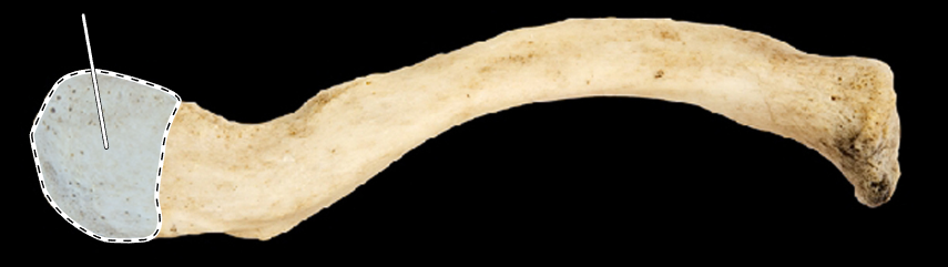
Name the highlighted portion.
(Clavicle, superior view, left side).
Acromial end of clavicle.
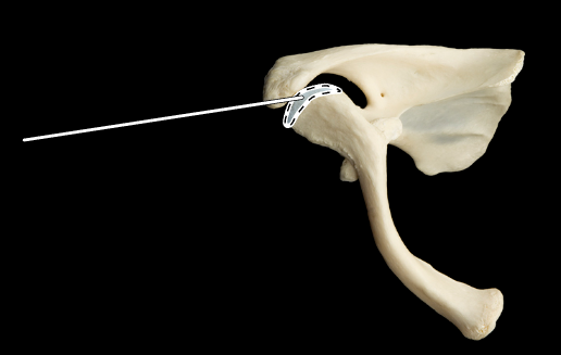
Name the highlighted portion.
(Superior view, right clavicle and scapula).
Acromial process (acromion).
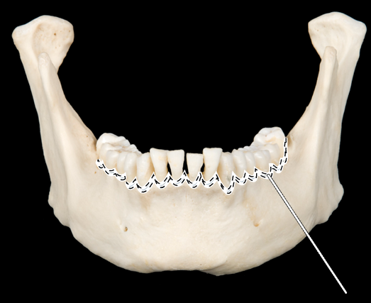
Name the highlighted portion.
(Mandible, anterior view).
Alveolar process of mandible.
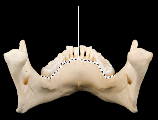
Name the highlighted portion.
(Mandible, posterior view).
Alveolar process of mandible.
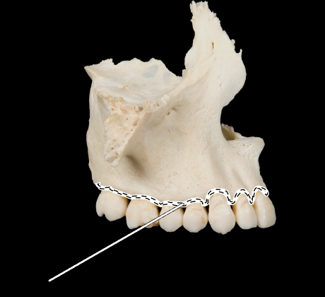
Name the highlighted portion.
(Maxilla, lateral view).
Alveolar process of maxilla.
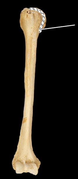
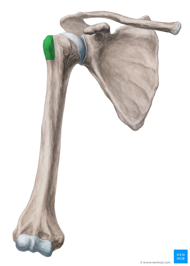
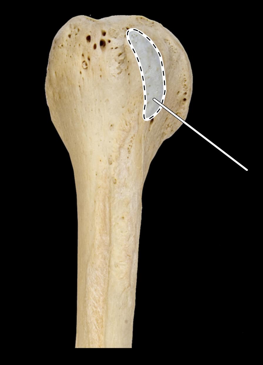
Name the highlighted portion.
(Humerus, anterior view, right side).
Anatomical neck of humerus.
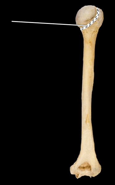
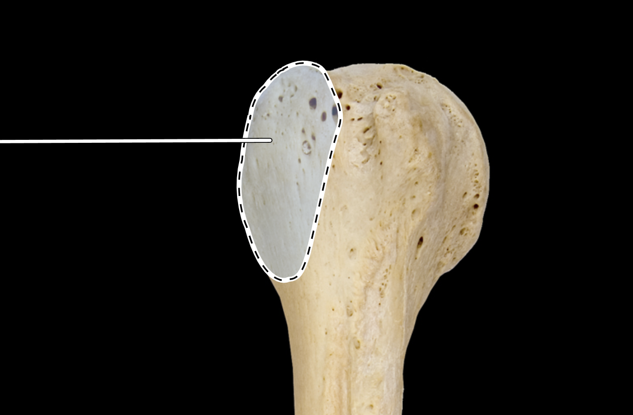
Name the highlighted portion.
(Humerus, posterior view, right side).
Anatomical neck of humerus.
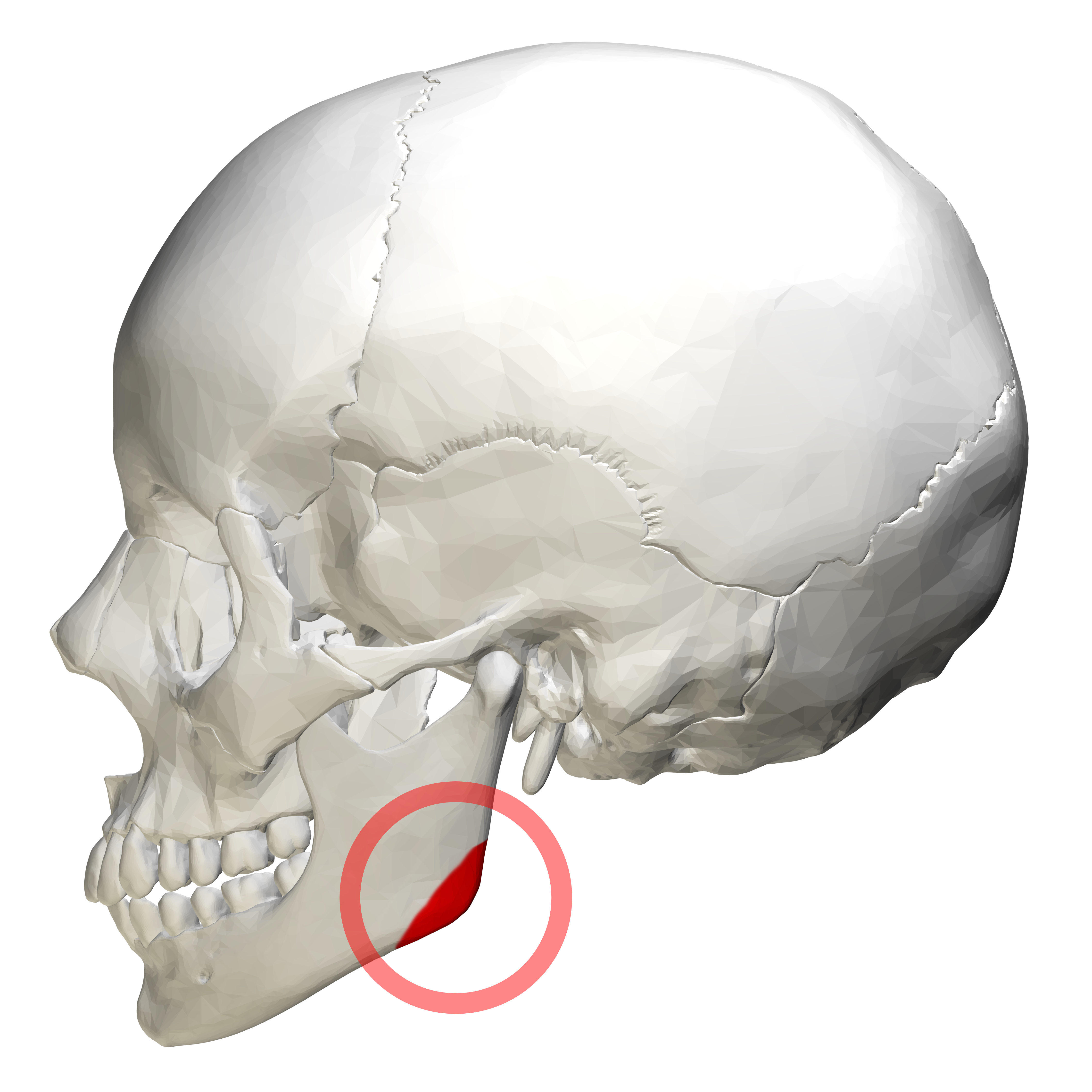
Name the highlighted portion.
(Skull, lateral view).
Angle of mandible.
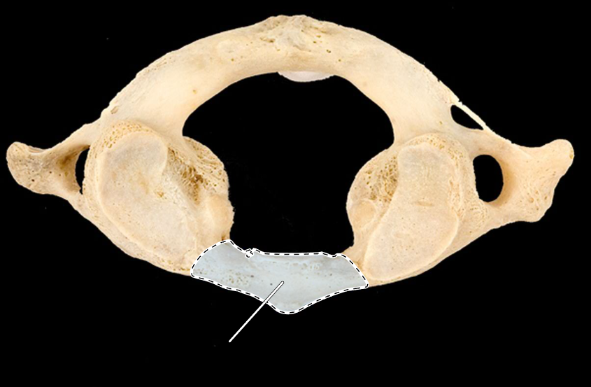
Name the highlighted portion.
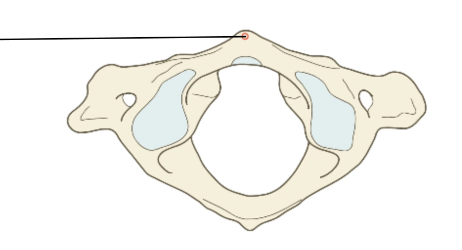
(Atlas, superior view).
Anterior arch.
Name the highlighted portion.
(Atlas, inferior view).
Anterior arch.
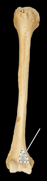
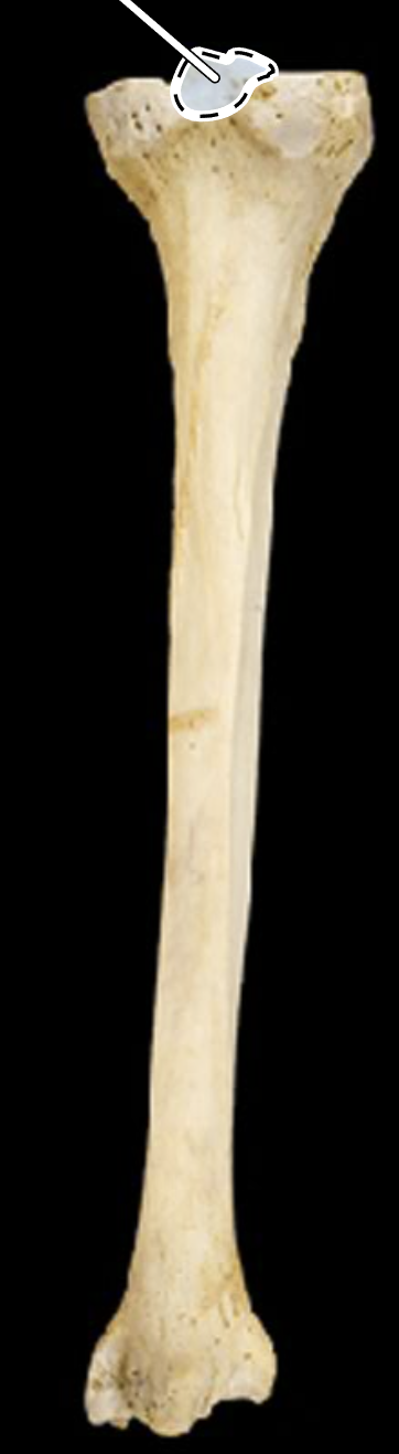
Name the highlighted portion.
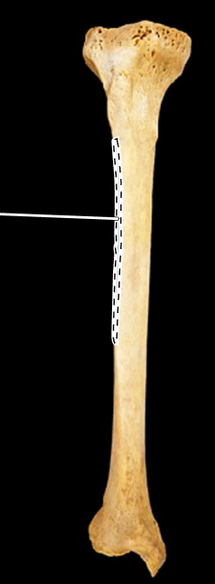
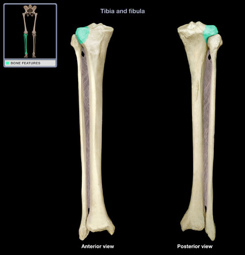
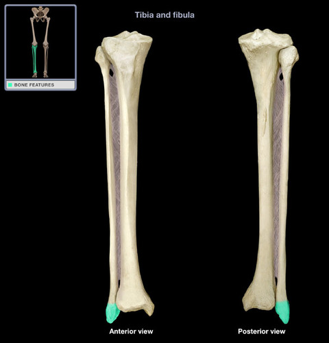
(Tibia, anterior view, right side).
Anterior crest of tibia.
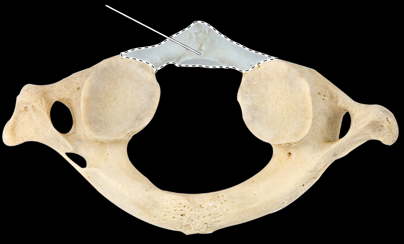
Name this process.
Anterior tubercle.
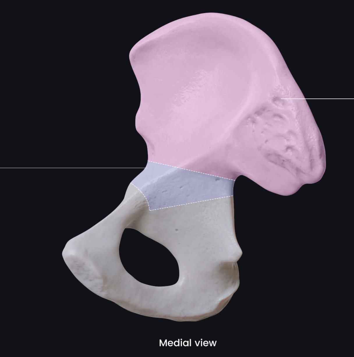
Name the bone highlighted in purple.
Body of ilium.
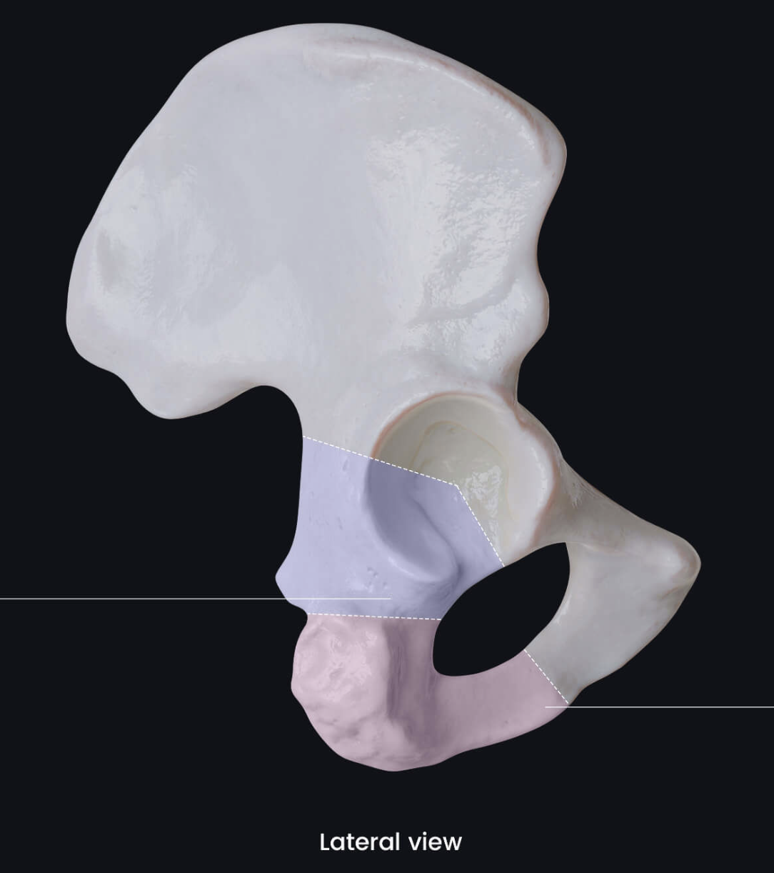
Name the bone highlighted in purple.
Body of ischium.
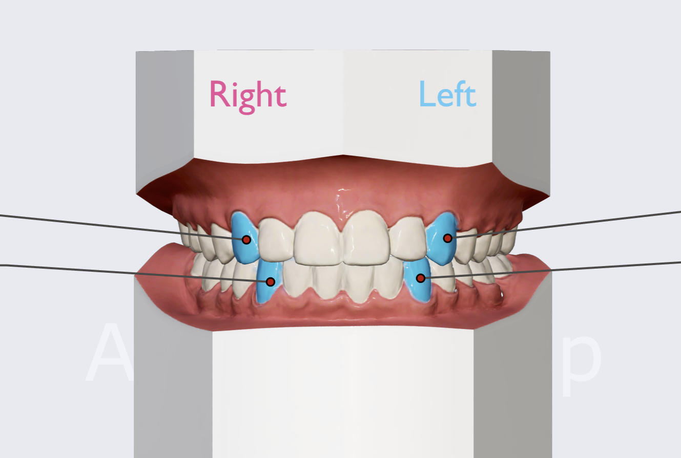
Name the teeth highlighted in blue.
Canines.
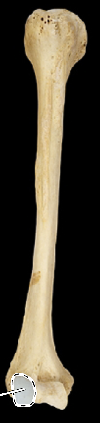
Name the highlighted portion.
(Anterior view).
Capitulum.
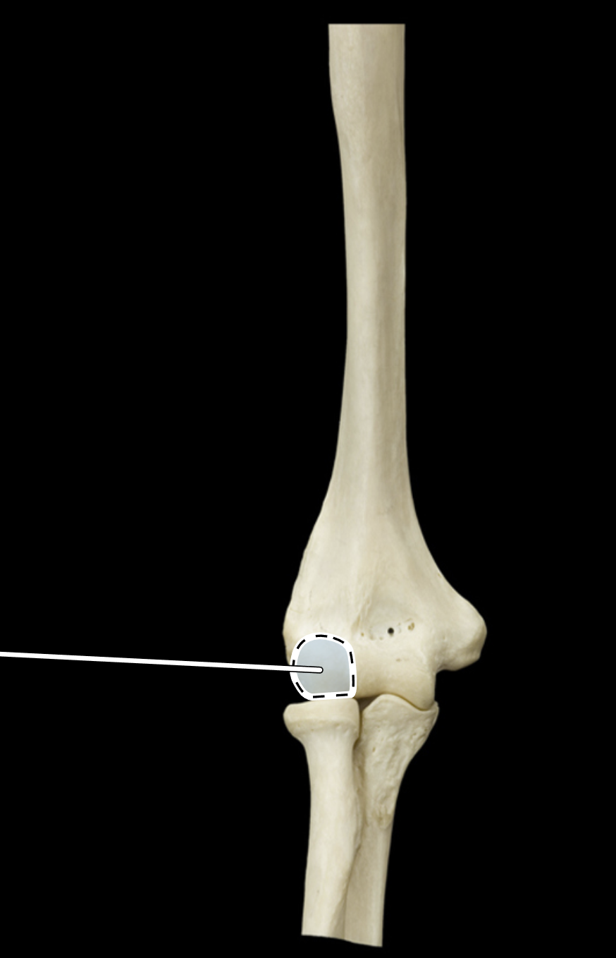
Name the highlighted portion.
(Anterior view).
Capitulum.
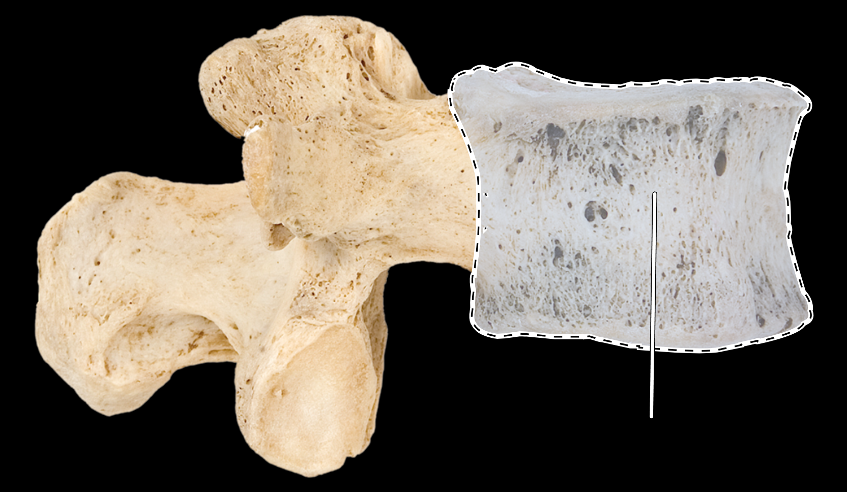
Name the highlighted portion.
(Lumbar vertebra, lateral view).
Centrum (body).
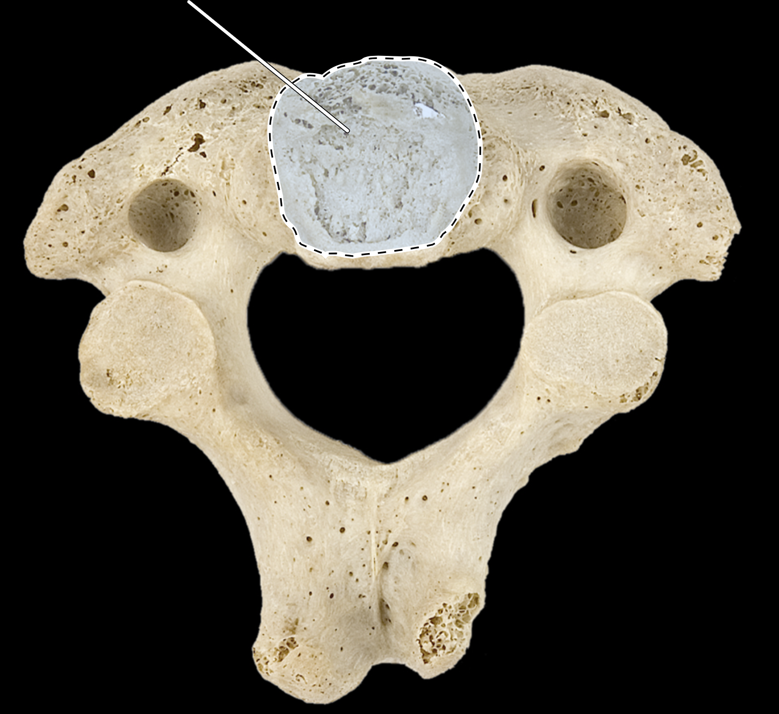
Name the highlighted portion.
(Axis, inferior view).
Centrum (body).
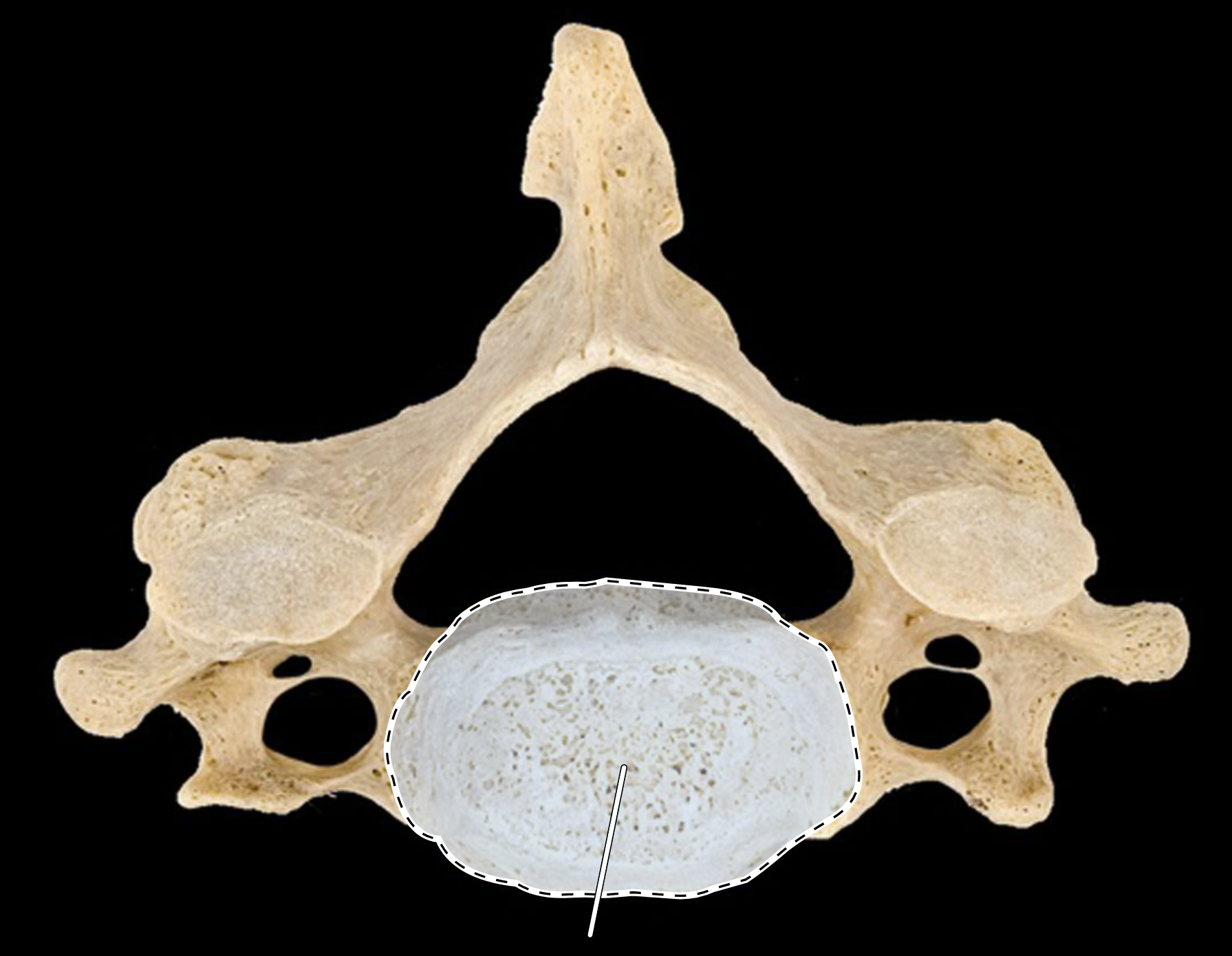
Name the highlighted portion.
(Cervical vertebra, superior view).
Centrum (body).
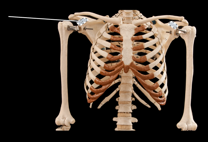
Name the highlighted portion.
(Thoracic cage, anterior view).
Coracoid process of scapula.
Name the highlighted portion.
(Anterior view).
Coronoid fossa of humerus.
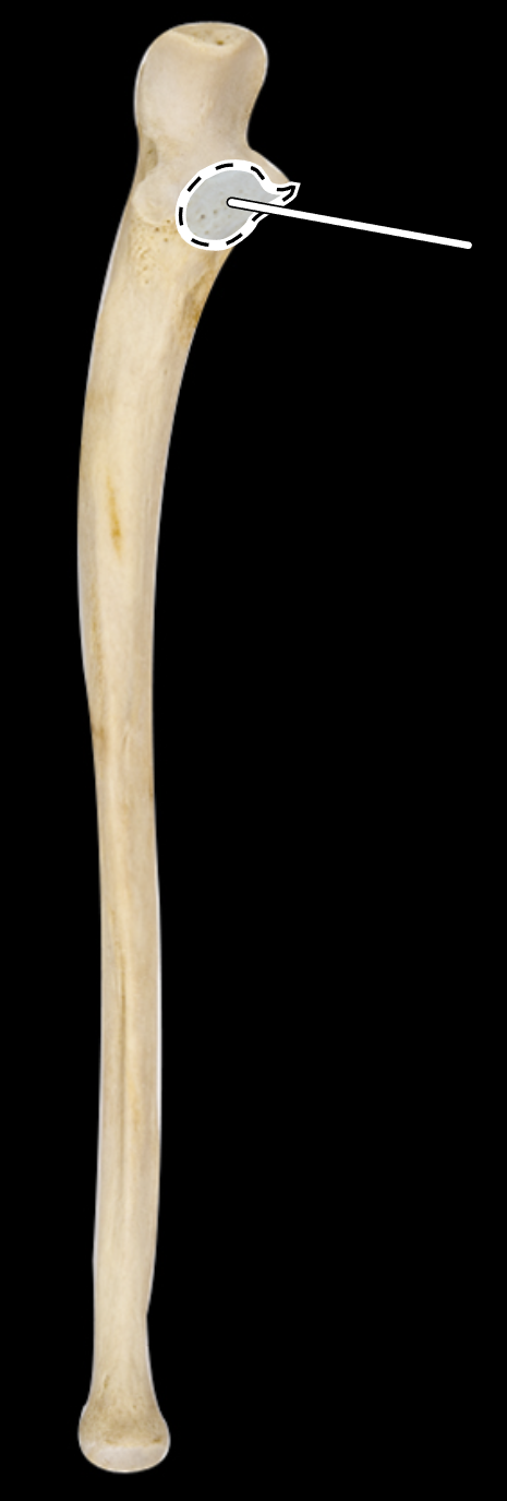
Name the highlighted portion.
(Anterior view, right side).
Coronoid process of ulna.
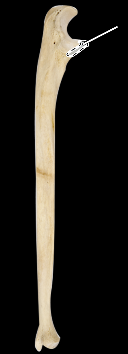
Name the highlighted portion.
(Lateral view, right side).
Coronoid process of ulna.
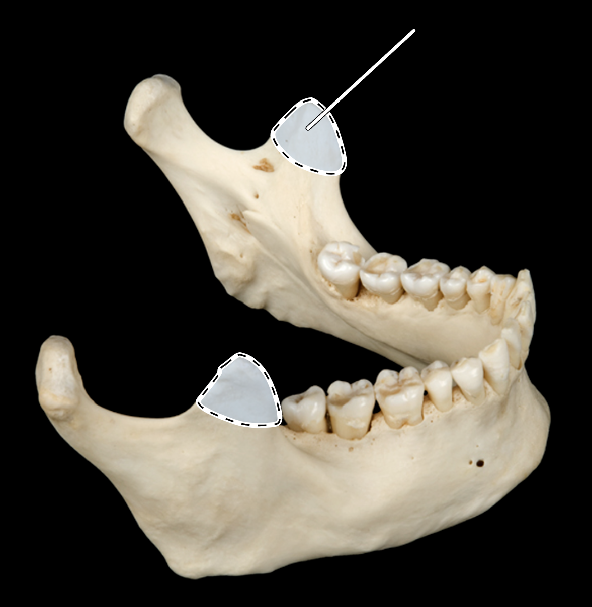
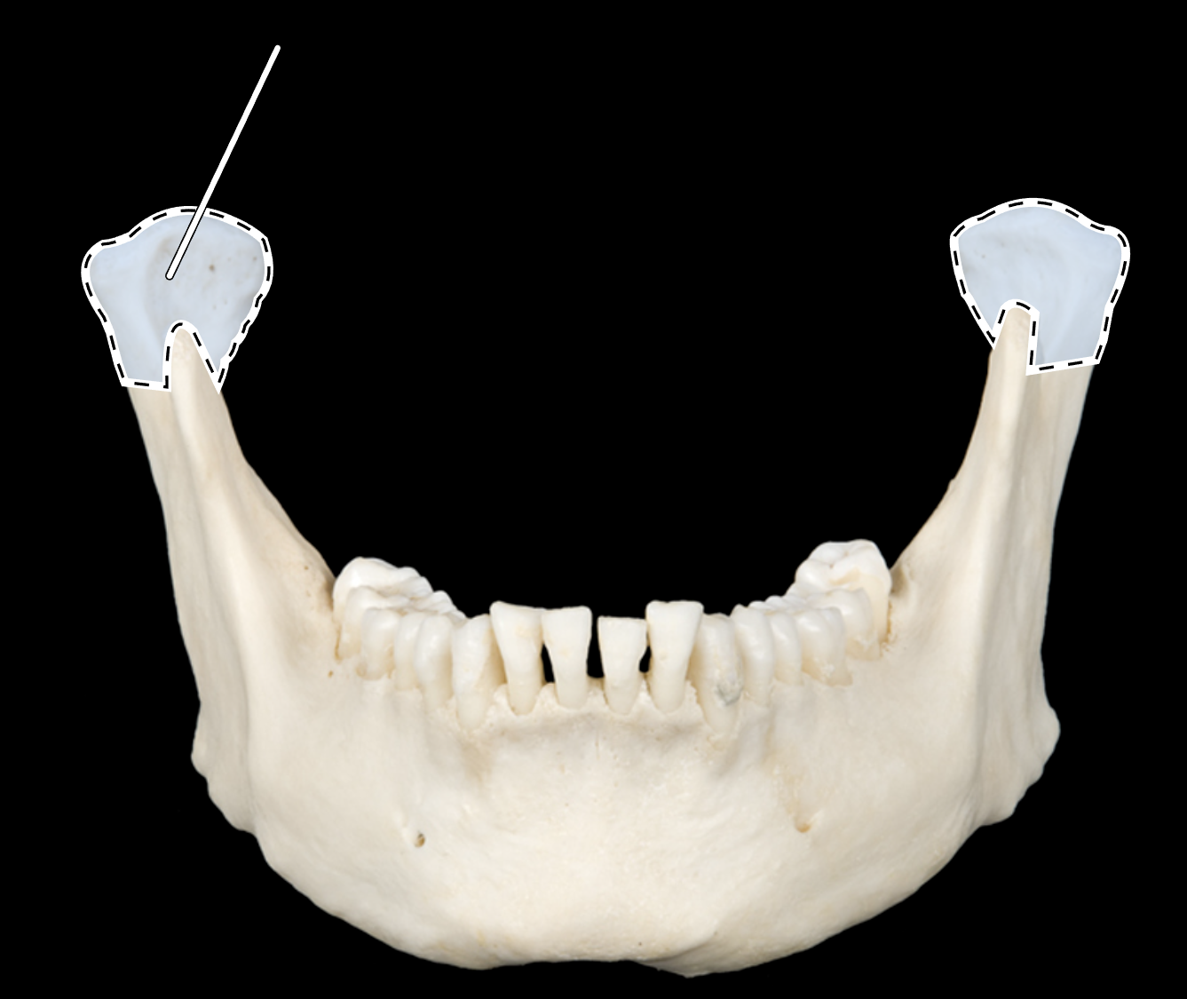
Name the highlighted portion.
Coronoid process of mandible.
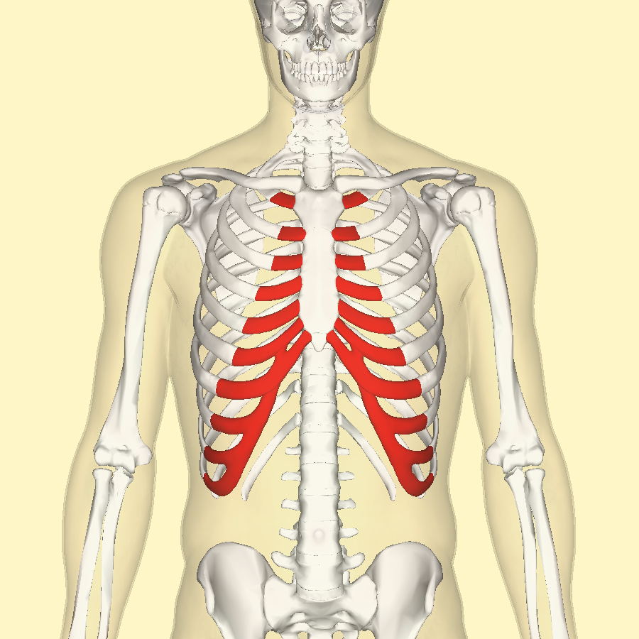
Name the portion highlighted in red.
Costal cartilage of rib.
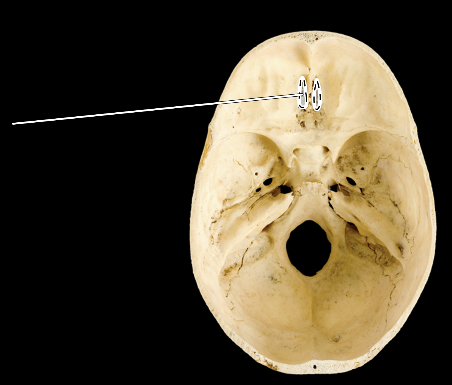
Name the highlighted portion.
(Superior view of cranial cavity).
Cribriform plate of ethmoid.
Name the highlighted portion.
(Posterosuperior view of cranial cavity).
Cribriform plate of ethmoid.
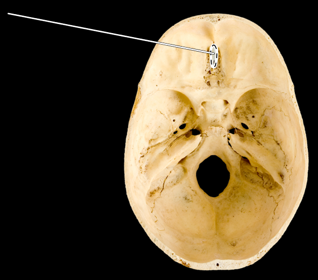
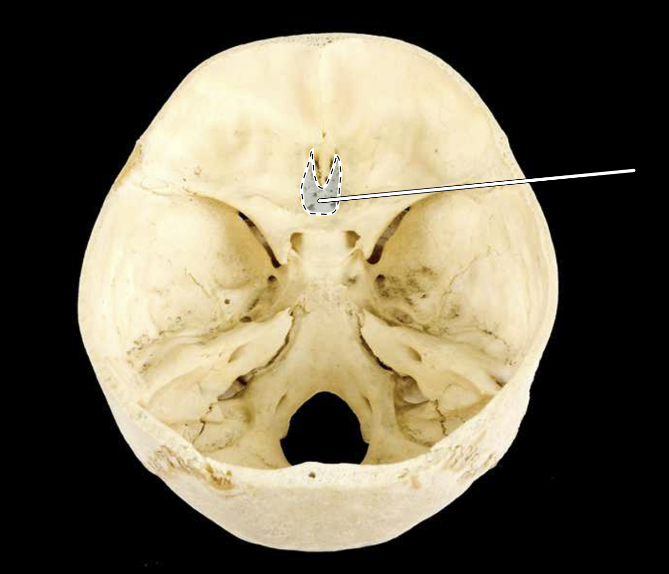
Name the highlighted portion.
(Skull, superior view of cranial cavity).
Crista galli.
Name the highlighted portion.
(Anterior view).
Deltoid tuberosity.
Name the highlighted portion.
(Posterior view).
Deltoid tuberosity.
Name the highlighted portion.
(Lateral view, right side).
Deltoid tuberosity.
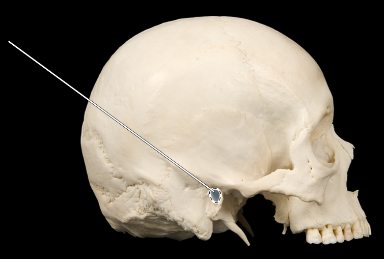
Name the highlighted portion.
(Skull, lateral view).
External auditory meatus.
Name the highlighted portion.
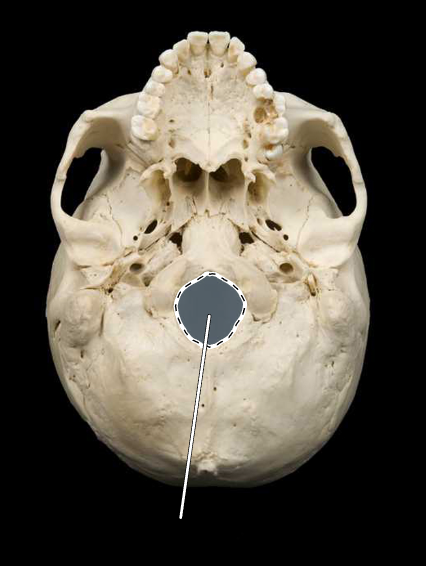
(Skull, inferior view).
Foramen magnum.
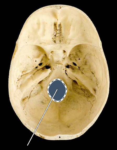
Name the highlighted portion.
(Skull, superior view of cranial cavity holes).
Foramen magnum.
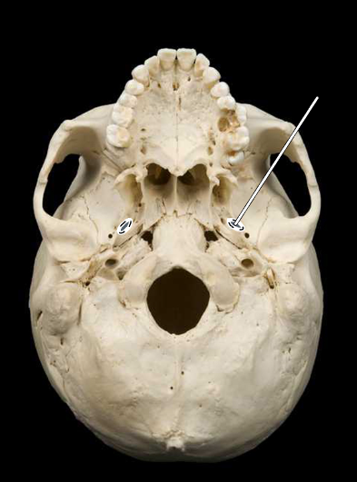
Name the highlighted portion.
(Skull, inferior view).
Foramen ovale.
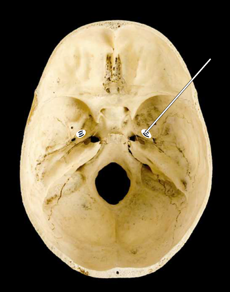
Name the highlighted portion.
(Skull, superior view of cranial cavity holes).
Foramen ovale.
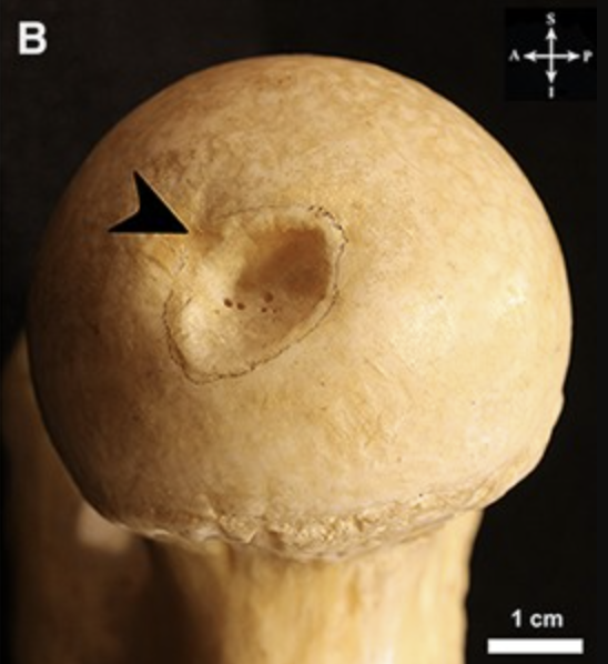
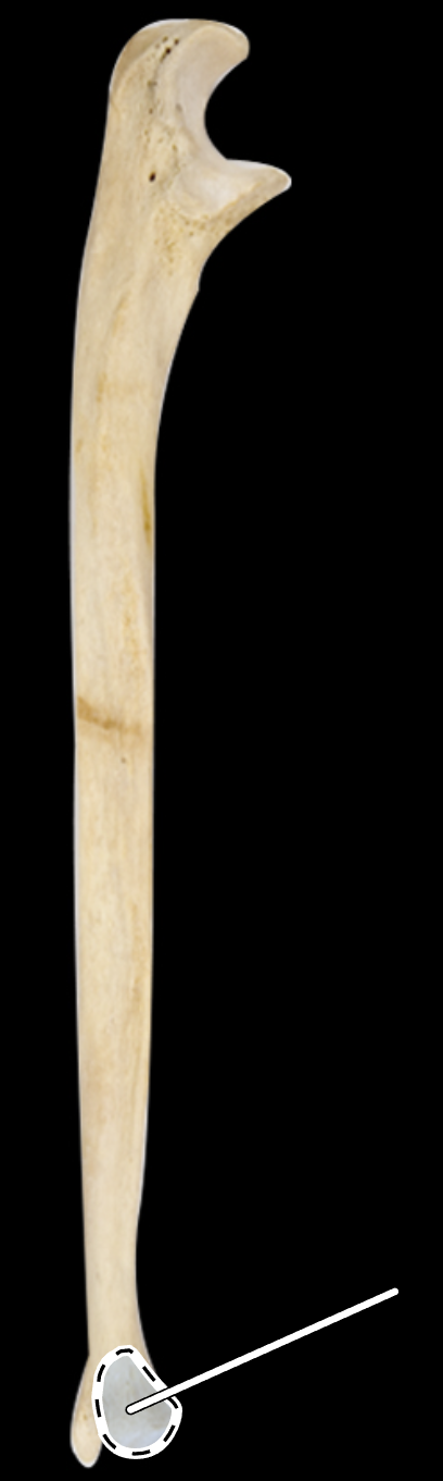
Name this indentation on the bone.
(Found on the head of the femur).
Fovea capitus.
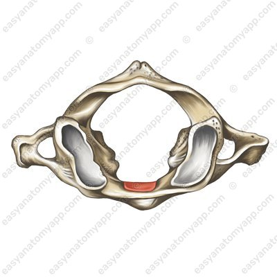
Name the portion of the bone highlighted in red.
(Found on the axis).
Fovea dentis.
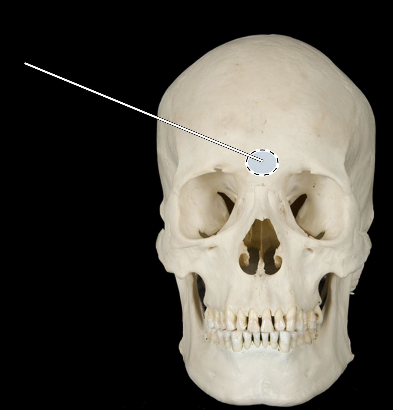
Name the highlighted portion.
(Skull, anterior view).
Glabella.
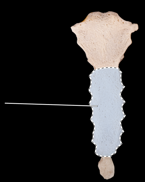
Name the highlighted portion.
(Sternum, anterior view).
Gladiolus (body).
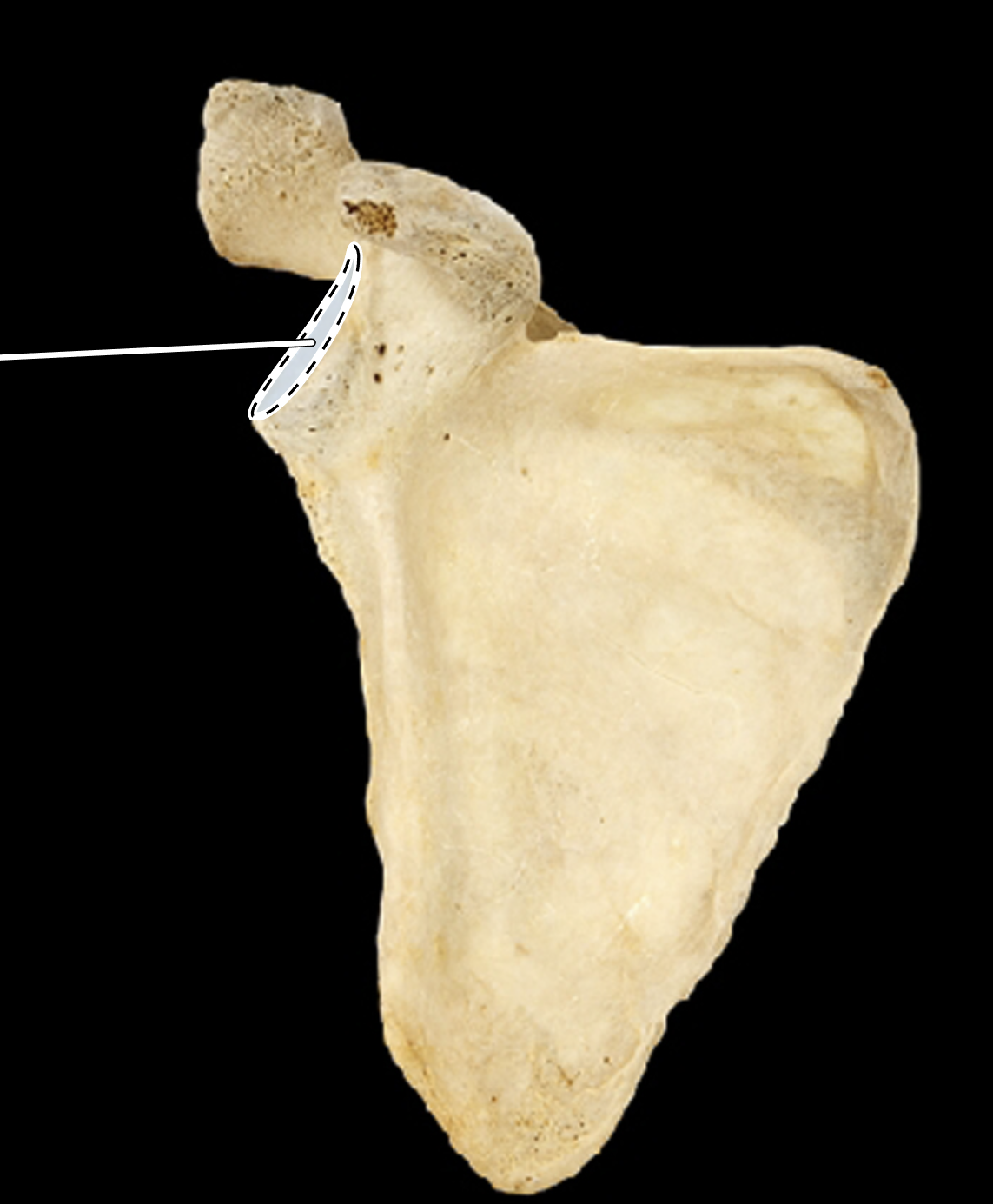
Name the highlighted portion.
(Scapula, anterior view, right side).
Glenoid fossa.
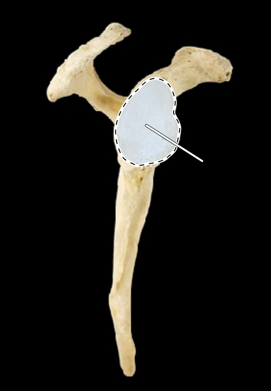
Name the highlighted portion.
(Scapula, lateral view, right side).
Glenoid fossa.

Name the highlighted portion.
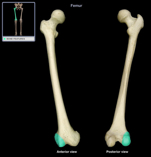
(Femur, posterior view, right side).
Gluteal tuberosity.
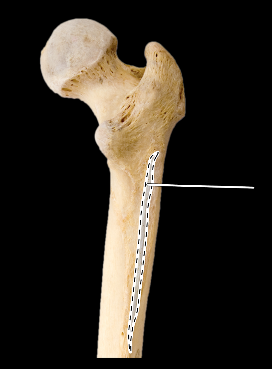
Name the highlighted portion.
(Femur, posterior view, right side).
Gluteal tuberosity.
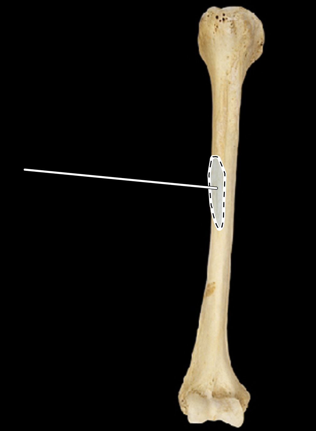
Name the highlighted portion.
(Humerus, anterior view, right side).
Greater tubercle (greater tuberosity).
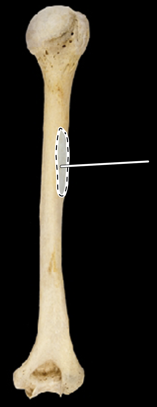
Name the highlighted portion.
(Humerus, anterior view, right side).
Greater tubercle (greater tuberosity).
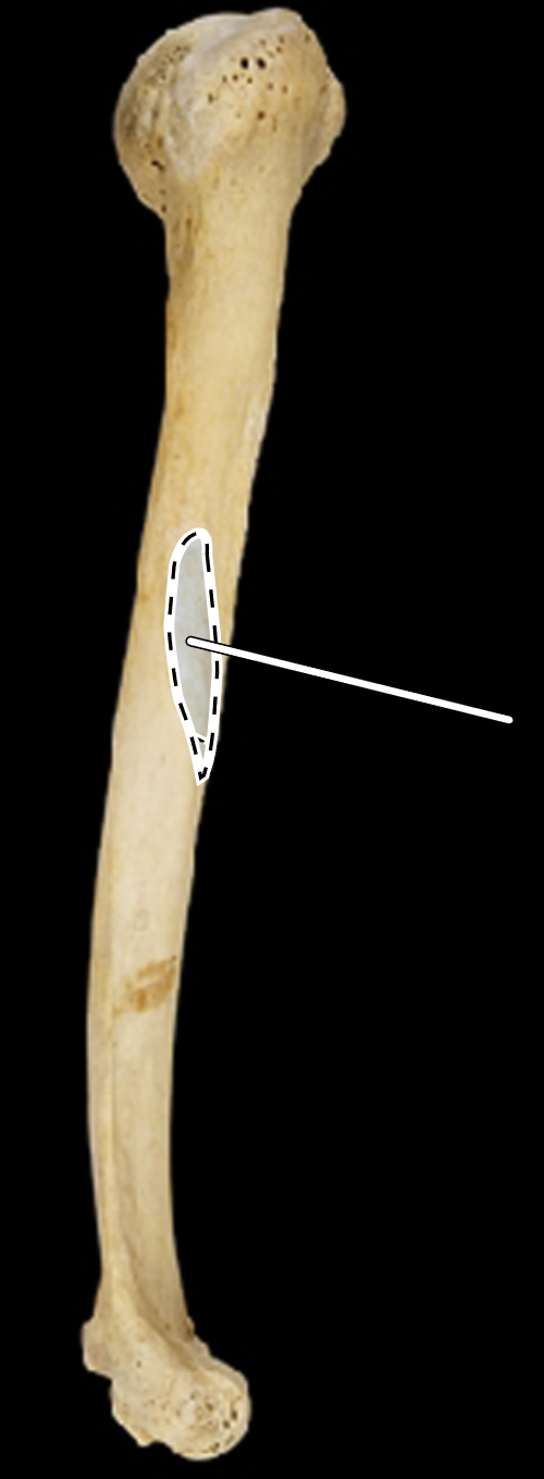
Name the highlighted portion.
Greater tubercle (greater tuberosity).
Name the highlighted portion.
(Femur, anterior view, right side).
Greater trochanter.
Name the highlighted portion.
(Femur, posterior view, right side).
Greater trochanter.
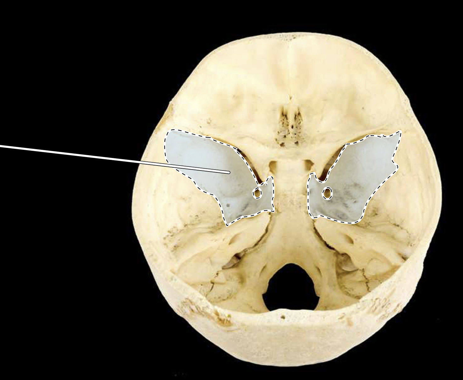
Name the highlighted portion.
(Posterosuperior view of cranial cavity).
Greater wing of sphenoid.
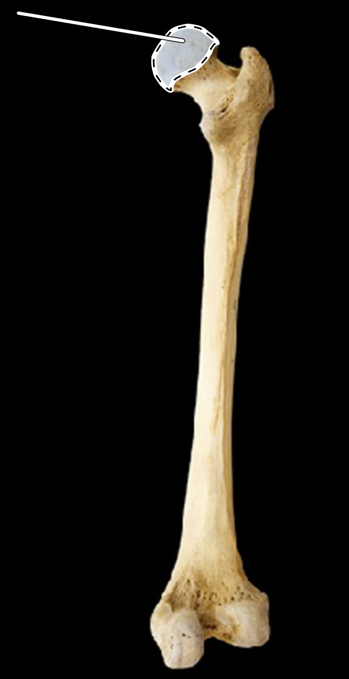
Name the highlighted portion.
(Femur, posterior view, right side).
Head of femur.
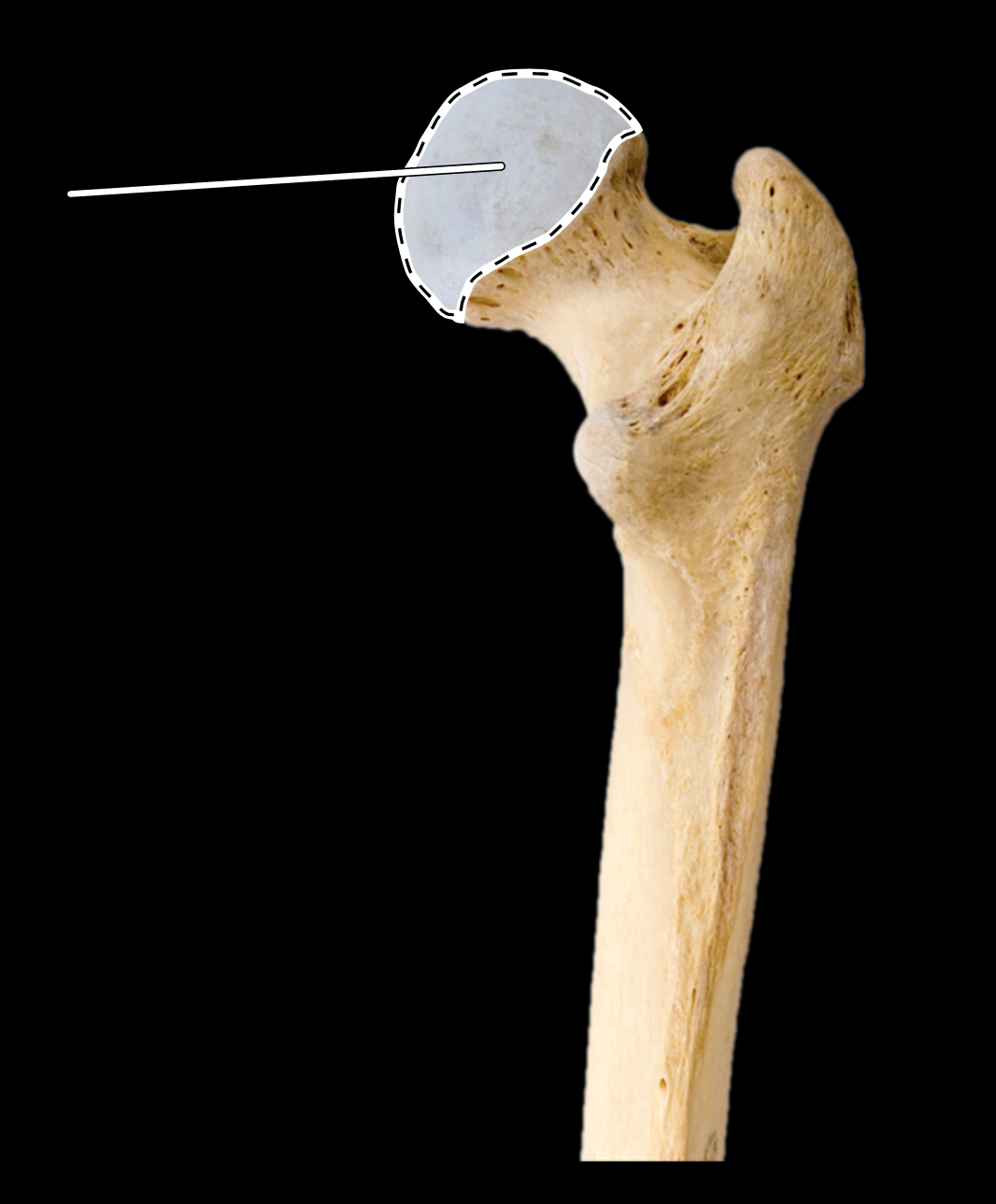
Name the highlighted portion.
(Femur, posterior view, right side).
Head of femur.
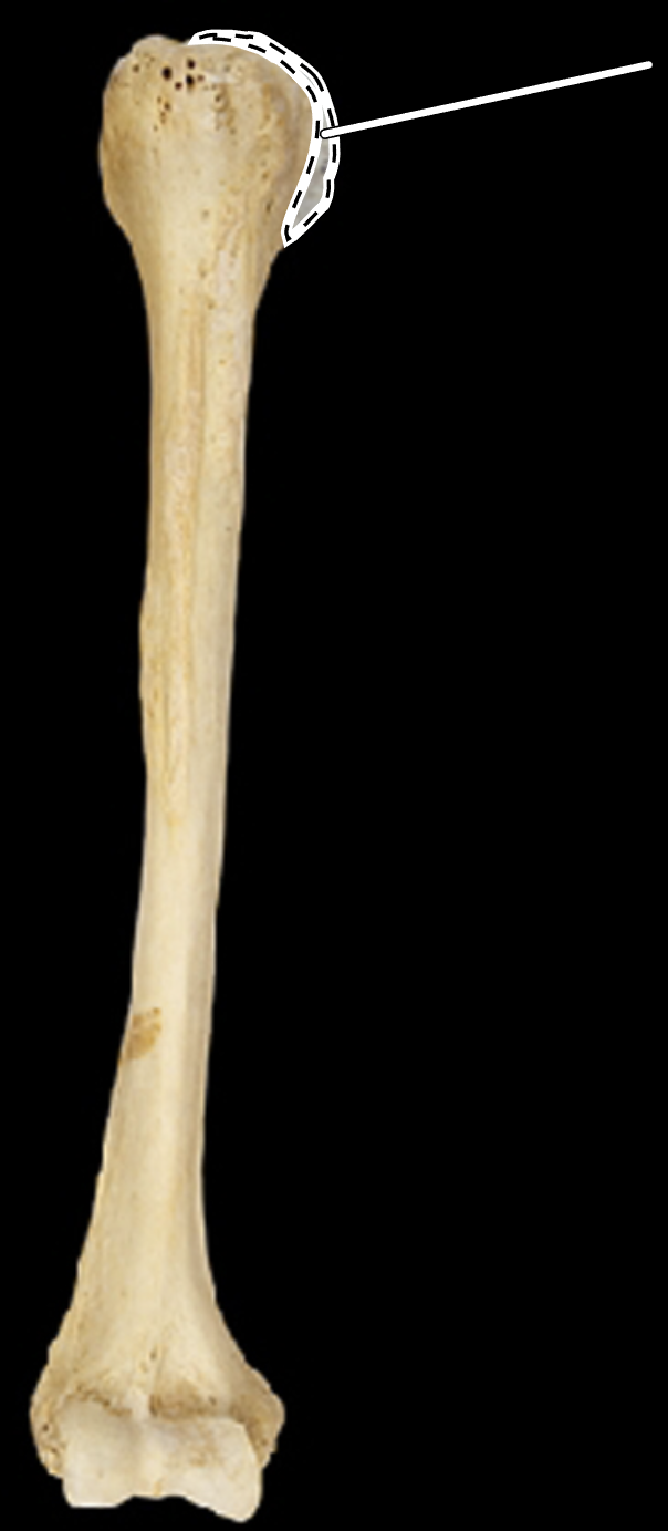
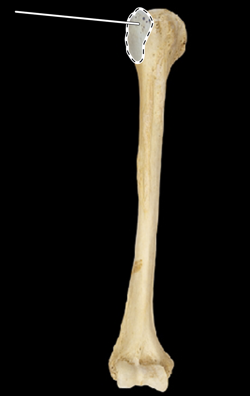
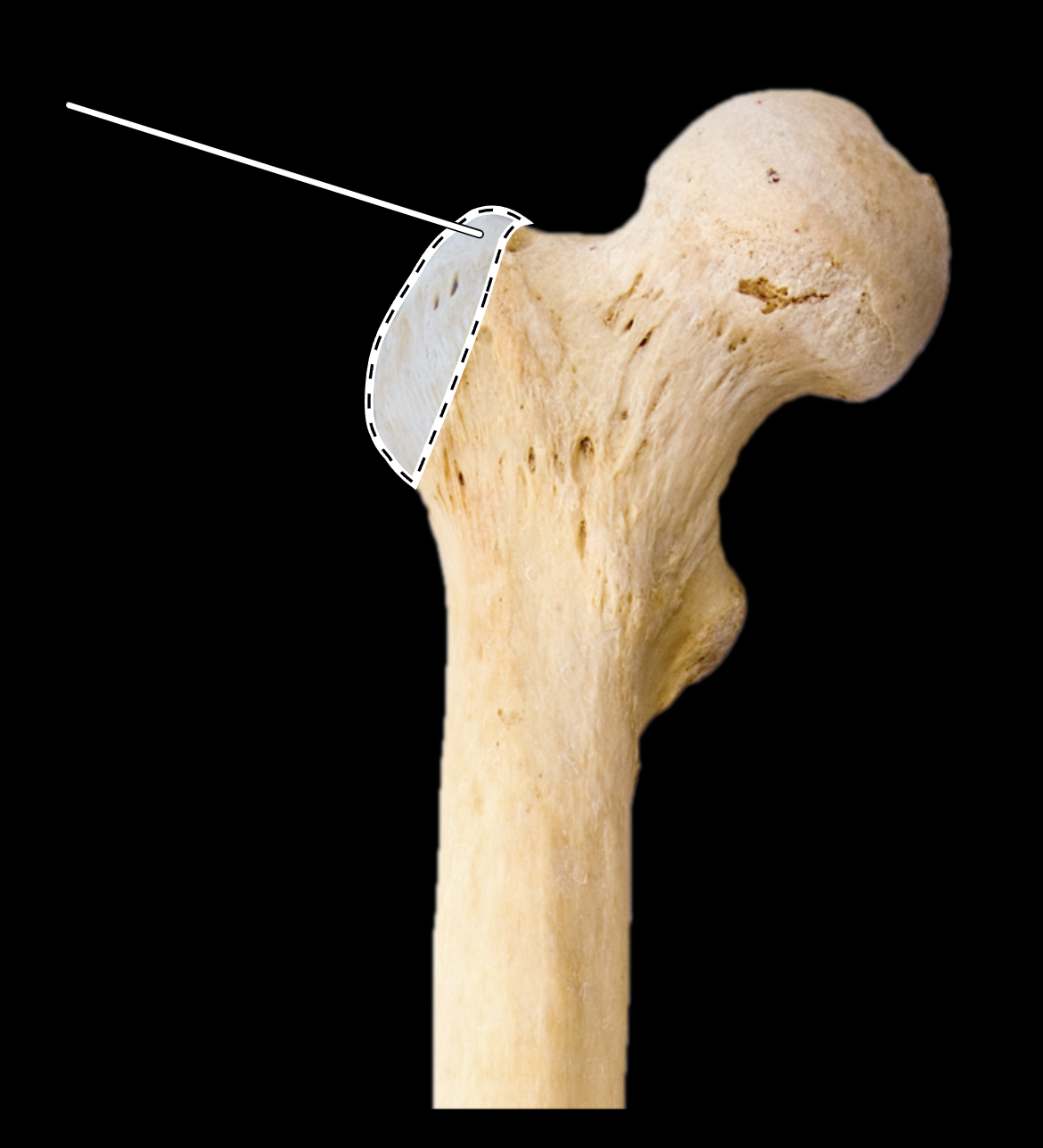
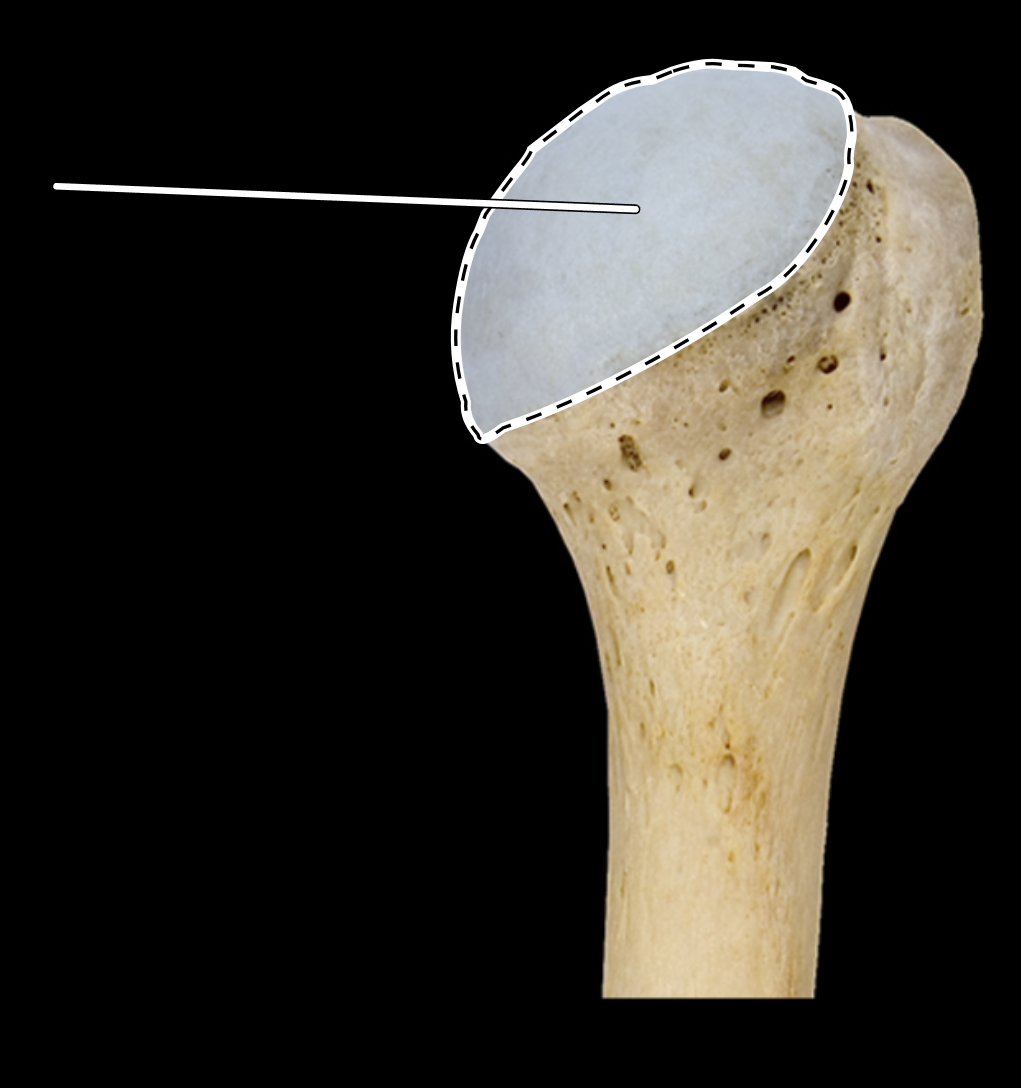
Name the highlighted portion.
(Anterior view, right side).
Head of humerus.
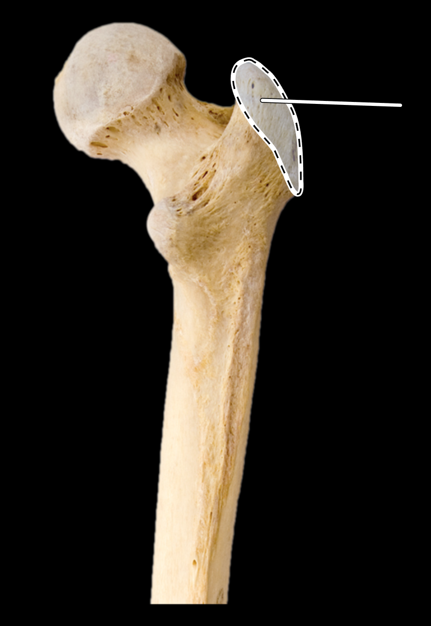
Name the highlighted portion.
(Humerus, posterior view, right side).
Head of humerus.
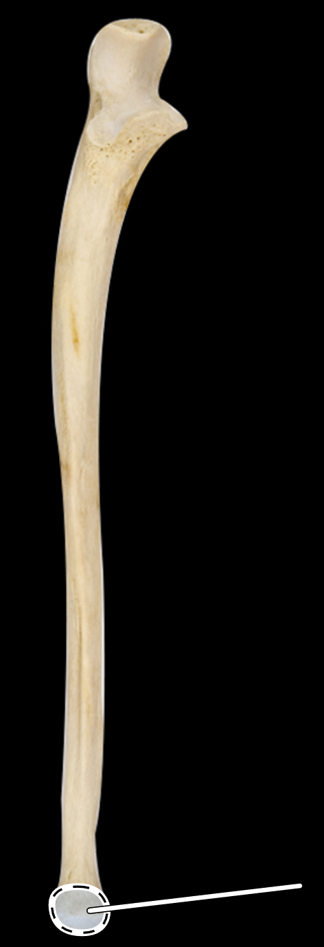
Name the highlighted portion.
(Anterior view, right side).
Head of radius.
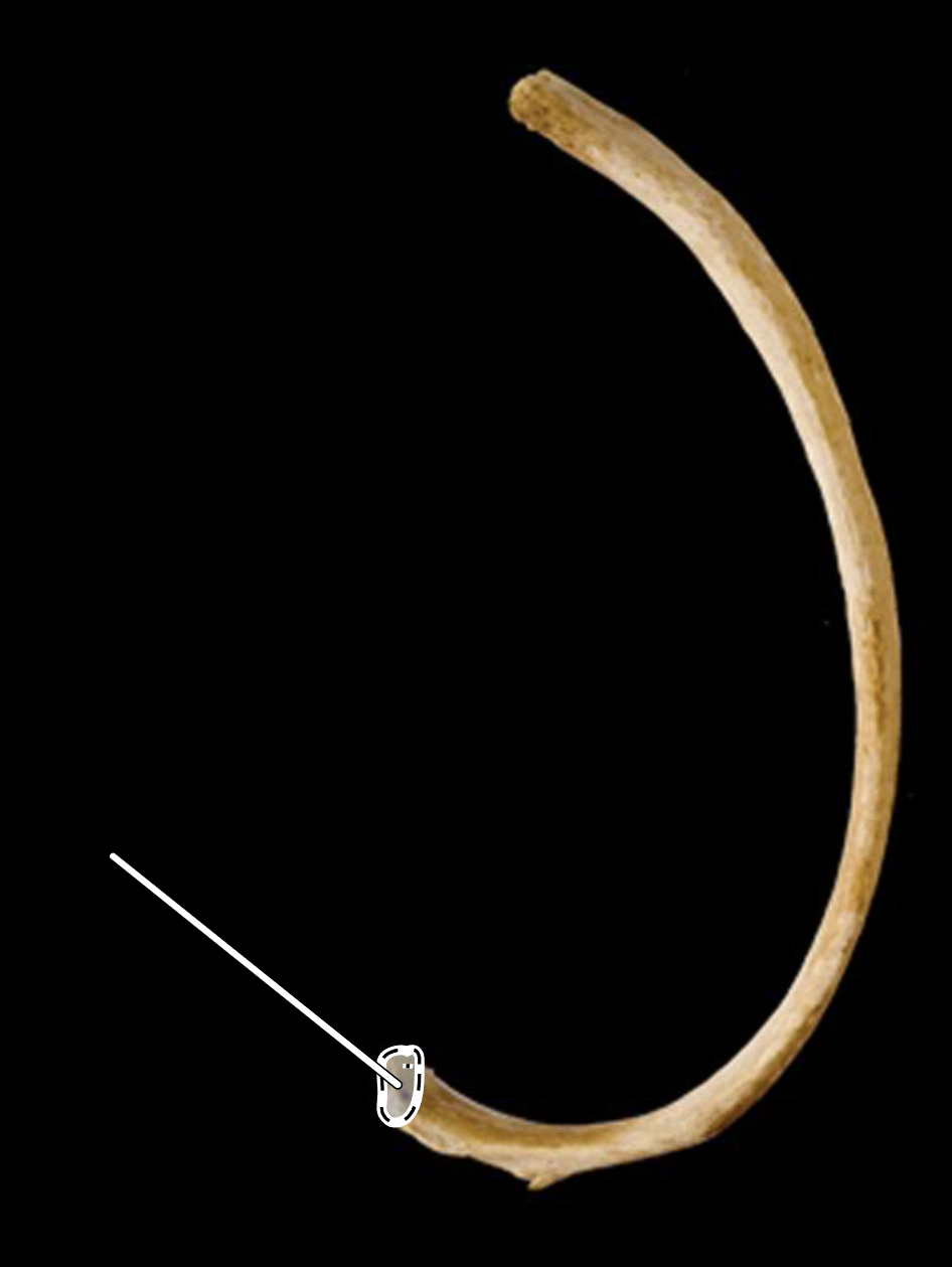
Name the highlighted portion.
(Superior view, right side).
Head of rib.
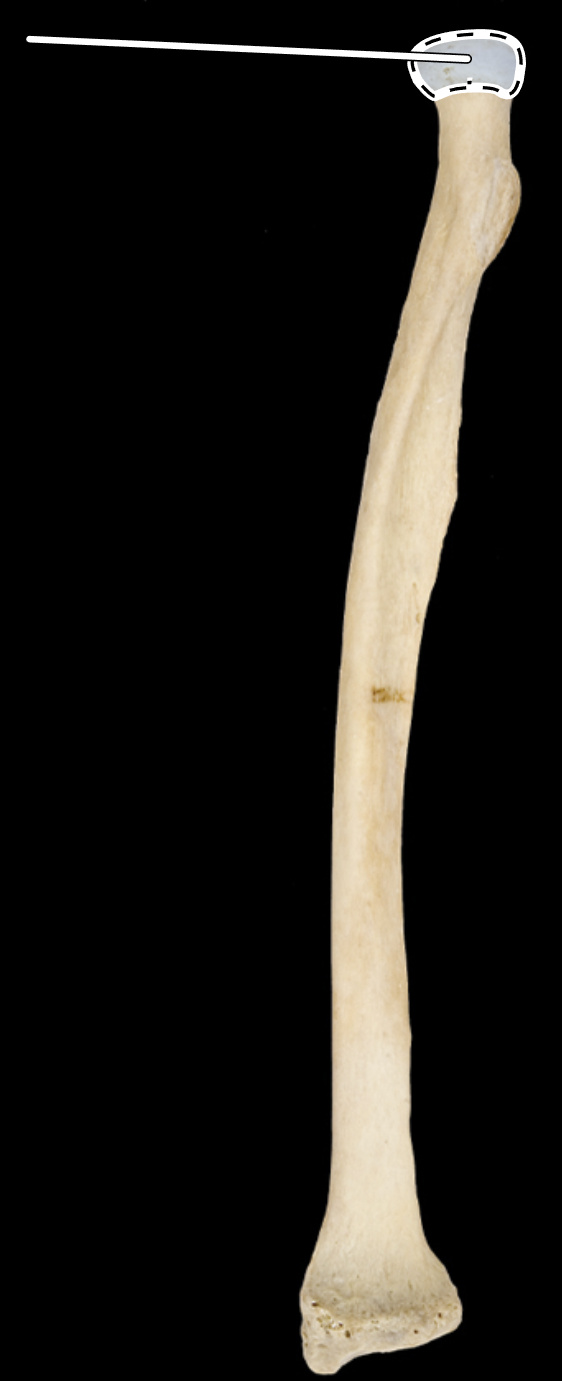
Name the highlighted portion.
(Anterior view, right side).
Head of ulna.
Name the highlighted portion.
(Lateral view, right side).
Head of ulna.
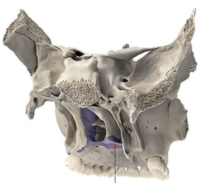
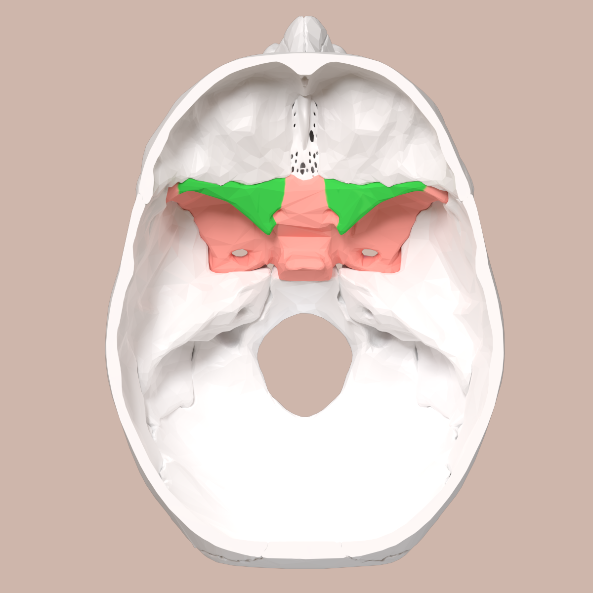
Name the flat portion of the area highlighted in purple.
Horizontal plate of palatine.
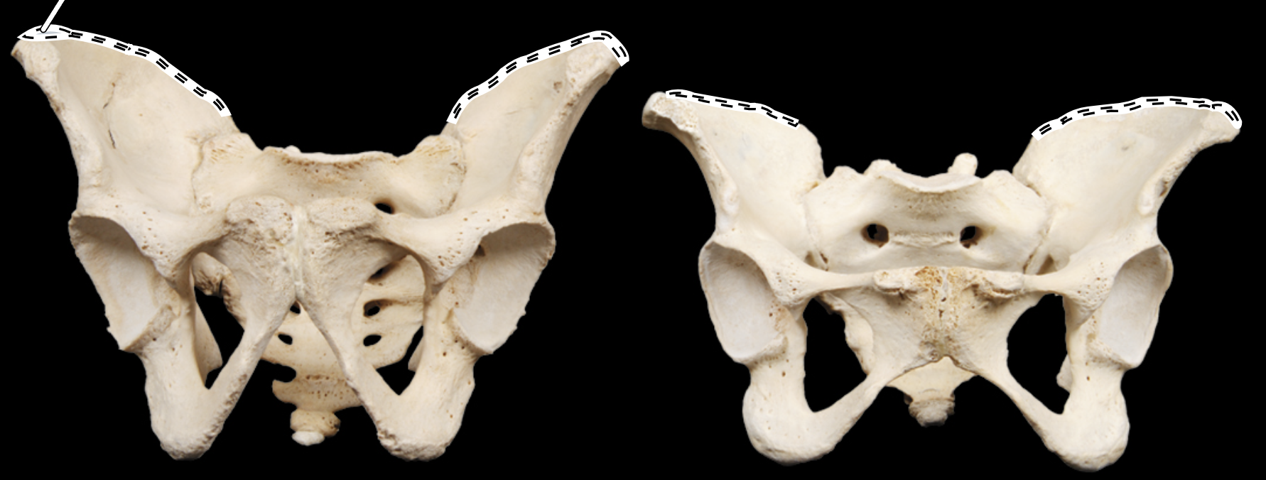
Name the highlighted portion.
(Pelvis, anterior view).
Iliac crest.
Name the highlighted portion.
(Pelvis, superior view).
Iliac crest.
Name the highlighted portion.
(Hip bone, medial view, right side).
Iliac fossa.
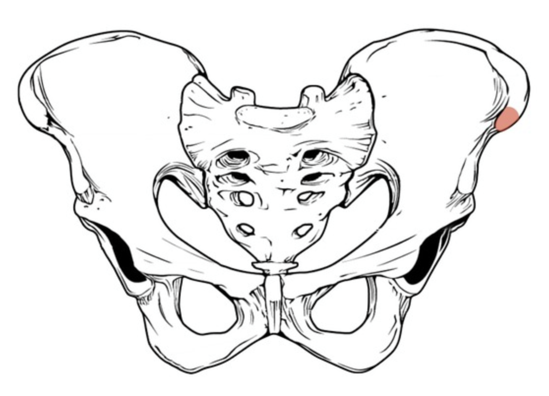
Name the highlighted portion.
Iliac spine (anterior superior iliac spine).
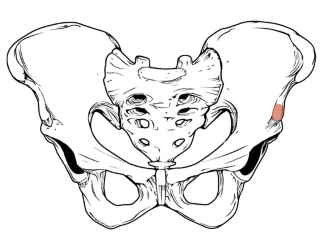
Name the highlighted portion.
Iliac spine (anterior inferior iliac spine).
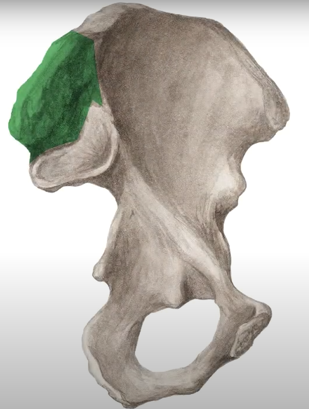
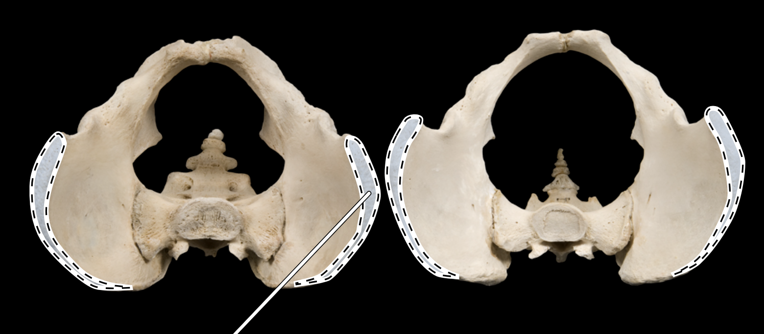
Name the highlighted portion.
Iliac tuberosity.
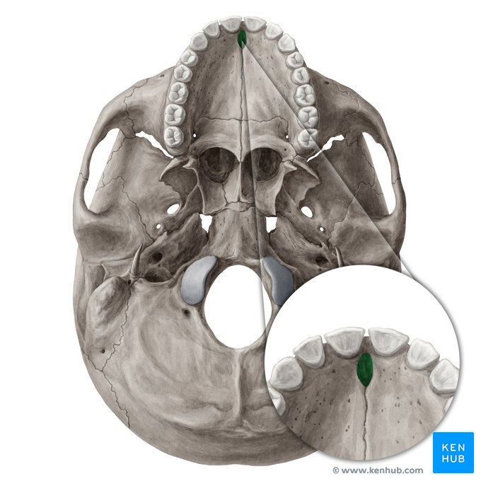
Name the highlighted portion.
Incisive foramen.
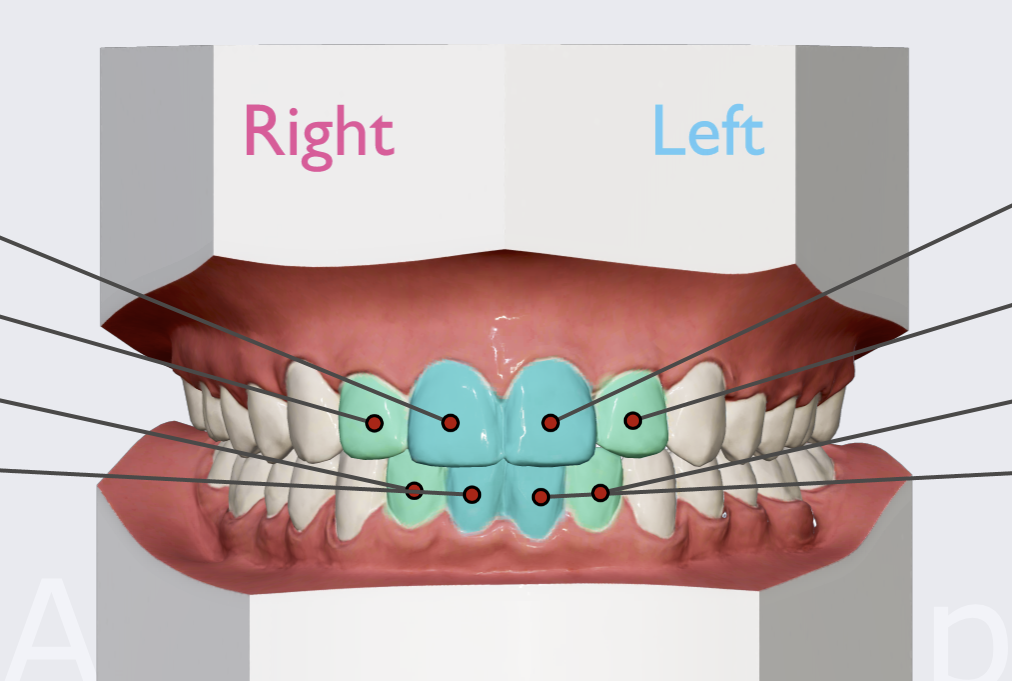
Name the teeth highlighted in blue and teal.
Incisors.
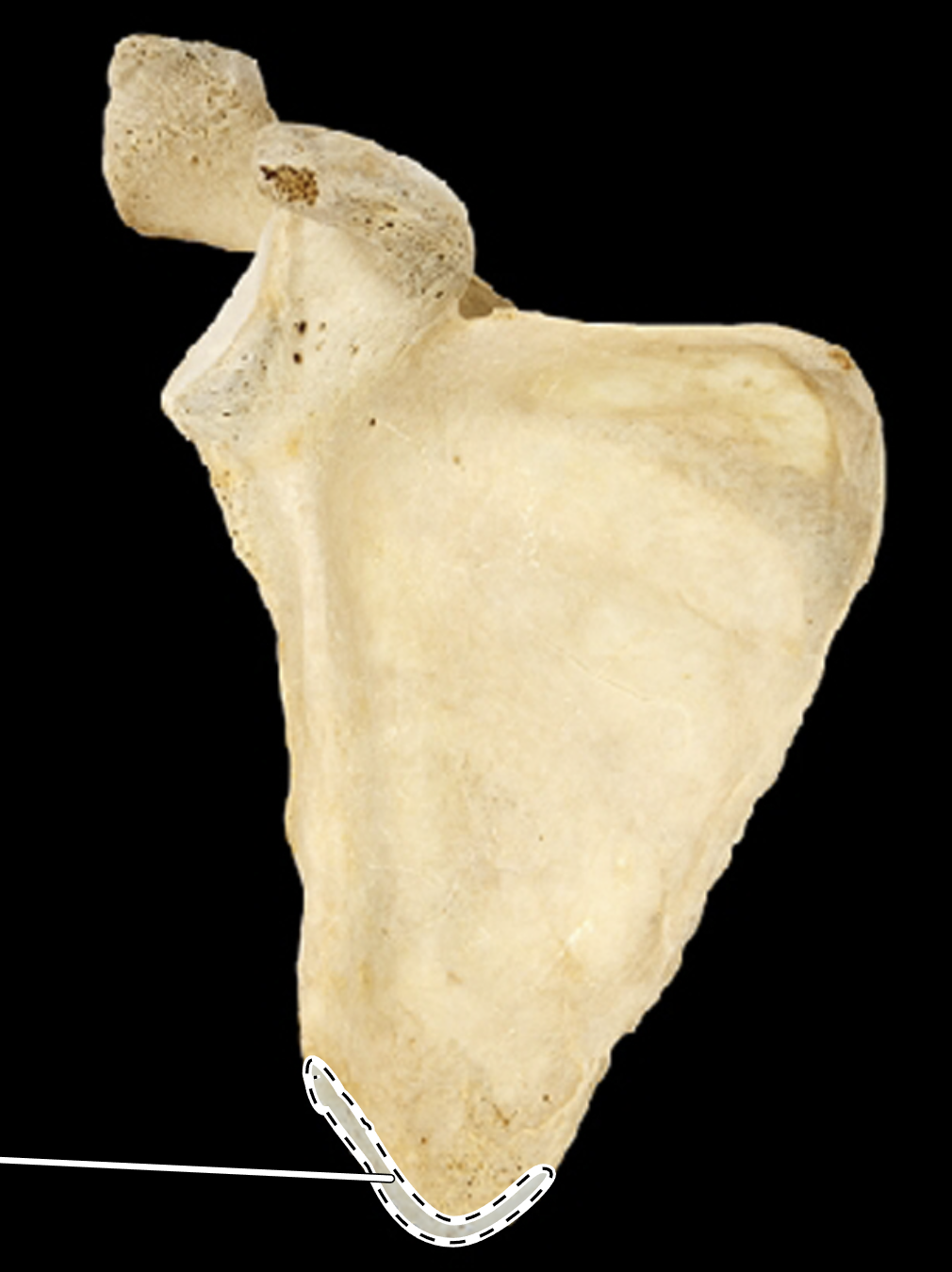
Name the highlighted portion.
(Anterior view, right side).
Inferior angle of scapula.
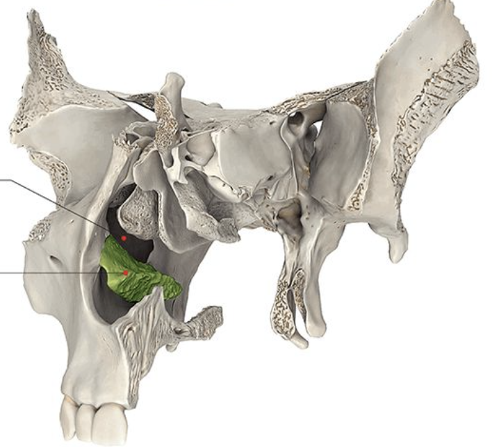
Name the bone highlighted in green.
Inferior nasal concha.
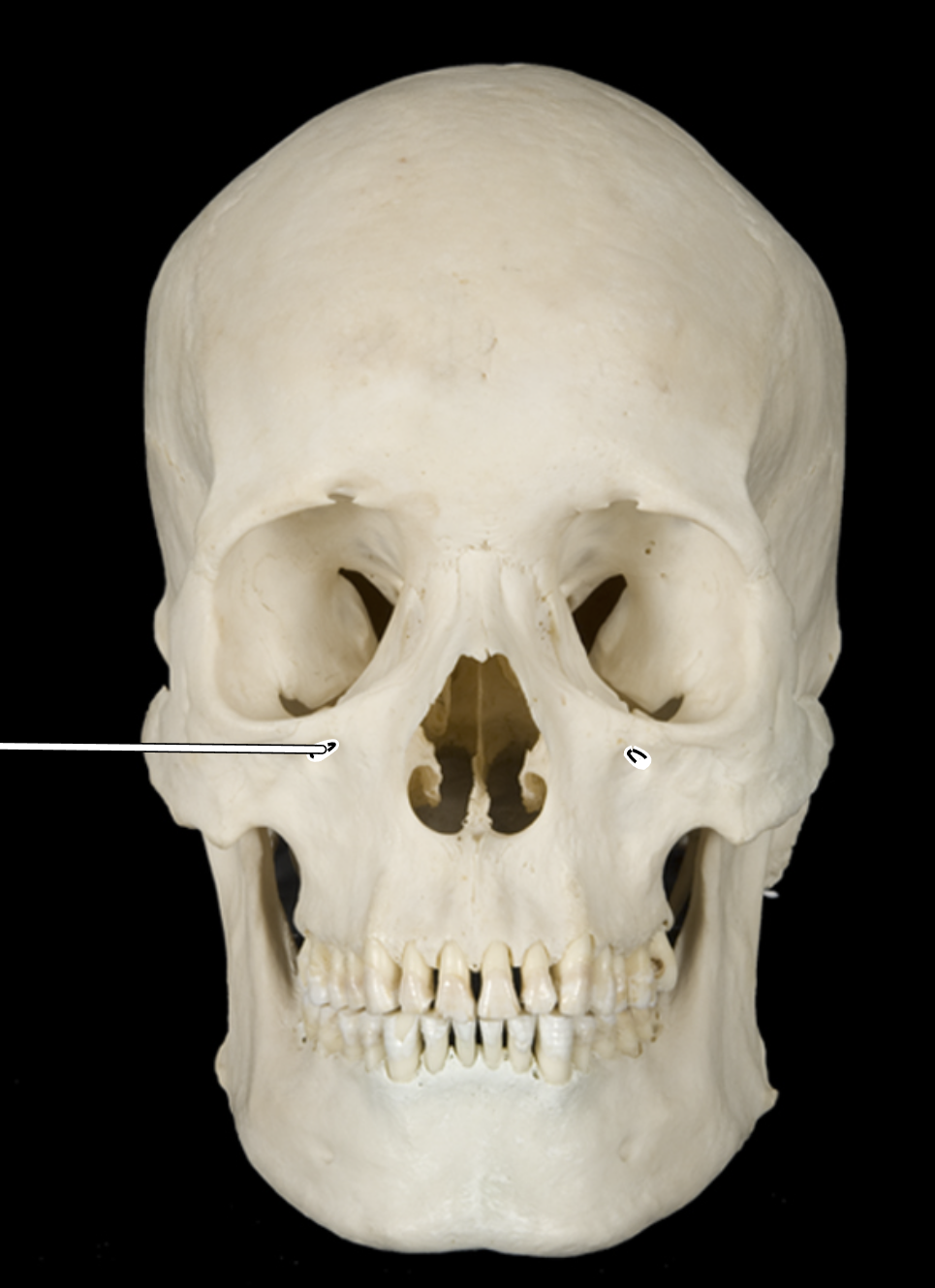
Name the highlighted portion.
(Skull, anterior view).
Infraorbital foramen.
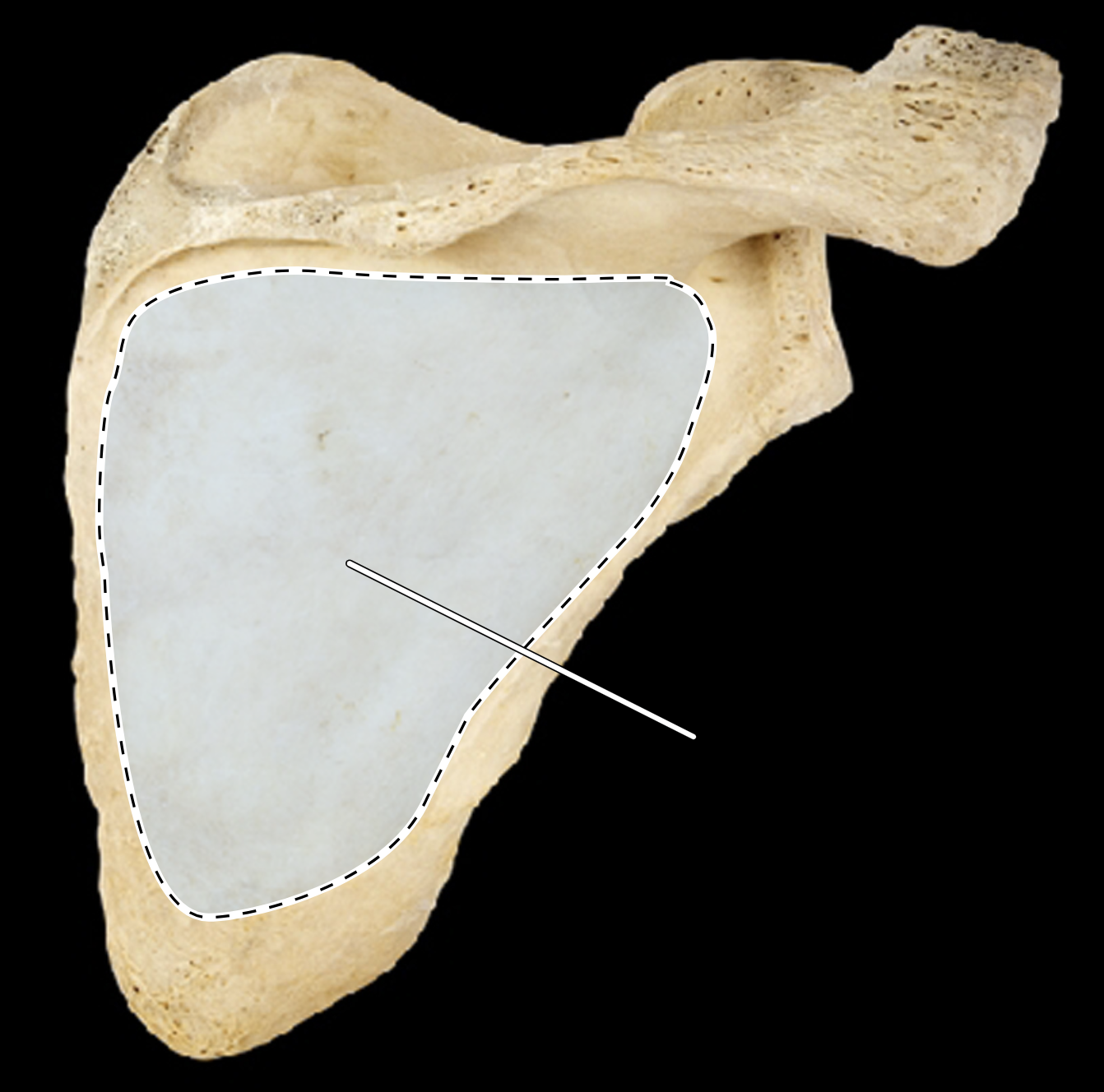
Name the highlighted portion.
(Scapula, posterior view, right side).
Infraspinous fossa.
Name the highlighted portion.
(Posterior view, right side).
Intercondylar eminence.
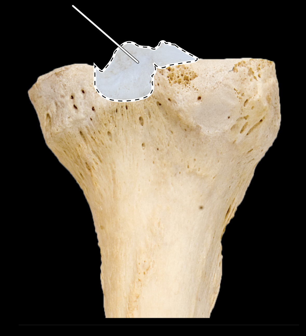
Name the highlighted portion.
(Posterior view, right side).
Intercondylar eminence.
Name the highlighted portion.
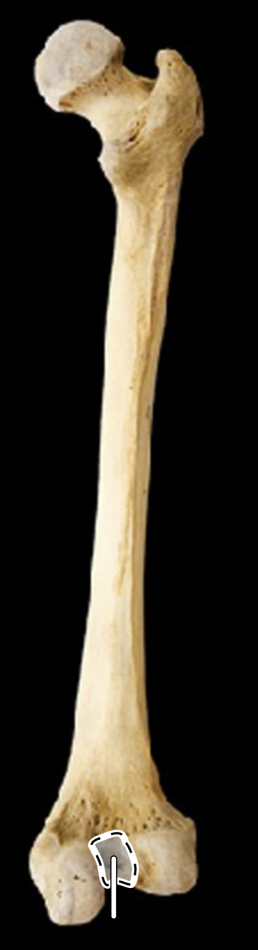
(Posterior view, right side).
Intercondyloid notch of femur (intercondylar fossa).
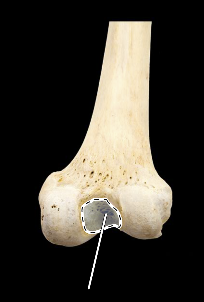
Name the highlighted portion.
(Posterior view, right side).
Intercondyloid notch of femur (intercondylar fossa).
Name the highlighted portion.
(Posterior view, right side).
Intertrochanteric crest.
Name the highlighted portion.
(Posterior view, right side).
Intertrochanteric crest.
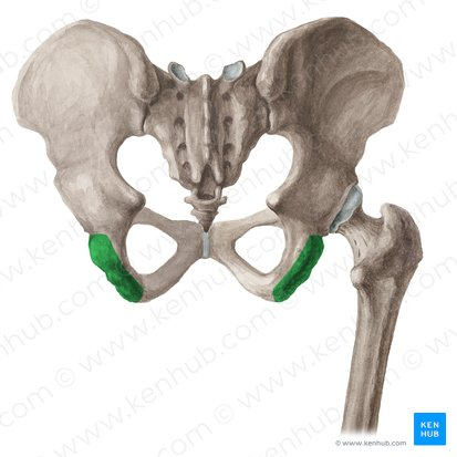
Name the highlighted portion.
Ischial tuberosity.
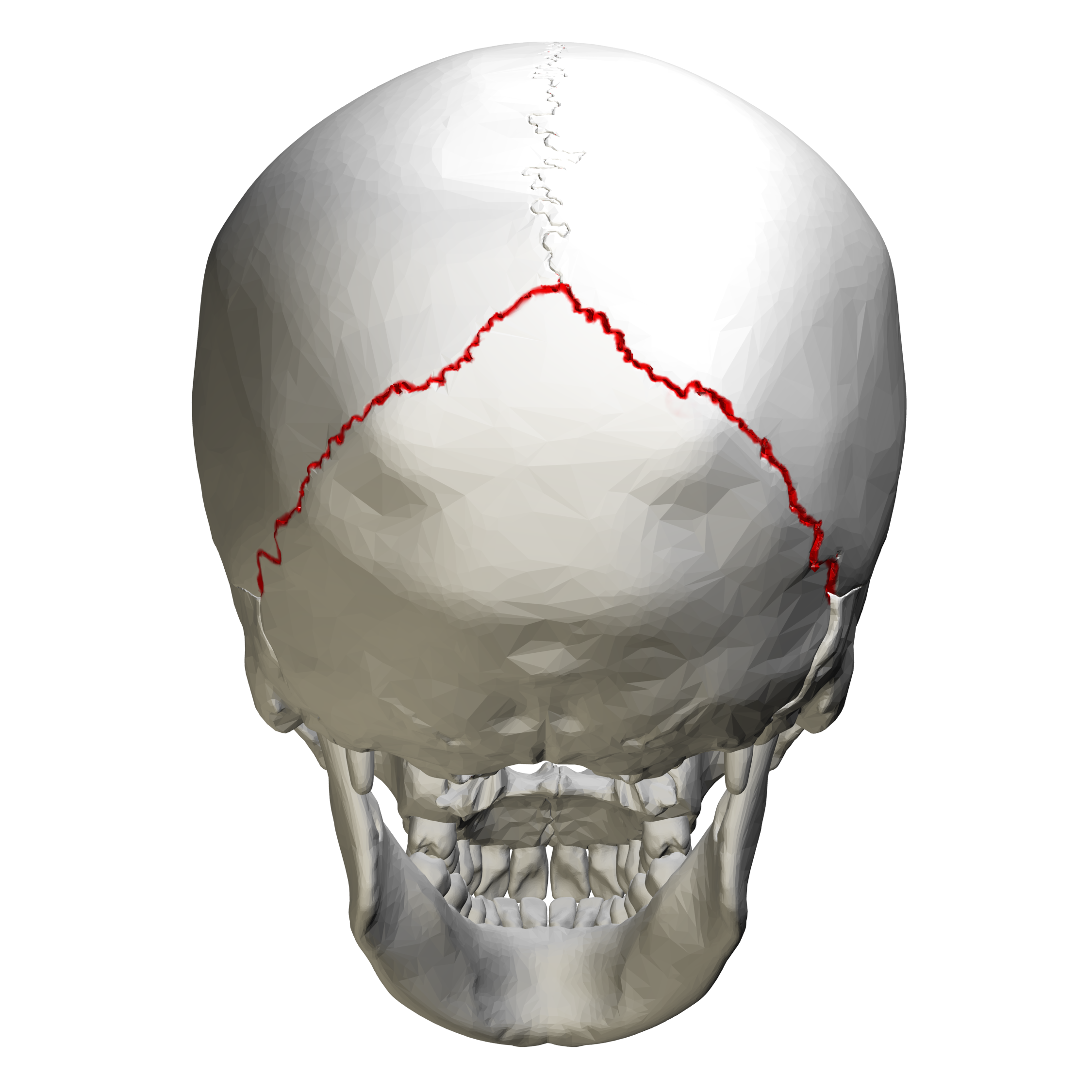
Name the highlighted portion.
Lambdoidal suture.
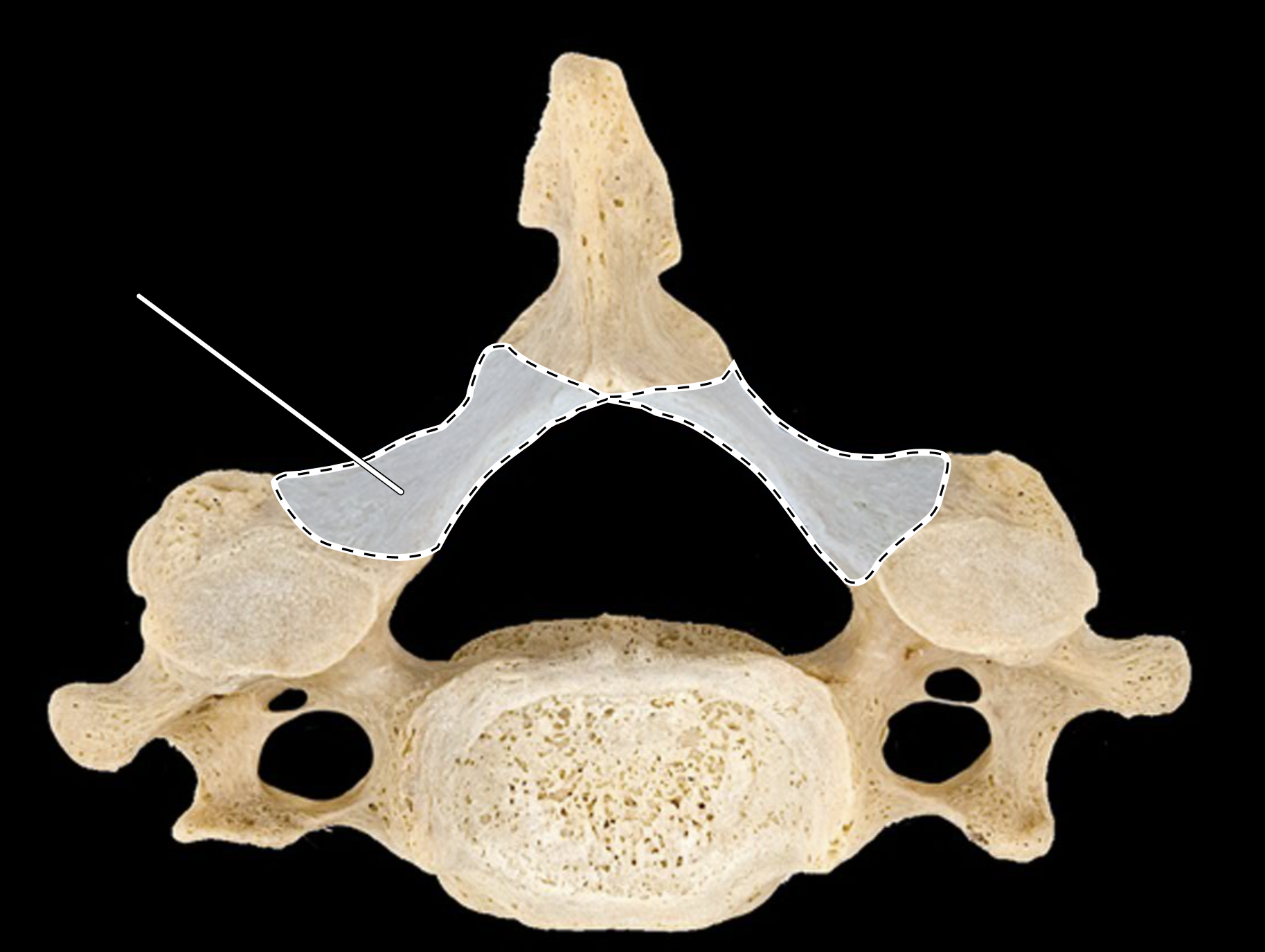
Name the highlighted portion.
(Superior view).
Lamina.
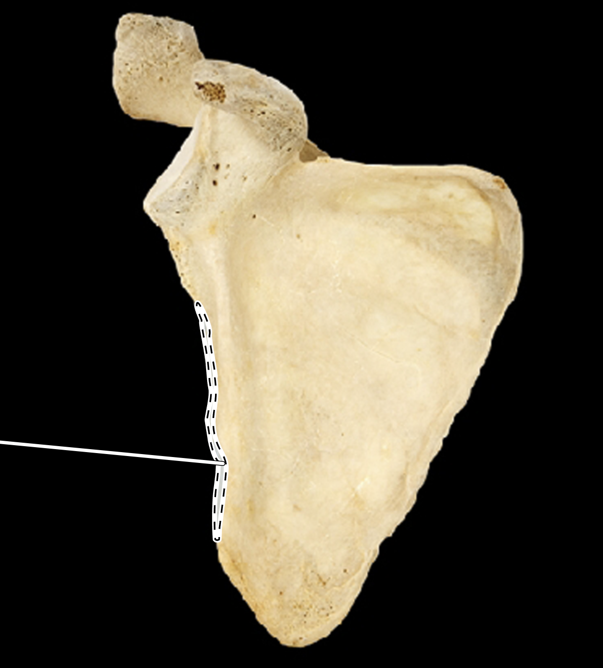
Name the highlighted portion.
(Anterior view, right side).
Lateral border of scapula (axillary border).
Name the highlighted portion.
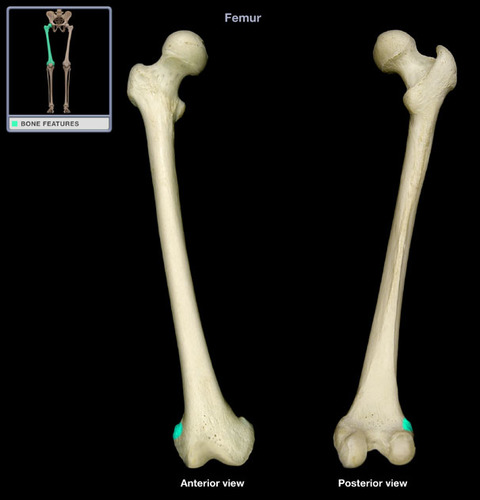
(Right side).
Lateral condyle (of femur).
Name the highlighted portion.
(Right side).
Lateral condyle (of tibia).
Name the highlighted portion.
(Right side).
Lateral epicondyle of femur.
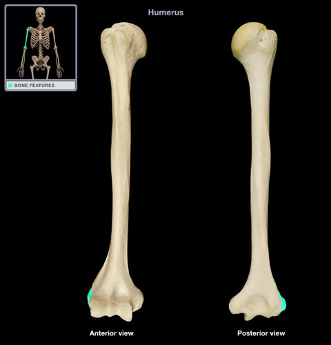
Name the highlighted portion.
(Right side).
Lateral epicondyle of humerus.
Name the highlighted portion.
(Right side).
Lateral malleolus (of fibula).
Name the highlighted portion.
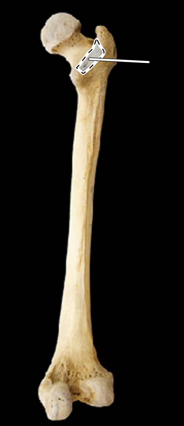
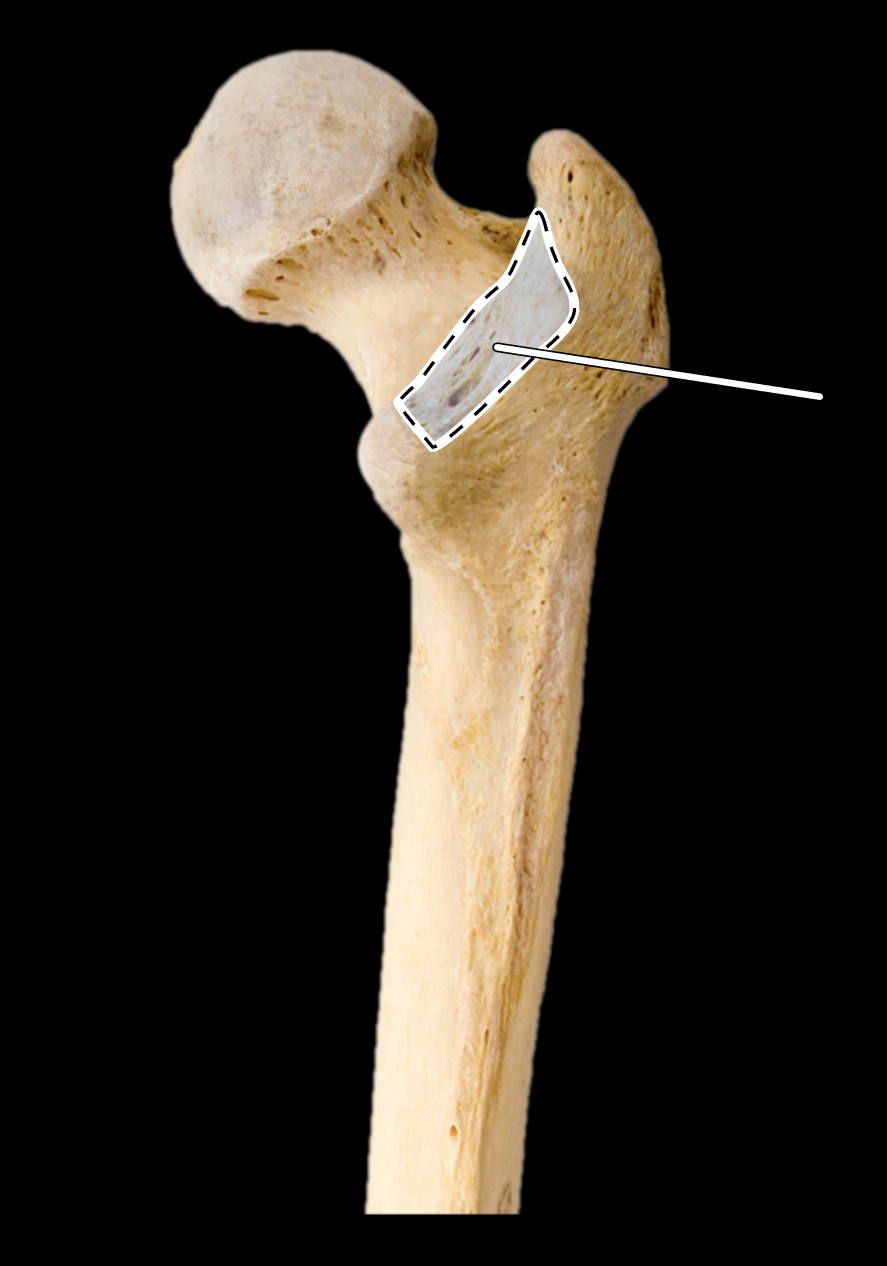
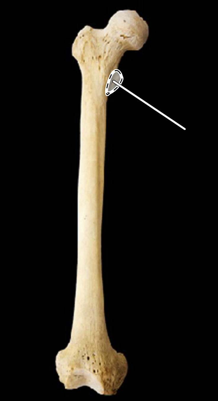
(Anterior view, right side).
Lesser trochanter.
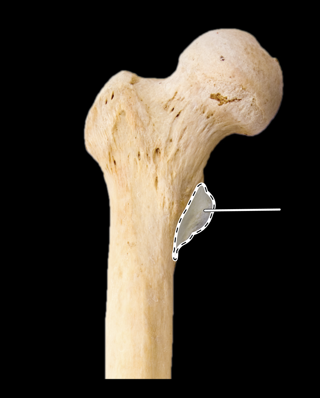
Name the highlighted portion.
(Anterior view, right side).
Lesser trochanter.
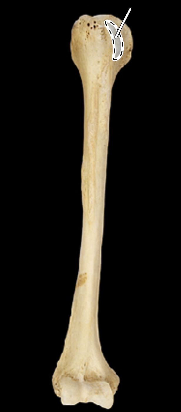
Name the highlighted portion.
(Anterior view, right side).
Lesser tubercle.
Name the highlighted portion.
(Anterior view, right side).
Lesser tubercle.
Name the portion highlighted in green.
Lesser wing of sphenoid.
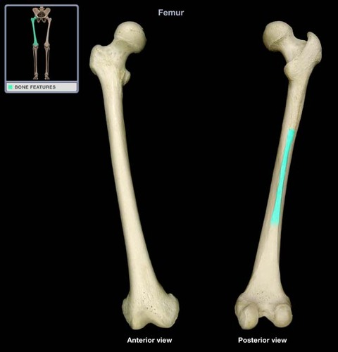
Name the highlighted portion.
(Rght side).
Linea aspera.
Name the highlighted portion.
Mandibular condyle.
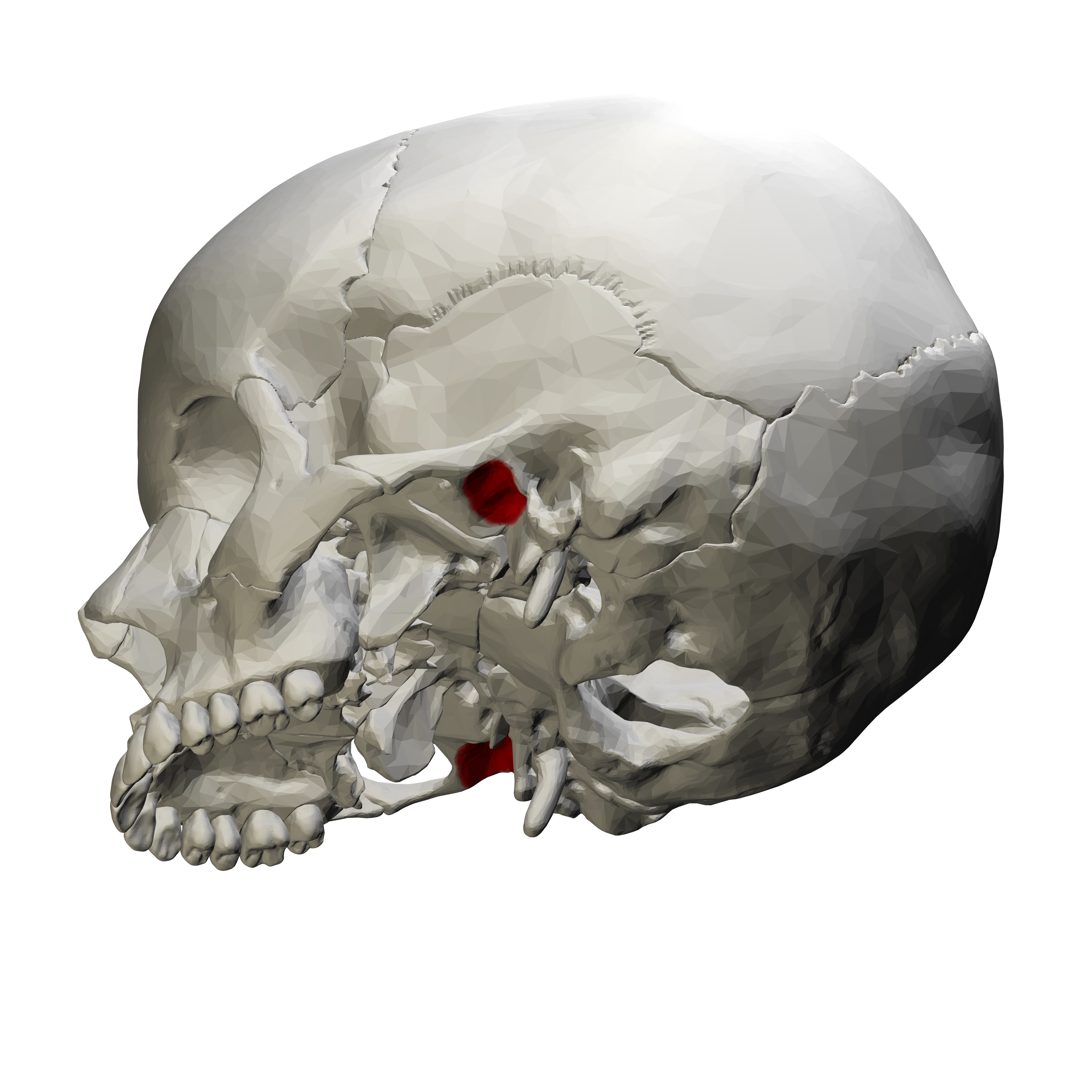
Name the highlighted portion.
Mandibular fossa.
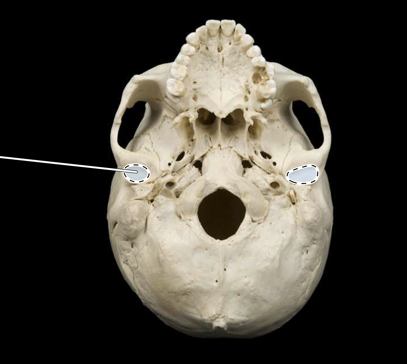
Name the highlighted portion.
Mandibular fossa.
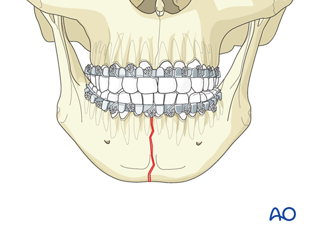
Name the portion highlighted in red.
Mandibular symphysis.