HP: Lec 14- neural control of SM, cardiac and smooth muscles
1/24
There's no tags or description
Looks like no tags are added yet.
Name | Mastery | Learn | Test | Matching | Spaced |
|---|
No study sessions yet.
25 Terms
Neural control of skeletal muscles- lower motor neurons
nervous system controls skeletal muscles
Lower motor neurons: cell bodies in the ventral horn of spinal cord or in the brain stem- influenced by:
stimulation or inhibition from upper motor neurons from brain
sensory feedback from muscles and tendons via reflexes
Skeletal Muscle Reflexes
skeletal muscles are usually referred to as voluntary and are controlled by descending motor pathways under conscious control
they can also contract unconsciously in response to certain stimuli- a reflex
monosynaptic reflex
disynaptic reflex involving two synapses
Sensory feedback on skeletal muscle
Golgi tendon organs: respond to tension a muscle puts on a tendon
Muscle spindle apparatus: respond to muscle length
muscles that require more control have more spindles
stretching a muscle causes spindles to stretch
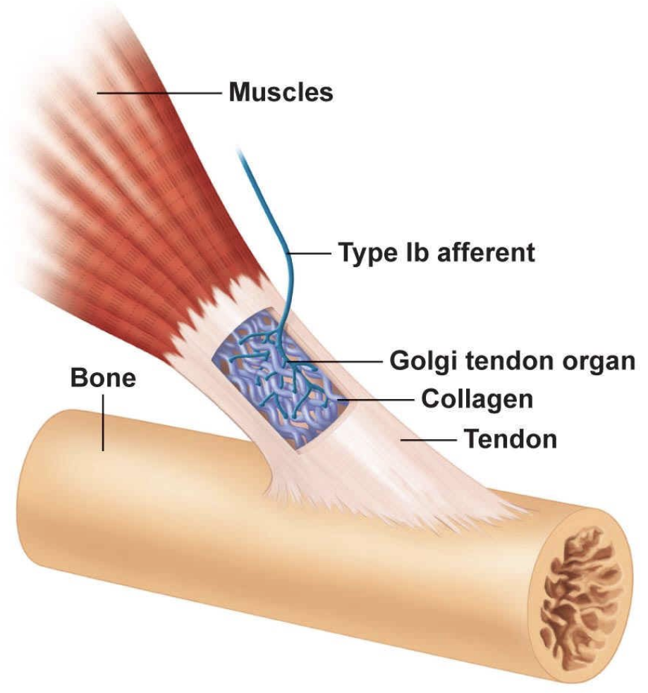
Golgi Tendon Organ
what is it?
where does it lie?
what are they stimulated with?
what does it cause?
function?
golgi tendon organ is a proprioceptive sensory receptor organ that sense changes in muscle tension
it lies at the origins and insertions of skeletal muscle fibers into the tendons of skeletal muscle
are stimulated with excessive tension during muscle contraction
cause a reflex inhibition of the muscle
function: to protect the muscle and connective tissue from injury
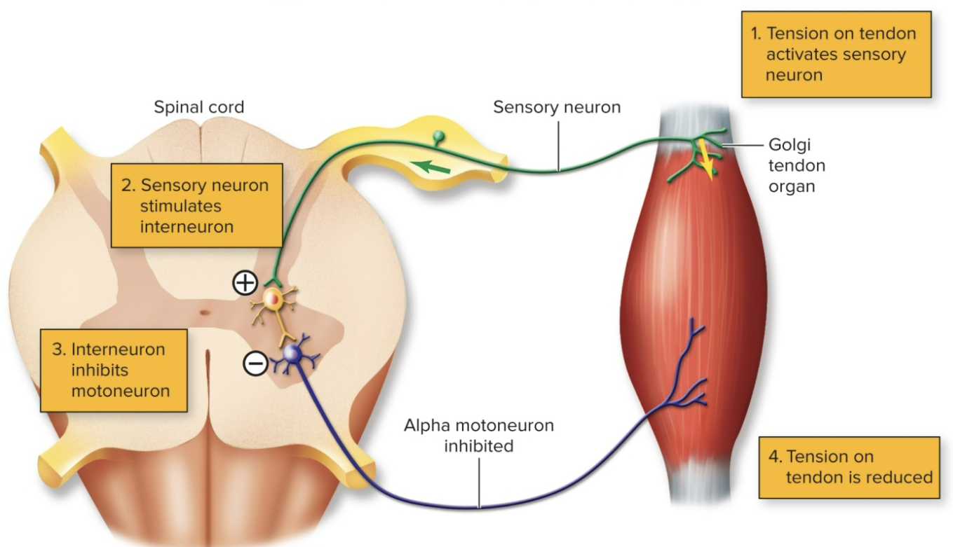
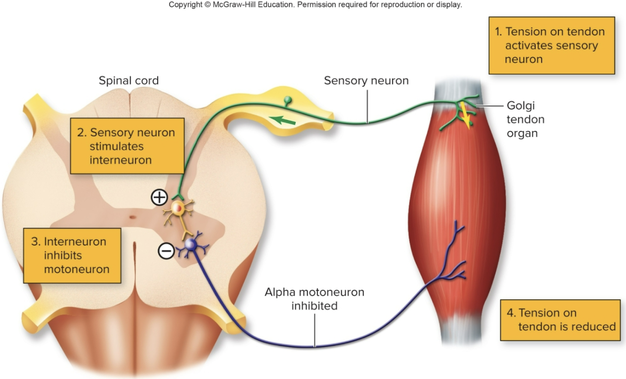
Inhibitory Golgi tendon reflex- how does the golgi tendon reduce tension?
constantly monitors tension in tendons
sensory neuron stimulates interneuron in spinal cord
interneuron inhibits motor neuron
tension in tendon is reduced
disynaptic reflex involving two synapses
Muscle spindle
what does it consist of?
what are they surrounded by?
these consist of very specialized muscle fibers called intrafusal fibers
the intrafusal fibers are surrounded by the “normal fibers, known as "extrafusal” fibers (typical skeletal muscle cell)
intrafusal fibers detect length and rate of change of the muscles thanks to specific receptors within the intrafusal muscle fibers called muscle spindle
What happens when a muscle spindle’s associated muscle is rapidly stretched?
the spindle can cause 2 things to happen
it may signal its muscle to contract to prevent it from going to far, too quickly in the stretch
it can inhibit the opposing muscle (the antagonist to the muscle being stretched) to prevent it from contracting so that it can’t contribute to any further stretching
What is the primary function of the muscle spindle?
the muscle spindle functions to alert the brain that nearby joints and soft tissues are in danger of being stretched too far
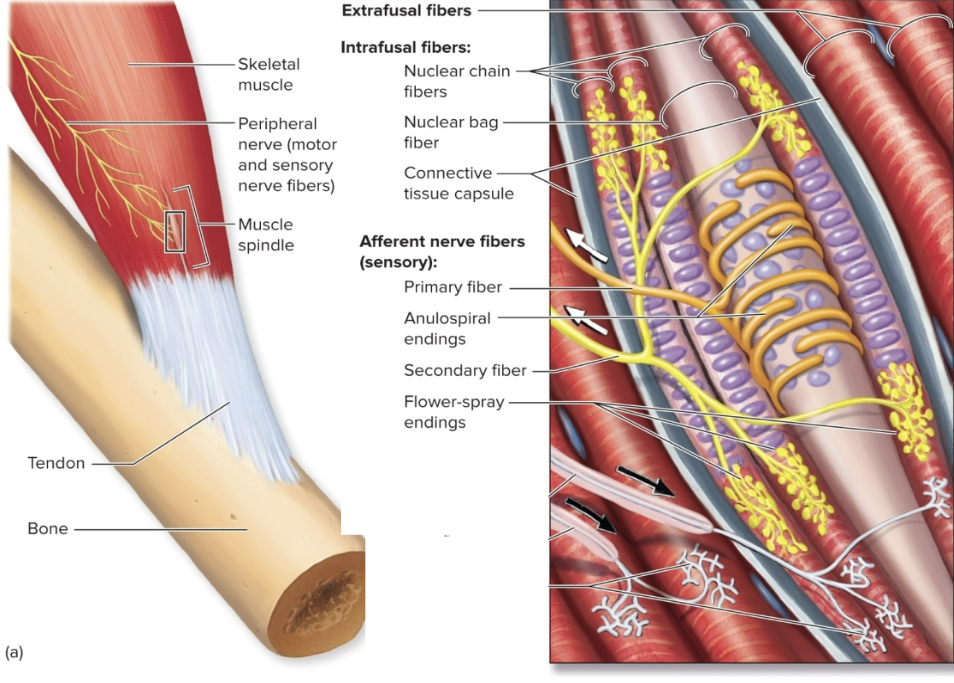
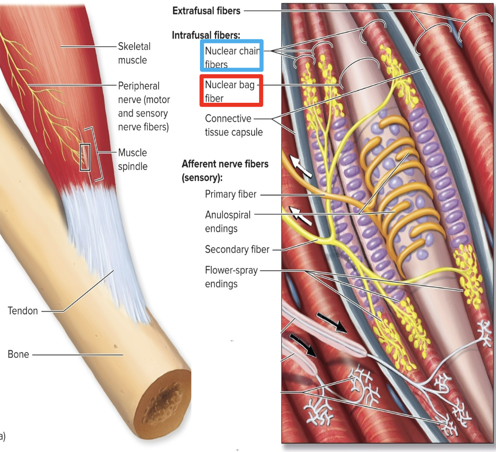
Types of Intrafusal fibers
Nuclear Bag Fibers: nuclei in loose central aggregates
Nuclear chain fibers: nuclei in rows
Types of sensory cells wrapped around the intrafusal fibers
Anulospiral endings: most stimulated at the beginning of the stretch (located in the nuclear bag fibers)
Flower Spray: respond more during sustained stretch (located in the nuclear chain fibers)
Monosynaptic Reflex
what is it?
what does it involve?
where is it loacted?
function?
simplest reflex
only involves a sensory neuron synapsing on a motor neuron in the spinal cord- ONE synapse monosynaptic
maintains optimal resting length of skeletal muscles- muscle stretch reflex
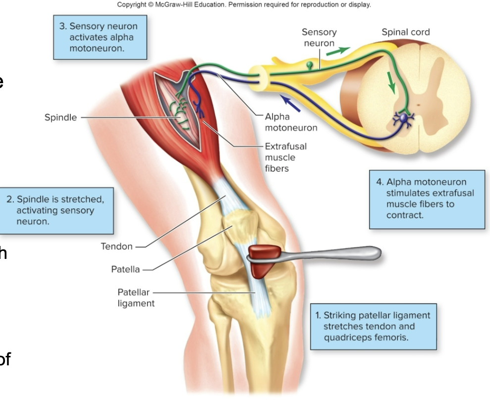
How can the monosynaptic reflex be stimulated? step 1-3
can be stimulated by striking the patellar ligament of the “knee-jerk reflex”:
passive stretch of a muscle (produced by tapping its tendon) stretches the spindle (intrafusal) fibers
stretching of a spindle distorts its central (bag or chain) region, which stimulates sensory neurons
action potentials are conducted by afferent (sensory) nerve fibers into the spinal cord on the dorsal roots of the spinal nerves
How can the monosynaptic reflex be stimulated? steps: 4-6
axons of sensory neurons, synapse with dendrites and cell bodies of somatic motor neurons located in the ventral horn grey matter of the spinal cord
efferent nerve impulses in the axons of motor neurons are conducted to the “ordinary” (extrafusal) muscle fibers
contraction of the muscle relieves the stretch of its spindles, thus decreasing activity in the spindle afferent nerve fibers
Monosynaptic Reflex Image
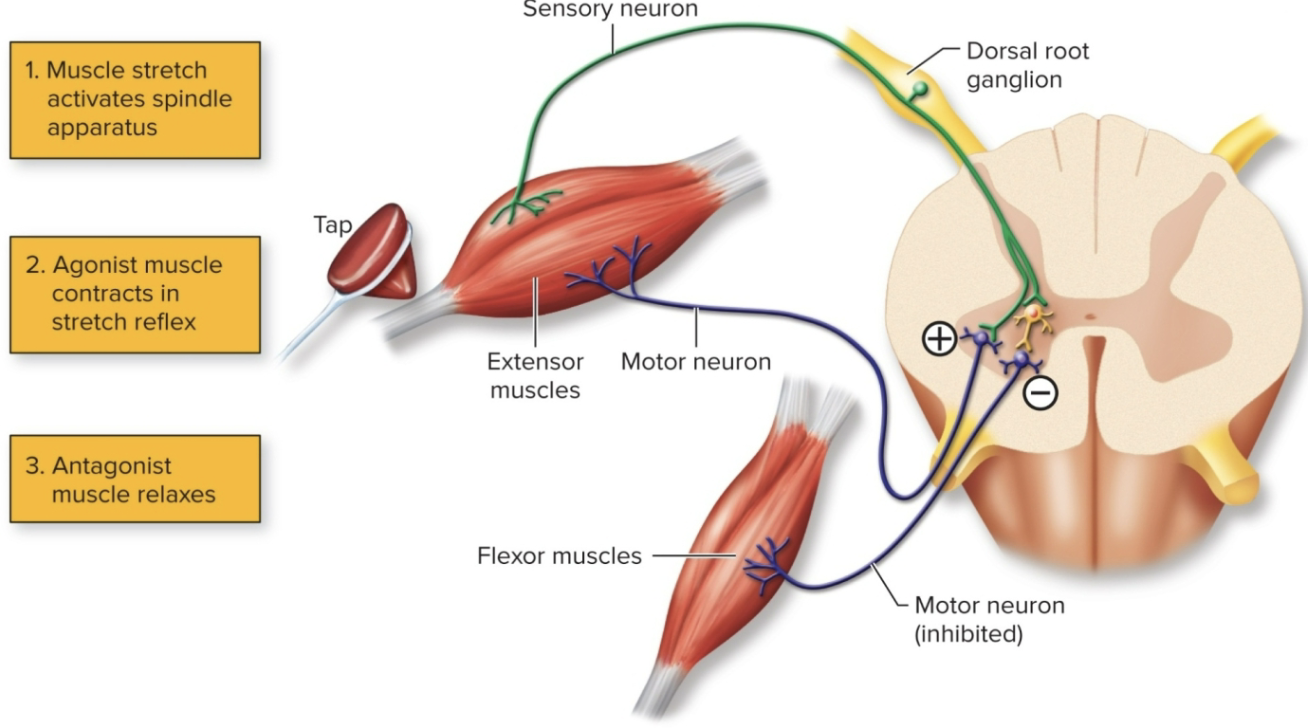
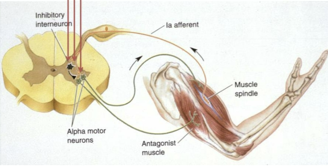
Skeletal muscle Reflexes: monosynaptic & disynaptic working together
in the knee jerk reflex, we have both the monosynaptic and disynaptic reflex working together… but why?
interneurons are also stimulated in the spinal cord to inhibit antagonistic muscles on that limb
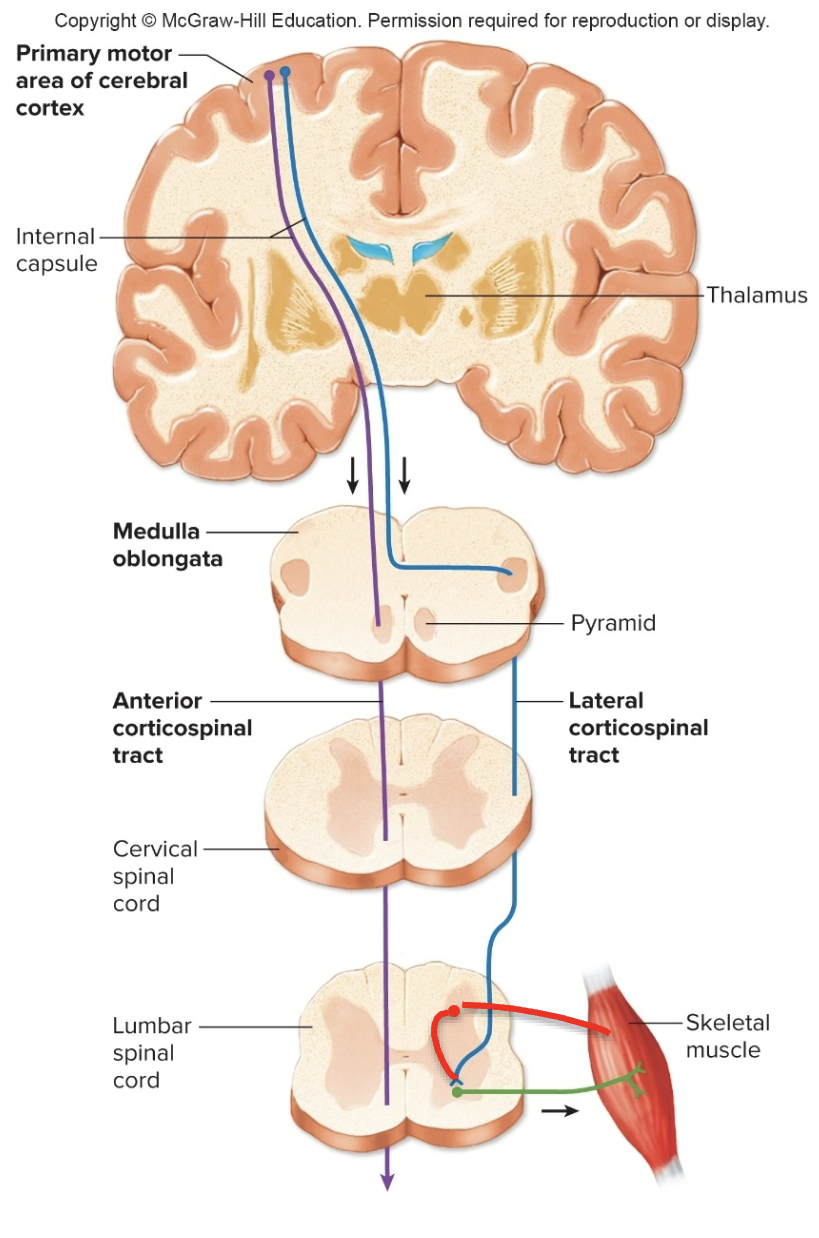
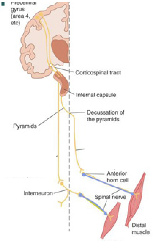
Upper Motor Neuron Control- 2 types of tracts
Upper motor neurons may stimulate or inhibit motor actions
Pyramidal tracts (contralateral) by the lateral and ventral corticospinal tracts
Extrapyramidal tracts: reticulospinal and vestibulospinal tracts- inhibition of lower motoneurons
important for posture and to prevent exaggerated stretch reflexes, and hyperactivity
What is Paralysis?
paralysis is a loss of strength in and control over a muscle or group of muscles- most of the time this is not due to a problem with the muscles themselves, it is more likely due to a problem somewhere along the nerve cells delivering the signals for your muscles to move
Types of paralysis (1)
Flaccid Paralysis: damage to lower motor neurons or spinal nerve
reduces muscle tone
depressed stretch reflexes
atrophy- muscles shrink and become flabby
Types of Paralysis (2)
Spastic Paralysis: damage to UPPER motor neurons or descending tracts
increased muscle tone
exaggerated stretch reflexes and hyperactivity- tight and hard
spasticity is probably caused by imbalance of signals from the upper motor neurons
ex: the removal of inhibitory influences exerted by the extrapyramidal tracts on the postural centers of the vestibular nuclei and reticular formation
Amytrophic Lateral Sclerosis
what is it?
what does it produce?
amytrophic lateral sclerosis (ALS-Lou Gehrig’s disease) is a neurodegenerative disease that affects both lower motor neurons in the spinal cord (leading to muscle wasting, or amyotrophy) and upper motor neurons
produces progressive muscle weakness and atrophy, and spastic paraylsis, death usually occurs from respiratory failure within 5 years from the onset of symptoms
Amytrophic Lateral Sclerosis and multiple sclerosis
ALS and MS (multiple sclerosis) are NOT the same disease, they sometimes are confused bc they have some similarities but they have more differences
Amytrophic Lateral Sclerosis
ALS: a disease that DESTROYS the motor neurons so that the body cannot communicate with the brain, symptoms of ALS progress continually, and result i paralysis, and death within a few years after the initial diagnosis
multiple sclerosis
MS is a disease that results in the destruction of the protective coating (myelin sheath) on the nerves of the CNS which causes a faulty relay of instructions from the brain to the body
many people have mild symptoms for years with periods of remission
Amytrophic Lateral Sclerosis and multiple sclerosis
MS is an autoimmune disease, while ALS is hereditary in up to 10 out of 100 people due to mutated proteins (in the rest-majority- is caused by a combination of environmental and genetic factors)
MS has more mental impairment and ALS has more physical impairment
Atrophy
what is it?
when is it most severe?
what are the two types of atrphy?
what are these types of atrophy caused by?
muscle atrophy (reduction in size), with its accompanying declines in strength, is most severe when it results from damage to the motor nerves, innervating the muscles
neurogenic atrophy may be the result of disease such as ALS or trauma
disuse atrophy can result when a person is bedridden for a period of time or is in a cast, so that the muscles are not working as they normally do to move a load against the force of gravity