ANHB2212 - Week 4 of Embryo Formation
1/30
There's no tags or description
Looks like no tags are added yet.
Name | Mastery | Learn | Test | Matching | Spaced | Call with Kai |
|---|
No analytics yet
Send a link to your students to track their progress
31 Terms
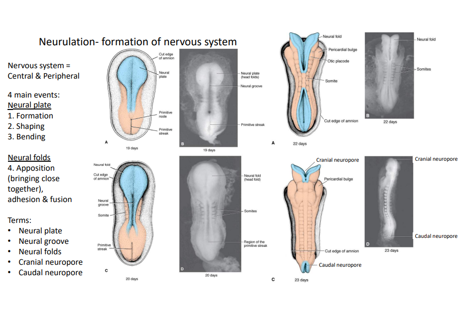
What are the 4 main events that occur during neurulation?
Neural plate:
1. Formation
2. Shaping
3. Bending
Neural folds:
4. Appositioning (bringing close together), adhesion & fusion
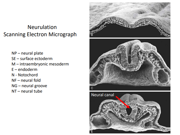
Neural plate
A thickened, flat layer of specialized ectoderm cells on the dorsal surface of a vertebrate embryo, forming during the third week of gestation as the first step in neurulation.
What induces neurulation?
Notochord
Neural Groove
A shallow, longitudinal median depression in the neural plate of an early vertebrate embryo.
Neural Folds
Elevated ridges of ectodermal tissue that rise on either side of the neural groove during early embryonic development.
Cranial Neuropore
The temporary, open cranial end of the developing neural tube in an embryo. It acts as a minute, fleeting, open communication between the neural tube and the amniotic cavity.
Caudal Neuropore
The temporary, open caudal end of the developing neural tube in an embryo. It acts as a minute, fleeting, open communication between the neural tube and the amniotic cavity.
Describe the process of primary neurulation.
At 20 days, the notochord induces the neural plate to fold towards the midline, forming the neural fold, neural groove and immature somites. By day 22, the neural folds rise and the neural groove deepens, causing the neural folds to adhere and fuse. By day 22, there is complete appositioning of the neural folds except for the cranial and caudal neuropore, which remain open. By now, somites have developed and the formation of the neural tube from the neural plate is complete.
What does the neural tube develop into in the future via secondary neurulation?
Sacral and coccygeal regions.
Describe the process of secondary neurulation?
After day 26 when the caudal neuropore closes, the tail bud develops a medullary cord. This cord develops a lumen. The lumen, medullary cord and neural tube merge and become continuous. This forms the basis of the central nervous system.

When does the cranial neuropore close?
Day 24
When does the caudal neuropore close?
Day 26
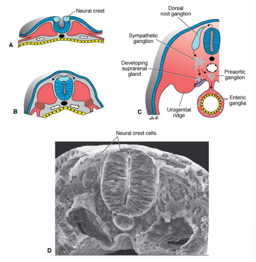
Neural Crest Cells
Form at the tips of the neural folds, migrate away when neural tube closure is complete to form the peripheral nervous system.
Somatopleure
Ectoderm + lateral plate mesoderm
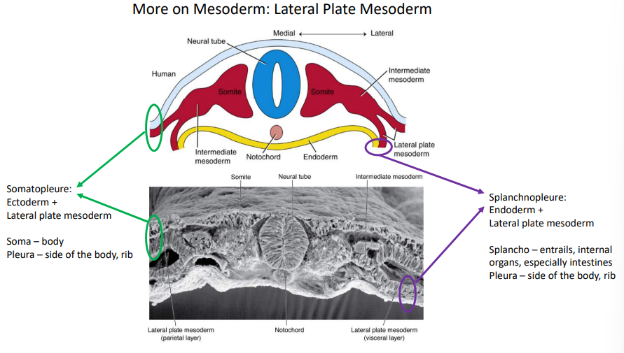
Splanchnopleure
Endoderm + lateral plate mesoderm

Etymology: Soma
Body
Etymology: Pleura
Side of the body, rib
Etymology: Splancho
entrails, internal organs
Describe the process of lateral folding.
At day 17, the lateral ends of the embryo begin to fold towards the midline. By day 22, the intraembryonic coelom forms as a space between the two layers of lateral plate mesoderm that’s continuous with the primitive body cavity. The rapid growth of the amnion surrounds and pinches the yolk sac to form the vitelline duct.
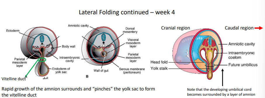
What is the last region of the embryo to undergo complete lateral folding?
Vitelline duct
Vitelline duct
Small channels connect the intraembryonic and extraembryonic coeloms and persist until the ventral body wall is completely sealed.
Describe the process of craniocaudal folding.
At day 17, the cranial and caudal ends of the embryo begin to fold towards each other. Beginning at day 22, there is repositioning of the primordial heart, septum transversum, oropharyngeal membrane and intraembryonic coelum from superior to ventral to inferior to the brain.
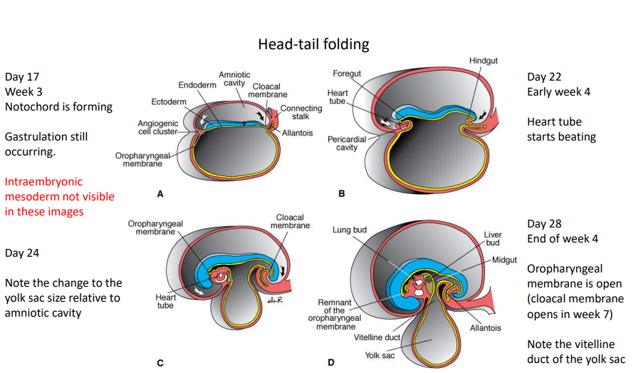
When does the heart tube start beating?
Day 22
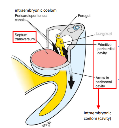
Septum Transversum
The immature diaphragm allows communication between the primitive pericardial cavity and the intraembryonic coelom via the pericardioperitoneal canals.
What are 3 defects in primary neurulation?
spina bifida
anencephaly
teratomas
Spina Bifida
Failure of caudal neuropore closure
Anencephaly
Failure of cranial neuropore closure
Teratomas
Appear in the sacrococcygeal regions due to remnants of primitive streak that cause the presence of muscle, cartilage, fat, hair and glandular tissue that arise from all germ layers.
What are 3 lateral folding defects?
Failure to close the ventral abdominal wall cranial-umbilicus
Failure to closure the ventral abdominal wall caudal-umbilicus
Uncovered amnion
Meckel’s Cartilage
A feature arising cranial to the pharyngeal arches that acts as jaw support during early development and gives rise to 2 ear ossicles: malleus and incus (not stapes).
Rathke’s Pouch
Marks the boundary between the surface ectoderm and endoderm.