Cranium - Module 7
1/39
There's no tags or description
Looks like no tags are added yet.
Name | Mastery | Learn | Test | Matching | Spaced |
|---|
No study sessions yet.
40 Terms
Cranial bones of the skull
Cranial bones
- Temporal
- Parietal
- Occipital
- Frontal
Ethmoid
Sphenoid

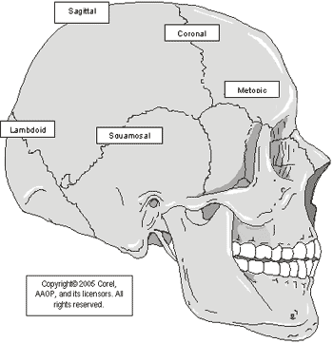
Cranial Sutures
Lateral
Superior
- Coronal suture
- Sagittal suture
Posterior
- Lambdoid structure
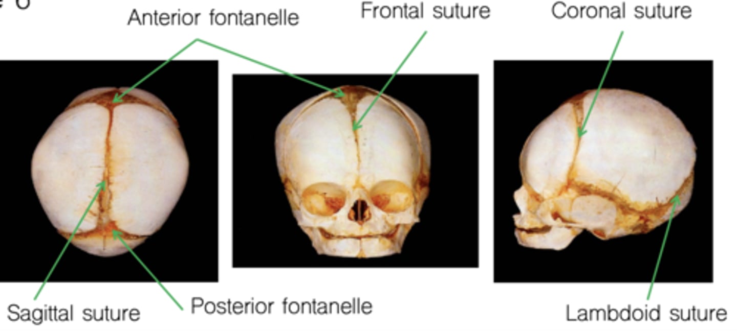
Infant Skull: Pre-sutures
"Soft spots" in infant skulls
Close at various times
- Posterior fontanelle (sagittal & lambdoid suture): by 6 months
- Anterior fontanelle (frontal & coronal sutures): ~middle of second year
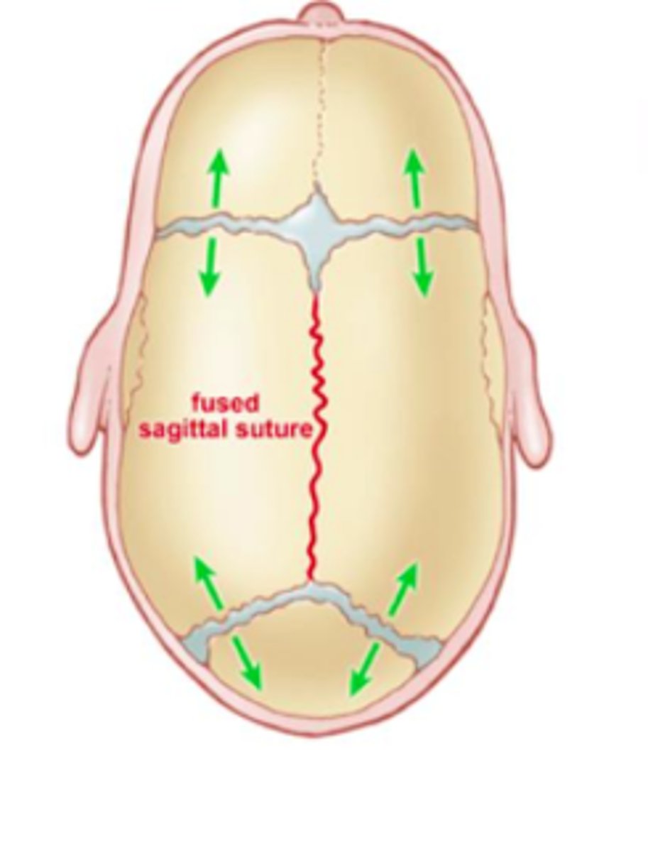
Craniosynostosis
Premature fusing of sutures
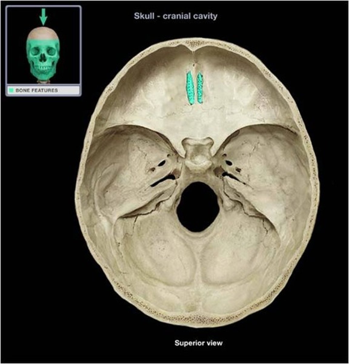
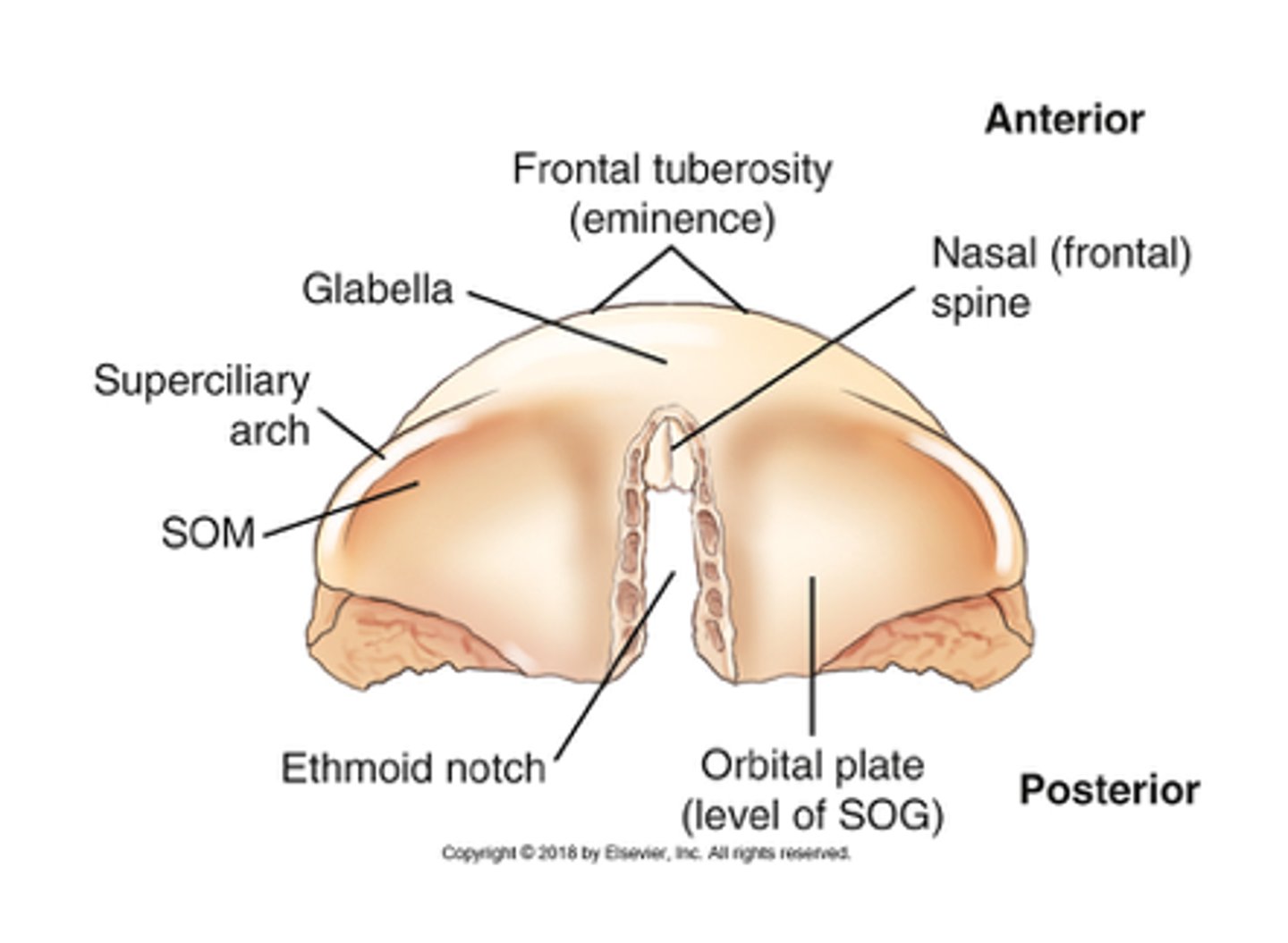
Frontal Bone Description
Forms anterior part of brain case
Vertical plate contributes to forehead
Horizontal plate contributes to roof of orbit and nasal cavities
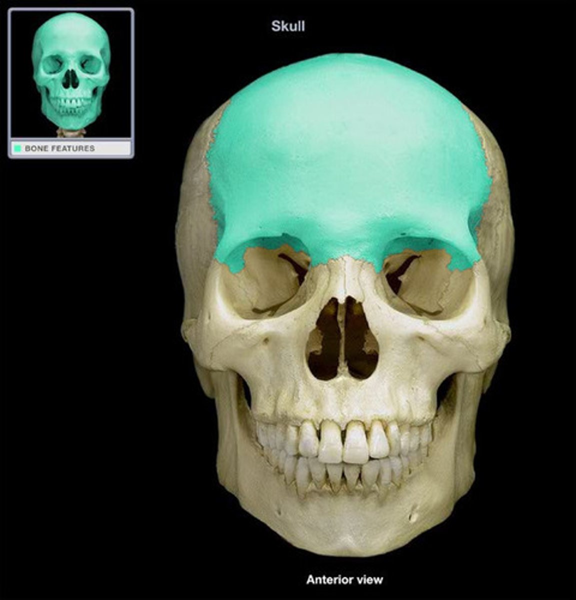
Zygoamtic process
Lateral to orbital surfaces for articulation with zygomatic bone
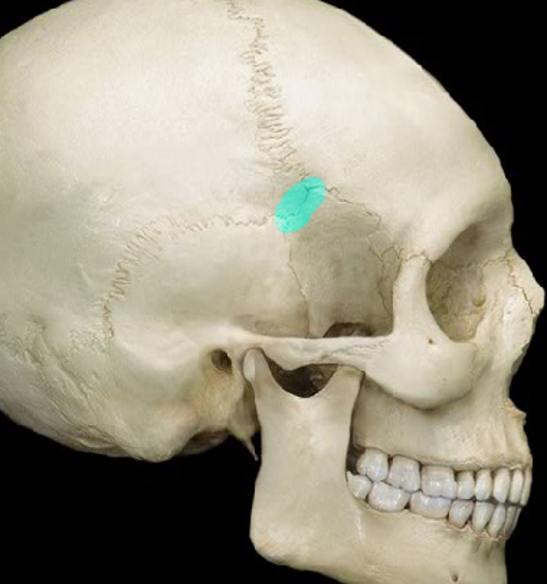
Frontal Bone - Ethmoid Notch
Midline notch called ethmoid notch articulates with ethmoid bone
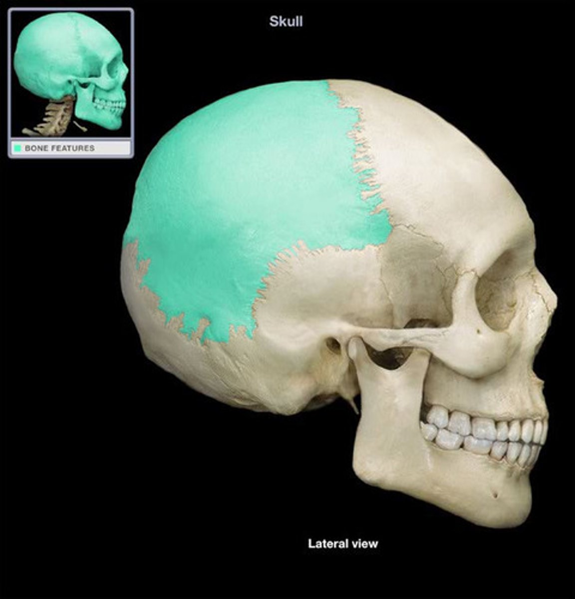
Parietal Bones Description
Form most of rounded roof of cranium
Each bone roughly quadrilateral
Attachments
- Anterior: frontal bone at coronal suture
- Medial: midline at sagittal suture with other parietal bone
- Posterior: occipital bone at lambdoid suture
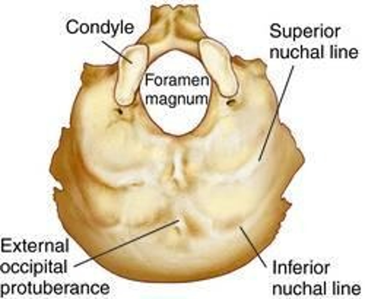
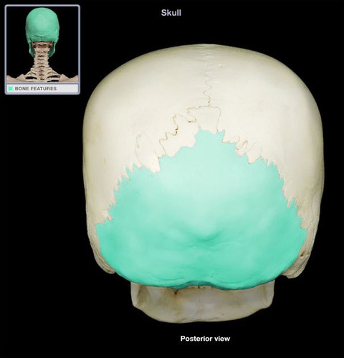
Occipital Bone Description
Inferior and posterior portion of cranium
- Forms lower and back portion of cranium
- Articulates with parietal bones at lambdoid suture
Includes foramen magnum
Includes occipital condyles

Occipital Bone Features
Occipital Condyles
- Lateral to foramen magnum
- Articulate with superior facets of
C1
Just posterior to each condyle
is a condylar fossa
- Recess for superior articular
processes of atlas when head
tilted sharply backward
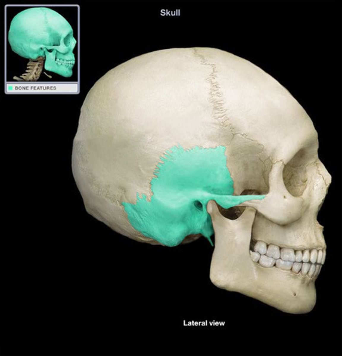
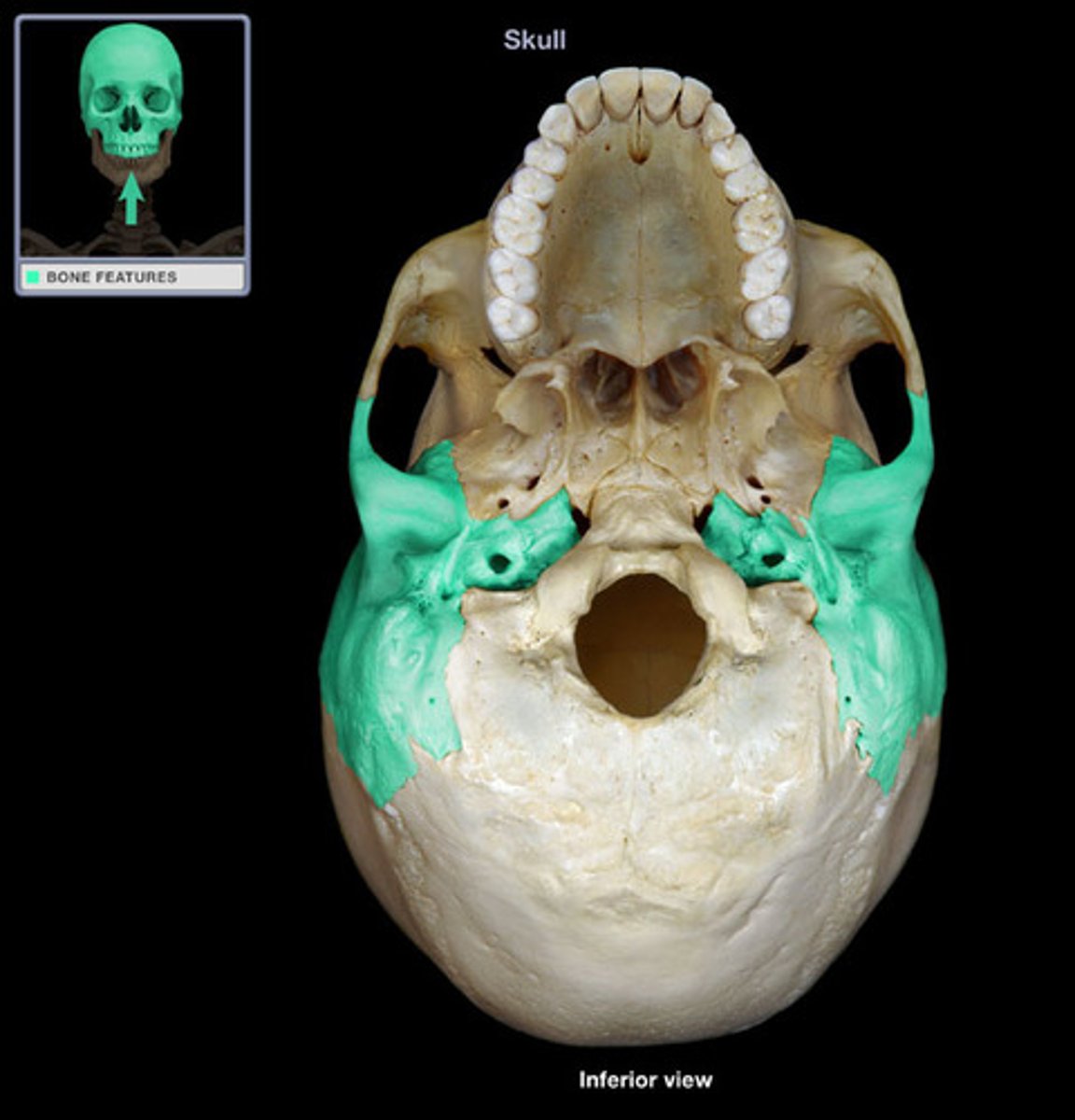
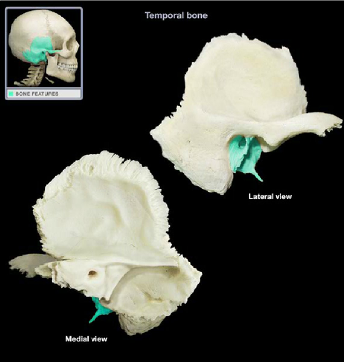
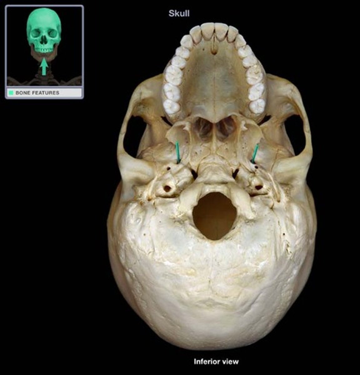
Temporal Bones Description
Paired bones forming most of lateral base and sides of brain case
Each bone has 3 parts
- Squamous part
- Includes zygomatic process
- Petrous part
- Tympanic part
- Includes styloid process

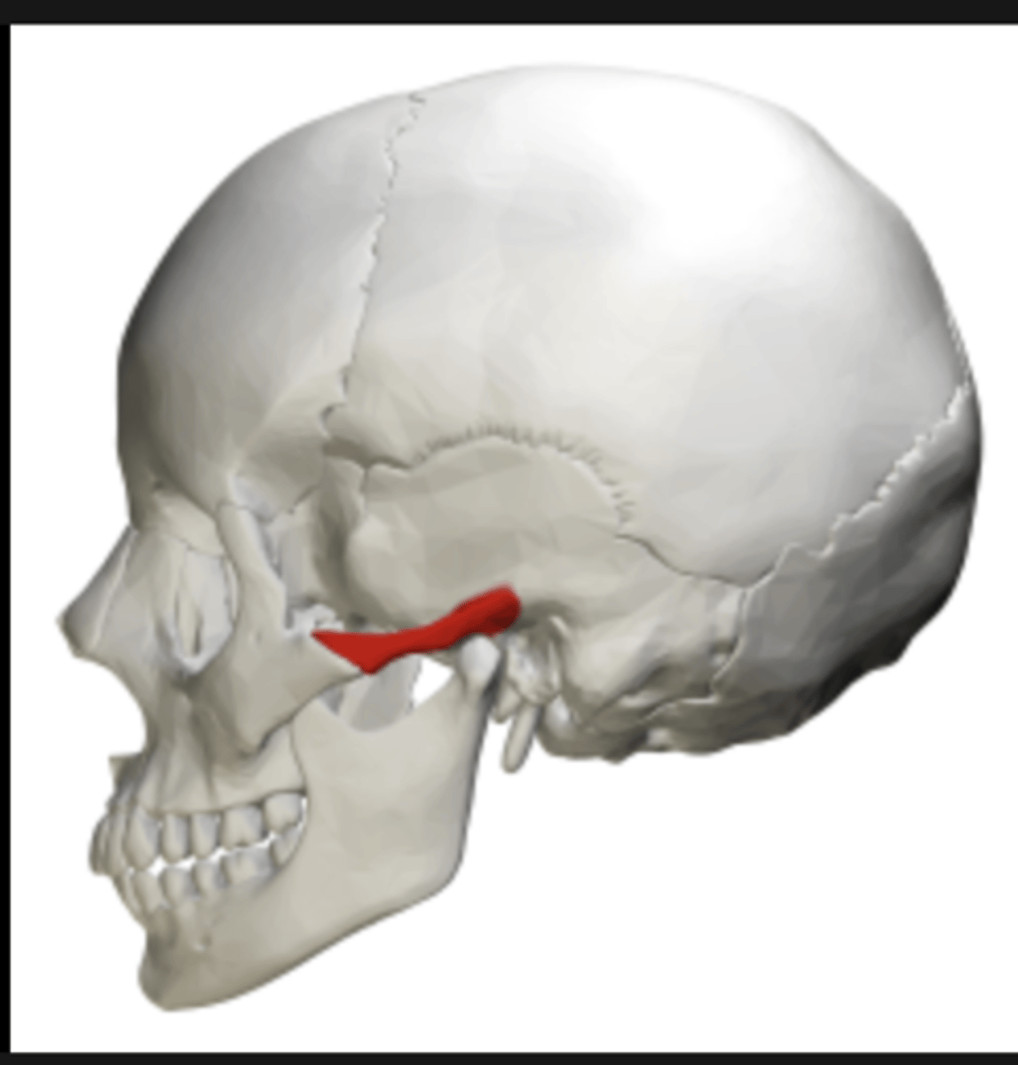
Temporal Bone Features
Zyogamtic process
External auditory meatus
Mastoid process
Temporomandibular joint
Temporal Bone - inferior view
Temporal Bone - Squamous Part
Lateral, anterior, superior aspect of bone
Zygomatic process
- Forms zygomatic arch with temporal
process of zygomatic bone
Mandibular fossa
- Articulates with condylar process of
mandible
- Part of temporomandibular joint (TMJ)
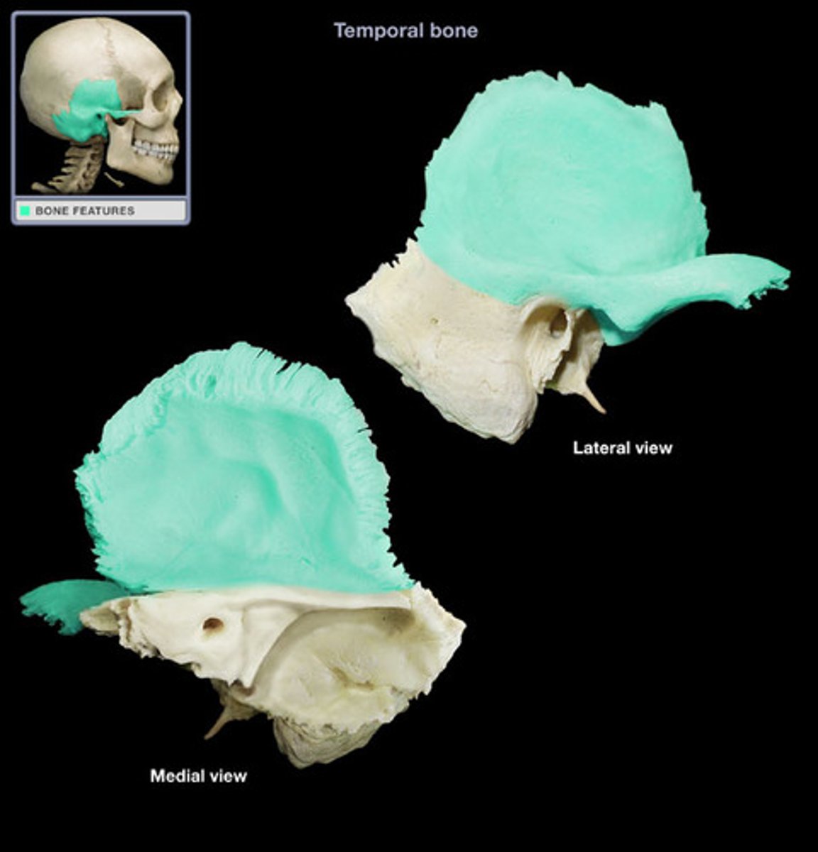
Temporal Bone - Petrous Part
Sometimes called petromastoid part
At base of skull
Houses cochlea and inner ear
Includes mastoid process
- Numerous muscle attachments
Temporal Bone - Tympanic Portion
Anterior to mastoid process and between squamous
and petrous portions
Contains part of external auditory meatus
- Essentially the area immediately around the opening
- Remainder in Petrous portion
Includes styloid process
- Numerous muscle attachments
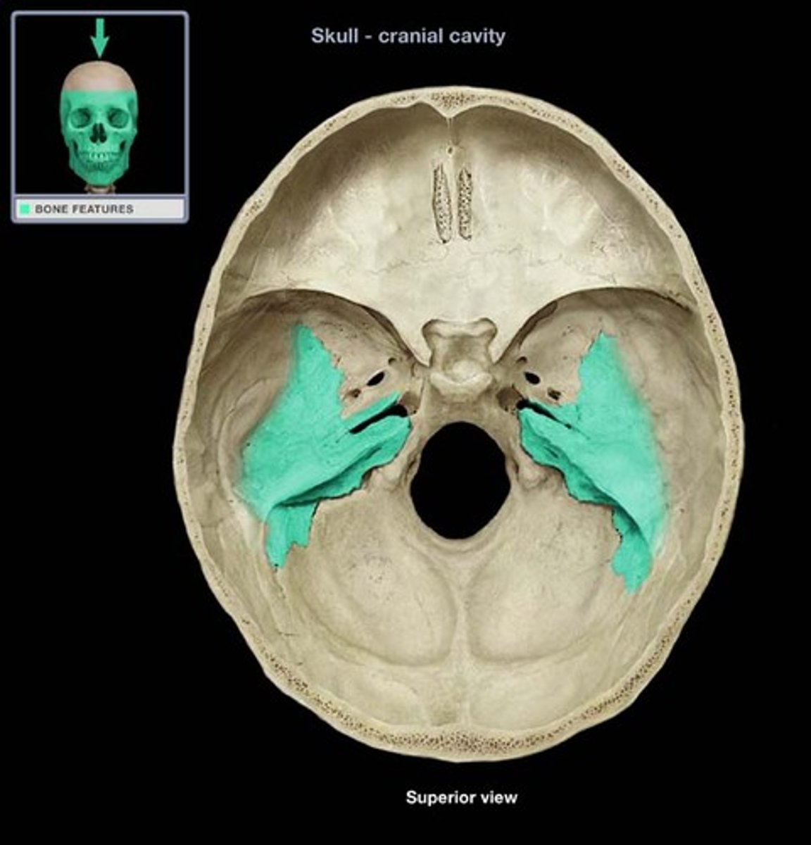
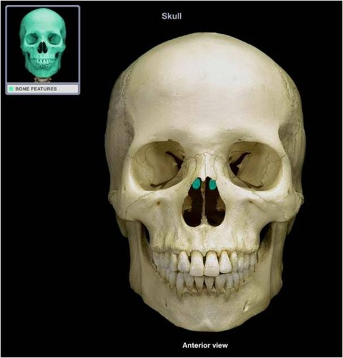
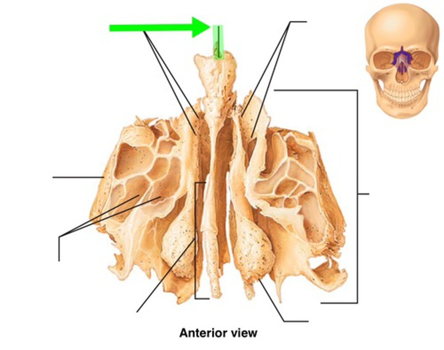
Ethmoid Description
Although regarded as cranial bone, also contributes to facial skeleton
Hangs down from ethmoid notch of frontal bone between orbital plates of frontal bone
Loosely behind and above the maxilla
Contributes to the
- walls of orbit
- lateral walls of nasal cavity
- bony nasal septum
Ethmoid - Frontal
Cribiform plate
Perpendicular plate
Superior and Middle Cochae

Cribiform plate
Horizontal plate that separates cranial and nasal cavities
Perpendicular plate
Thin plate directed inferiorly from underside of cribiform plate
Forms upper portion of bony nasal septum
Superior and Middle Conchae
Two lateral plates house superior and middle nasal concha
Form lateral walls of nasal
cavity
Complex curvature is to increase surface area to warm and humidify air
Ethmoid - Oblique Superior and Inferior
Cribiform plate attaches to
the ethmoid notch
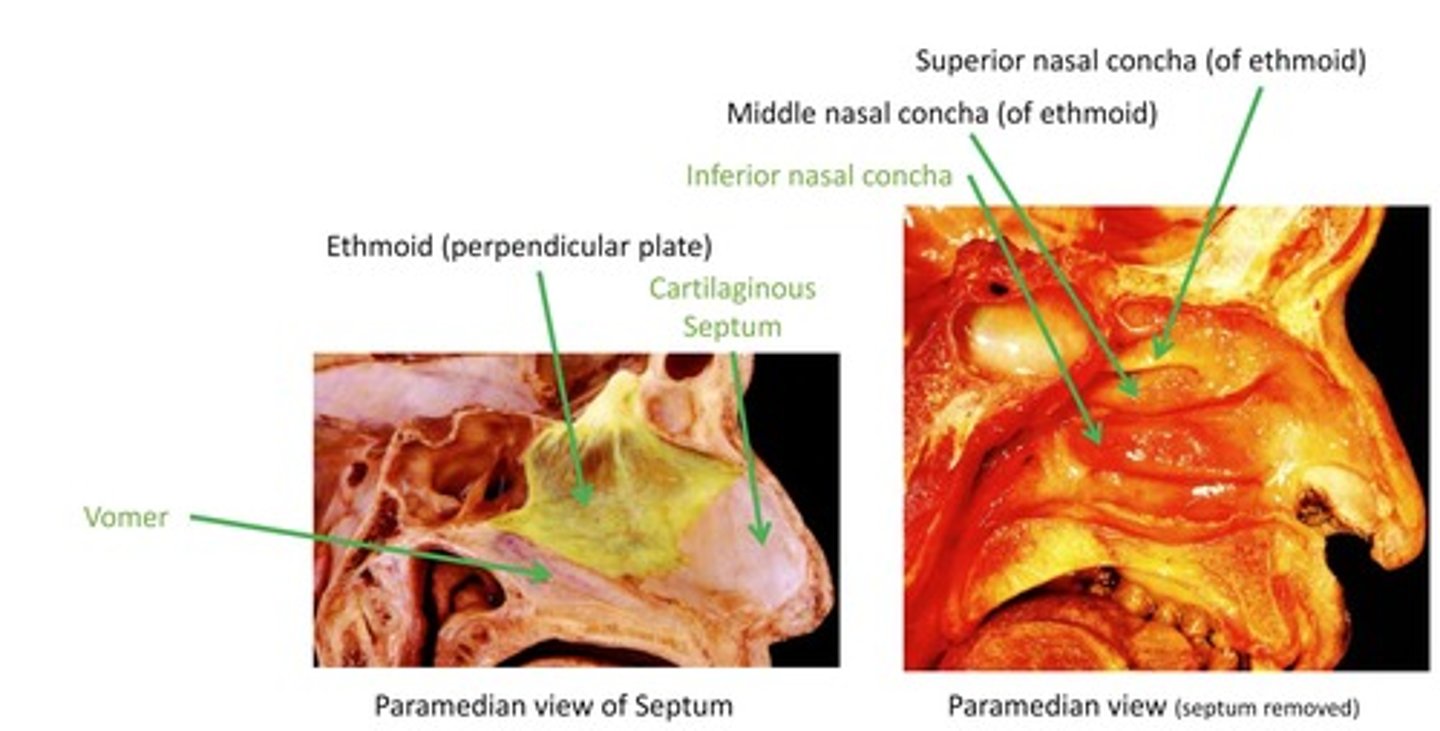
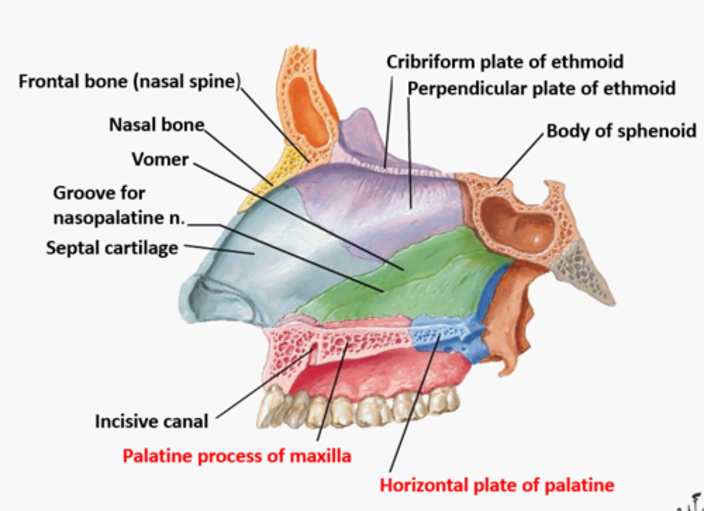
Ethmoid - Paramedian View of Nasal
Cavity
Vomer
Ethmoid (perpendicular plate)
Cartilaginous Septum
Inferior nasal concha
Middle nasal concha (of ethmoid)
Superior nasal concha (of ethmoid)
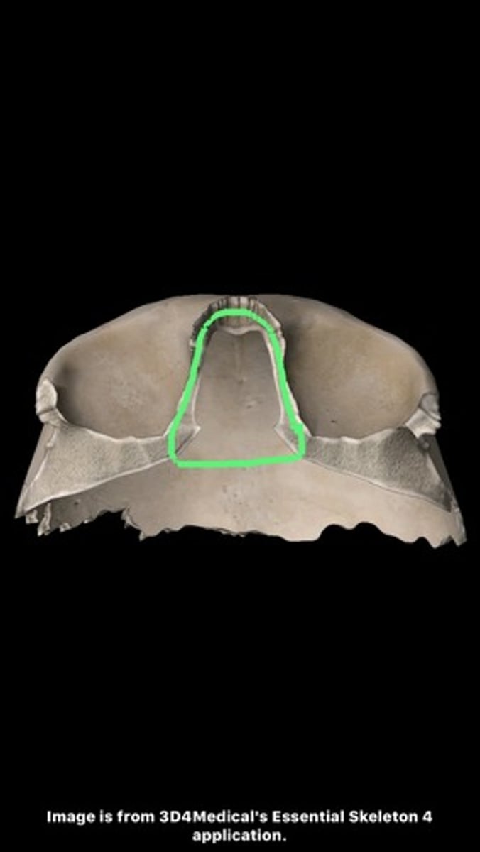
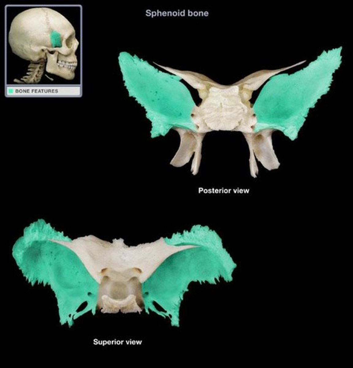
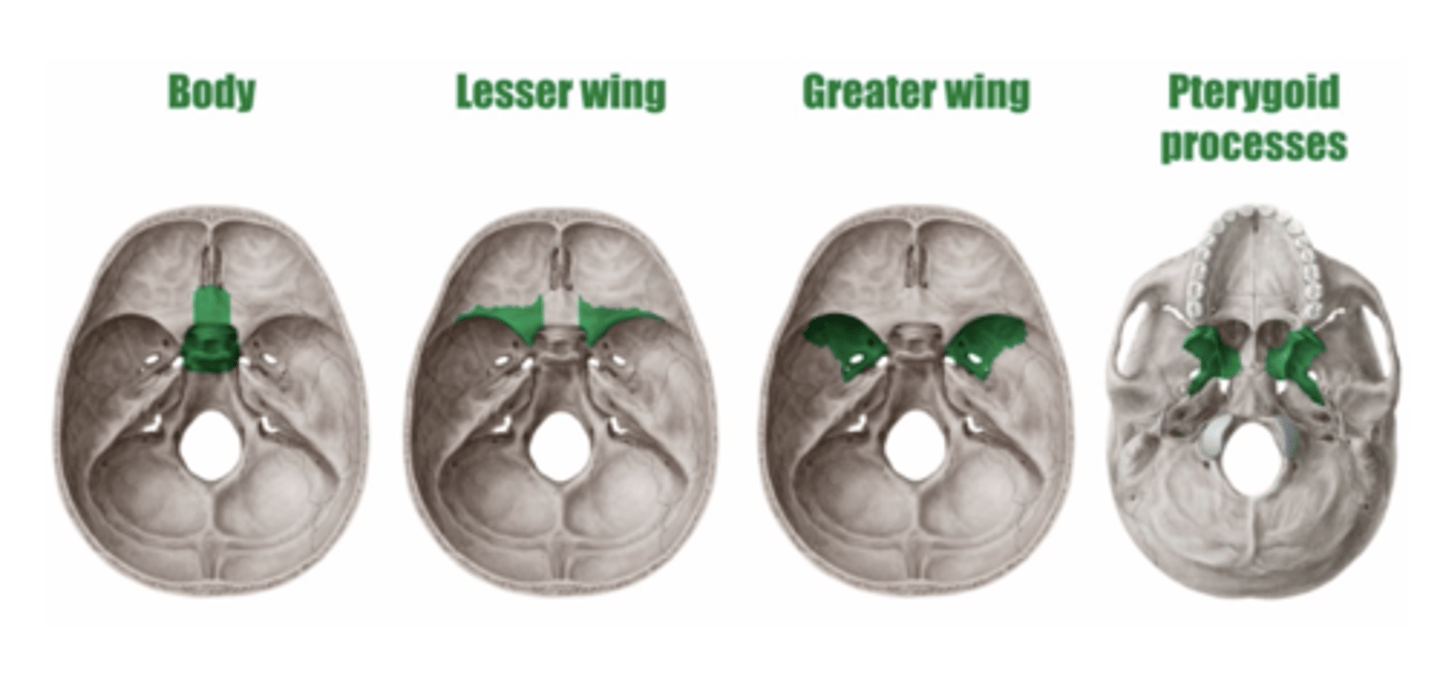
Sphenoid Bone Description
Located at base of skull, behind ethmoid, and in front of foramen magnum
Consists of
- Body
- Lesser wings
- Greater wings
- Pterygoid plates
Sphenoid articulates with all bones of the cranium and 3 facial skeleton bones
Sphenoid Bone Parts
Body
Lesser Wings
Greater Wings
Pterygoid Processes
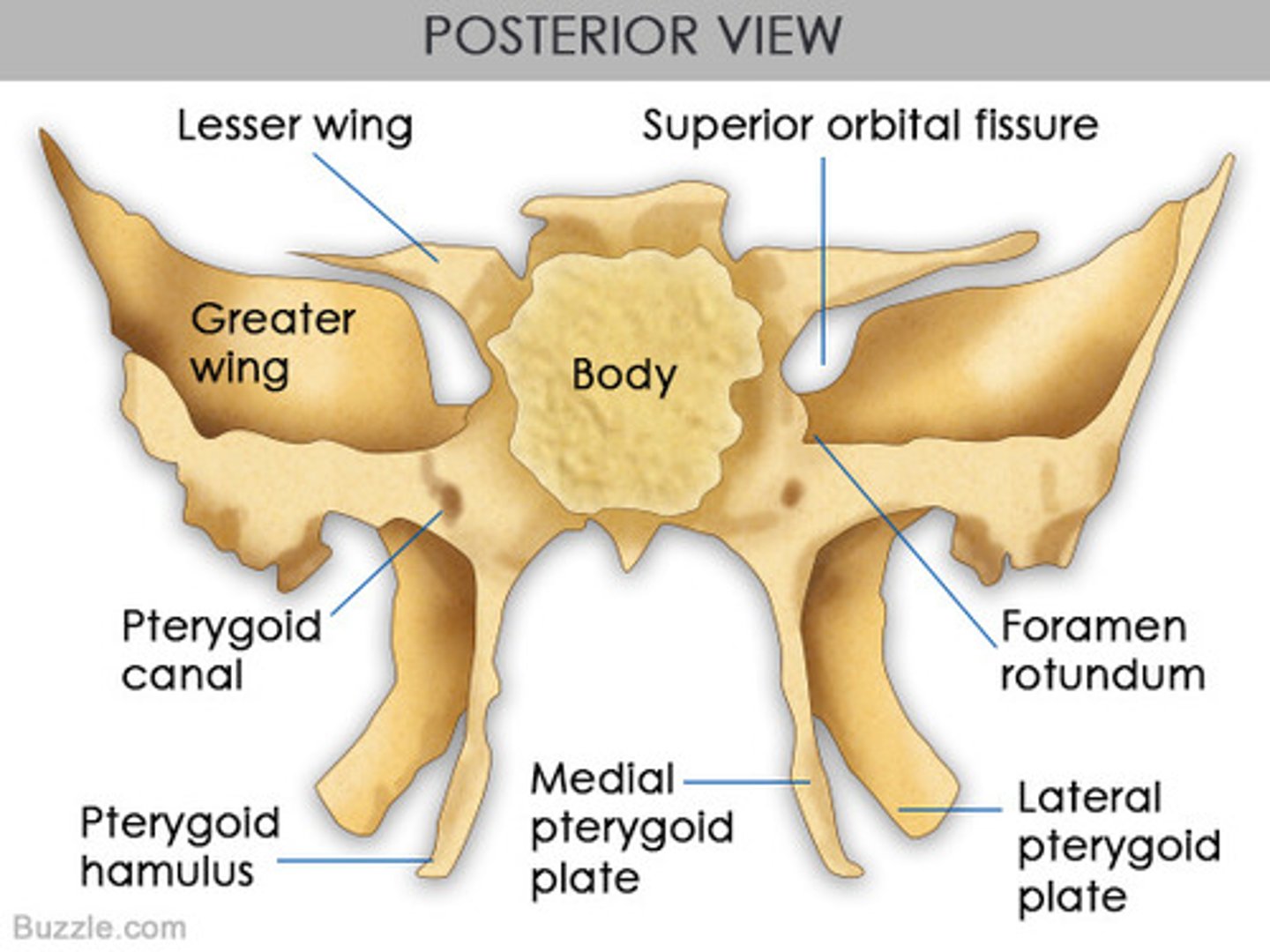
Body
Forms posterior wall of nasal cavity
Lesser Wings
Contribute to anterior cranial fossa and superior wall of orbit
Greater Wings
Contribute to middle cranial fossa and posterior ½ of lateral
wall of orbit
Pterygoid Processes
Medial and lateral
Attachment of pterygoid
muscles
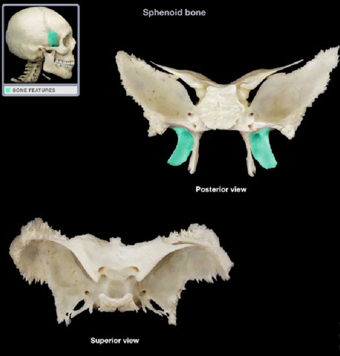
Sphenoid Bone - Lateral / Superior
Hamulus
Medial Pterygoid Plate
Lateral Pterygoid Plate
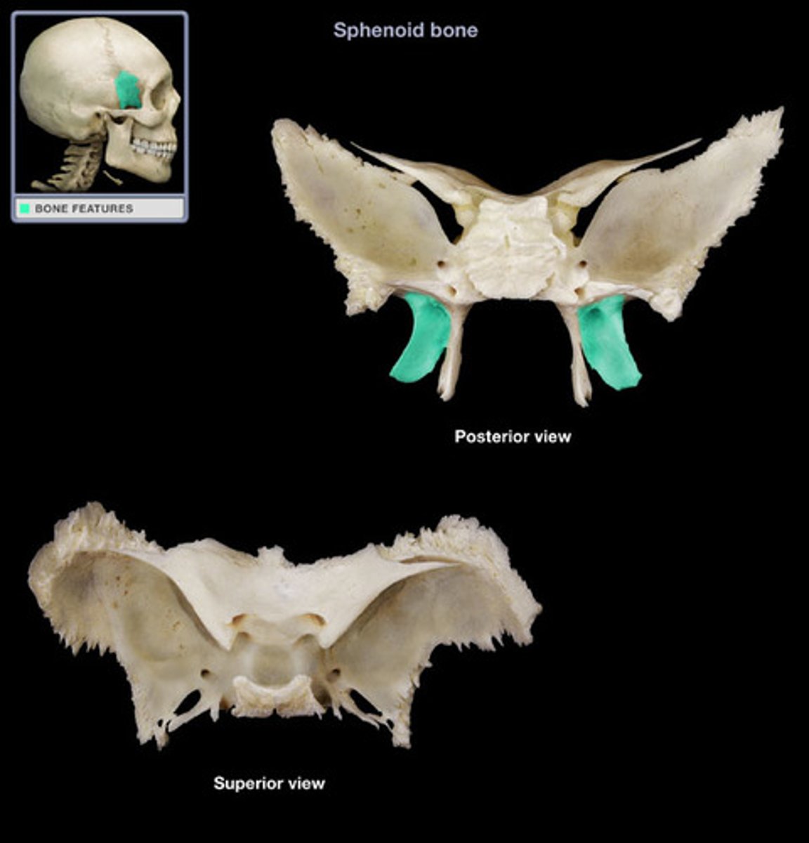
Sphenoid Bone - Superior/Inferior Views
Superior Views
- Body
- Lesser wing
- Greater wing
Inferior View
- Pterygoid processes
Sphenoid Bone - External View
Greater wing
Pterygoid process
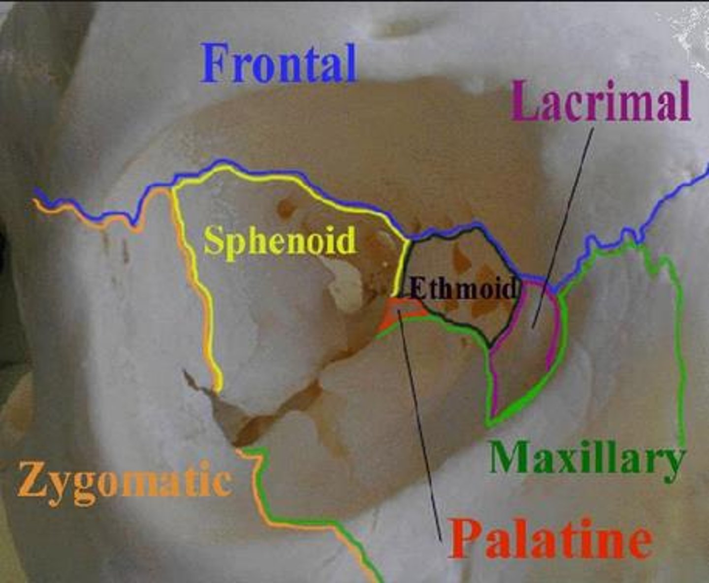
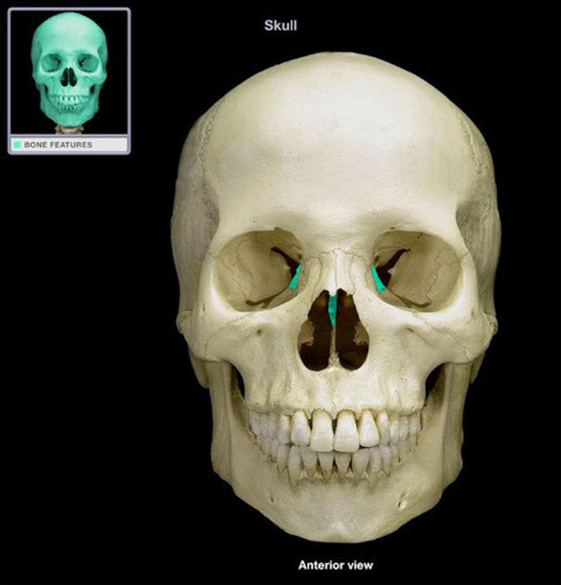
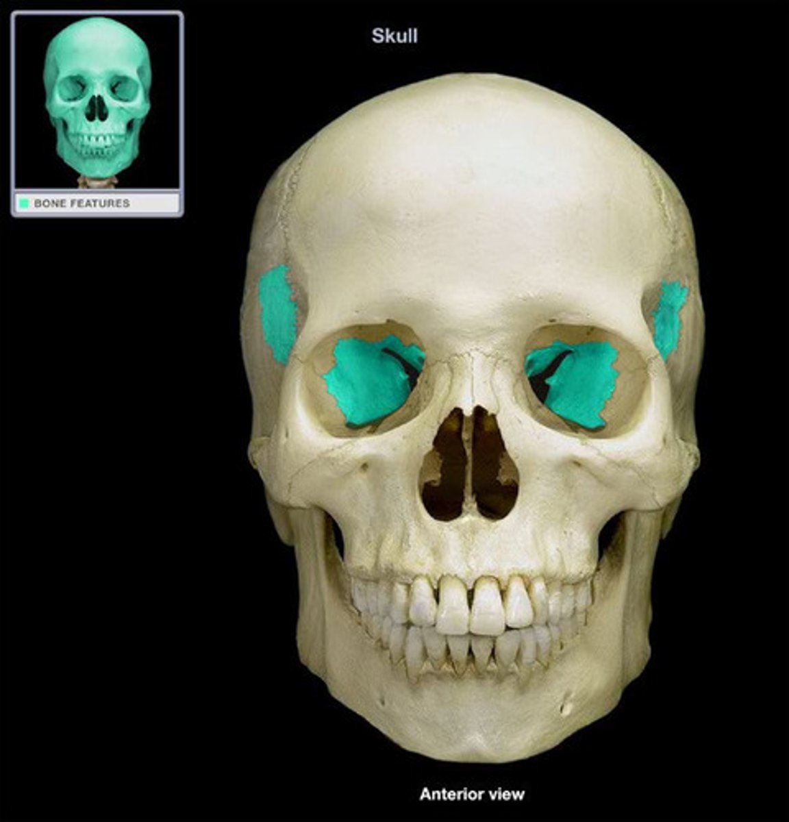
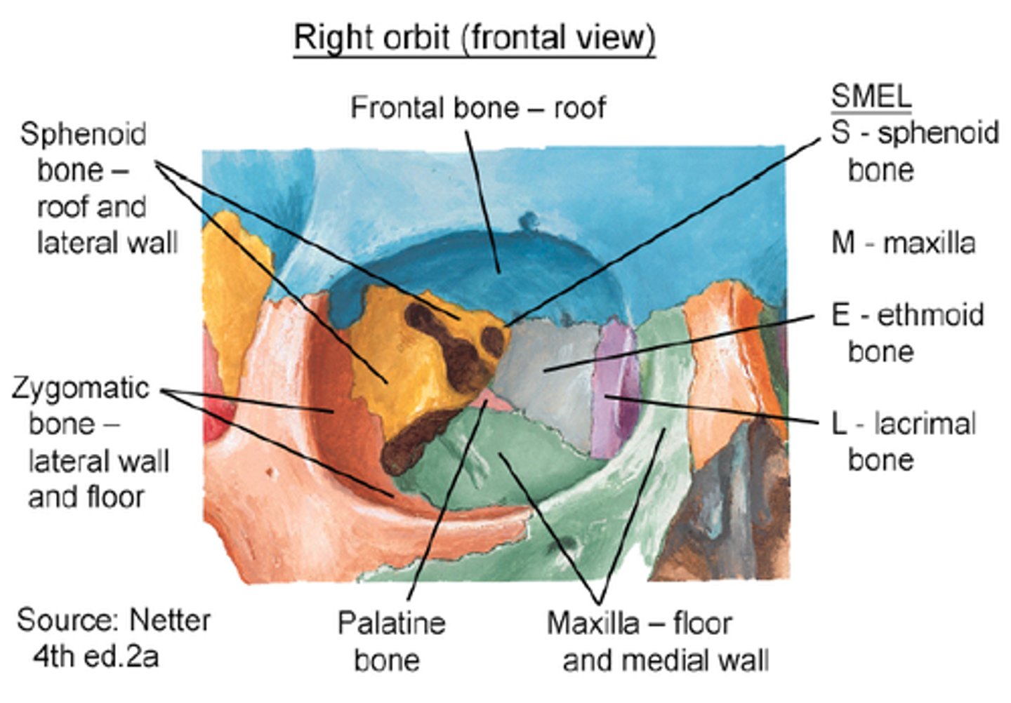
Bones of the Orbit
Ethmoid
Frontal
Sphenoid
Lacrimal
Maxilla
Zygomatic
Bones of the Nasal Cavity
Inferior nasal concha
Middle nasal concha (of ethmoid)
Superior nasal concha (of ethmoid)
What do the bones of the cranium contain?
a portion of the structure surrounding the brain
What are the ethmoid and sphenoid bones purpose?
define many of the complex passageways of the nasal cavity as well as some of the sinuses
What are the vomer, inferior concha, palatine, and maxilla purposes?
define the lower surfaces of the nasal cavity
How does the occipital bone articulate?
with the vertebral column at the occipital condyles
What are the sphenoid and temporal bones purpose?
have processes for muscular attachment
What is the orbit?
shaped by the frontal, ethmoid, sphenoid bones of the cranium as well as the lacrimal,
maxilla and zygomatic bones of the facial skeleton