HSC 214 Appendicular Skeleton: Lower Extremity, HSC 214 Articulations
1/88
There's no tags or description
Looks like no tags are added yet.
Name | Mastery | Learn | Test | Matching | Spaced | Call with Kai |
|---|
No analytics yet
Send a link to your students to track their progress
89 Terms
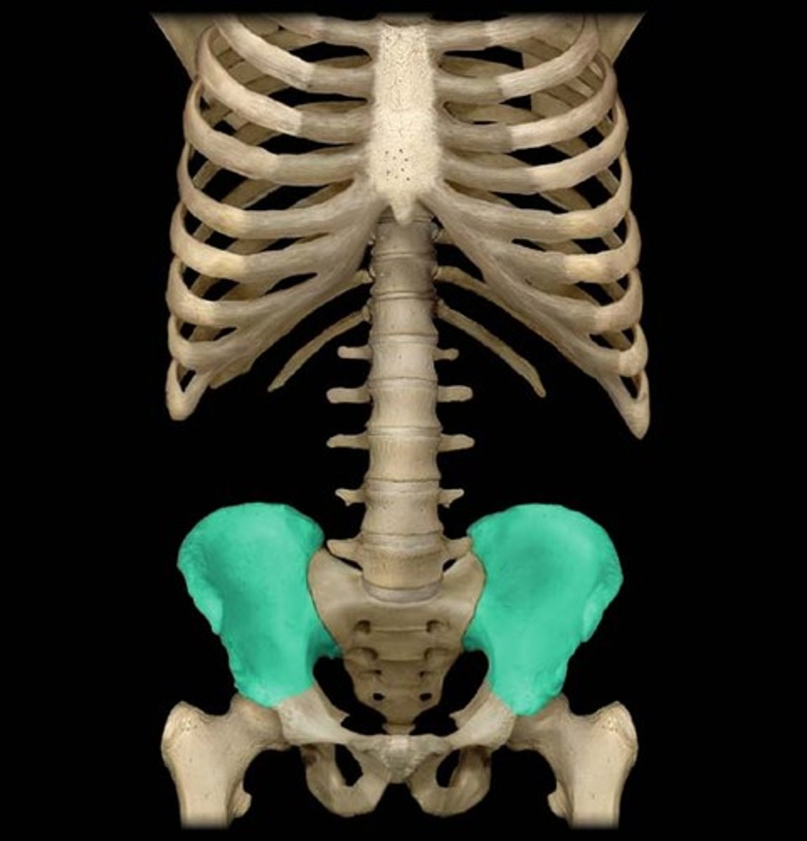
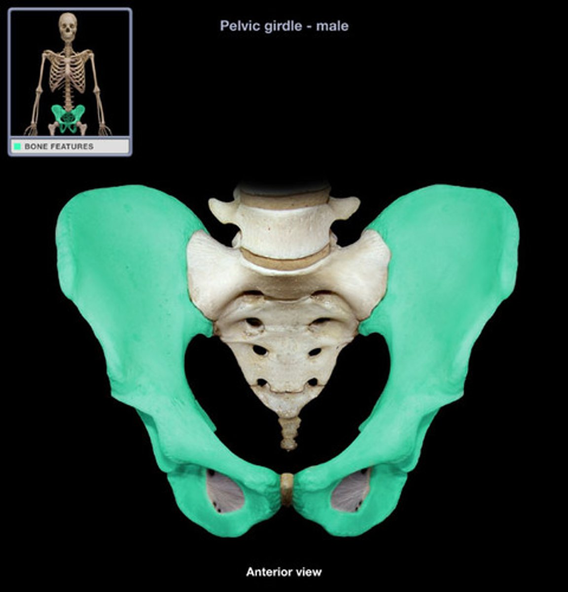
ilium
- fan-shaped superior part of the hip bone
- largest of the three coal (hip) bones
- articulates with the sacrum at the sacroiliac joint
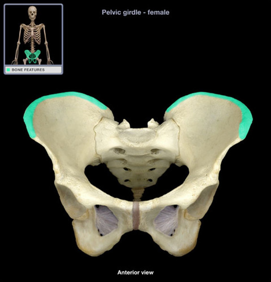
iliac crest
- prominent arching ridge on the superior aspect of the ilium
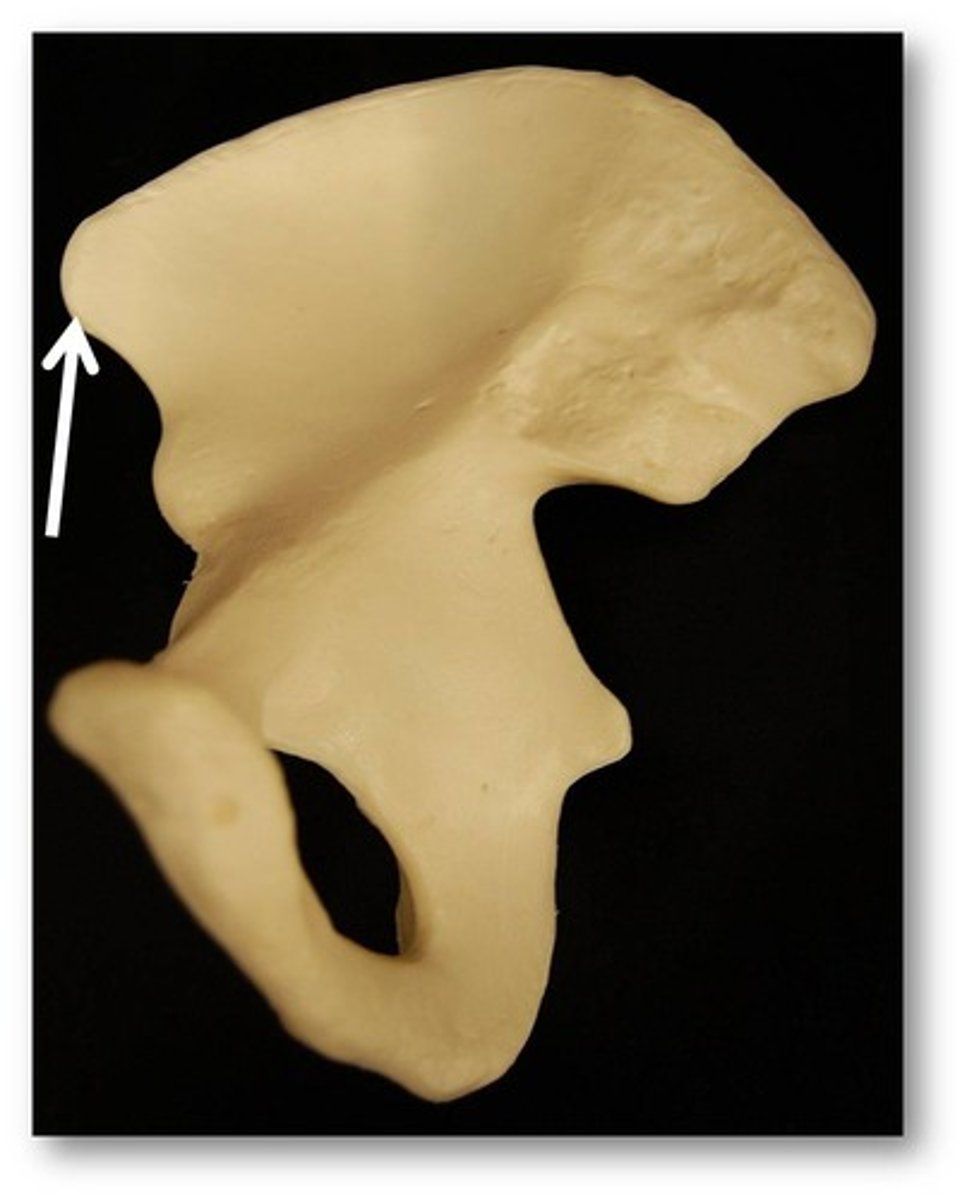
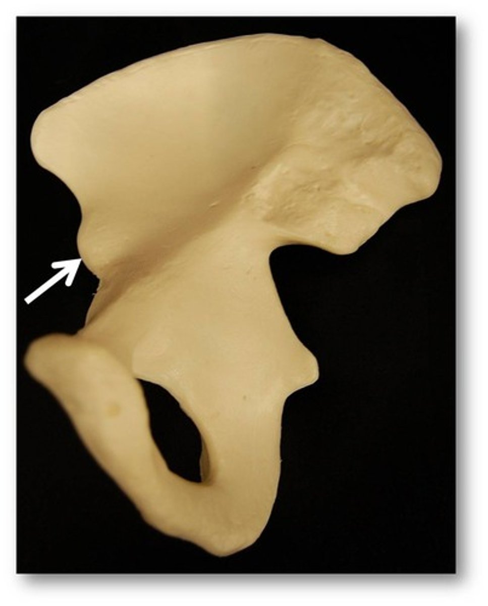
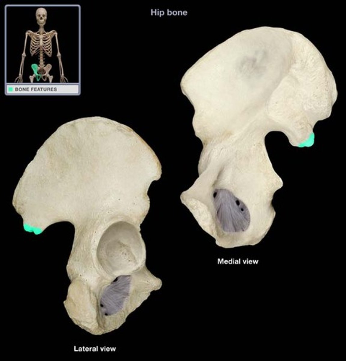
anterior superior iliac spine
- anterior point at the end of the iliac crest
- provides attachment for the sartorial and tensor fascia later muscles
anterior inferior iliac spine
- anterior roughened projection located directly inferior to the ASIS
- provides attachment for the rectus femoris muscle
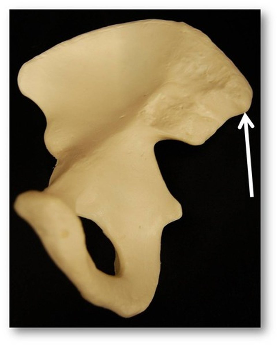
posterior superior iliac spine
- posterior point at the end of the iliac crest
- forms "dimples" in skin on the lower back
posterior inferior iliac spine
- posterior roughened projection located directly inferior to the PSIS
- low projection on the upper border of the greater sciatic notch
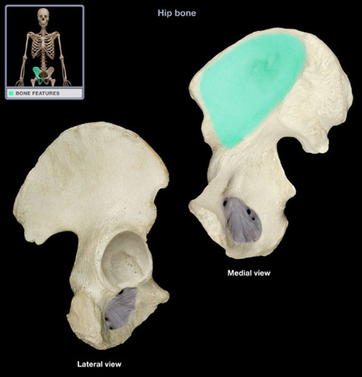
iliac fossa
- hollowed area on the anterior surface of the ilium
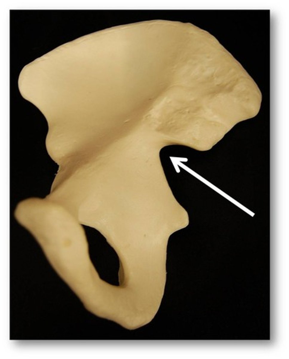
greater sciatic notch
- large, posterior indentation separating the posterior inferior iliac spine from the ischial spine
- space where the sciatic nerve and piriformis muscle pass
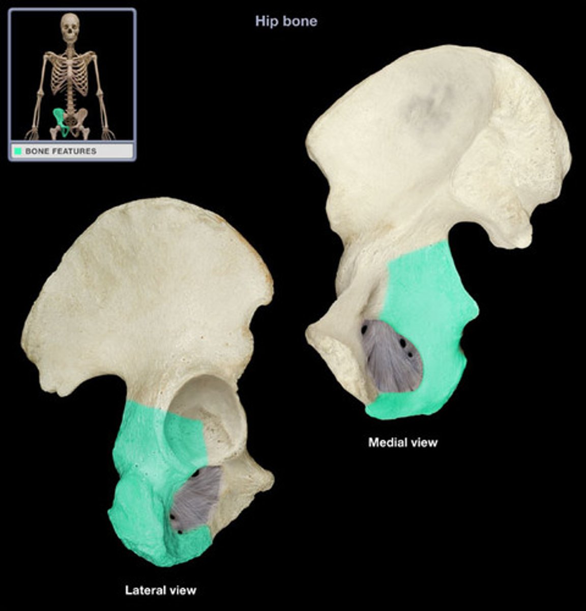
ischium
- U-shaped inferior, posterior part of the hip bone
- "sitting bone"
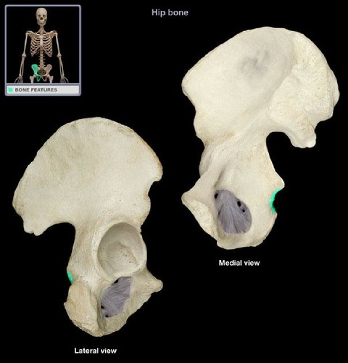
ischial spine
- posterior, pointed projection directly below the greater sciatic notch
- between the greater and lesser sciatic notches

lesser sciatic notch
- smal indent below the ischial spine
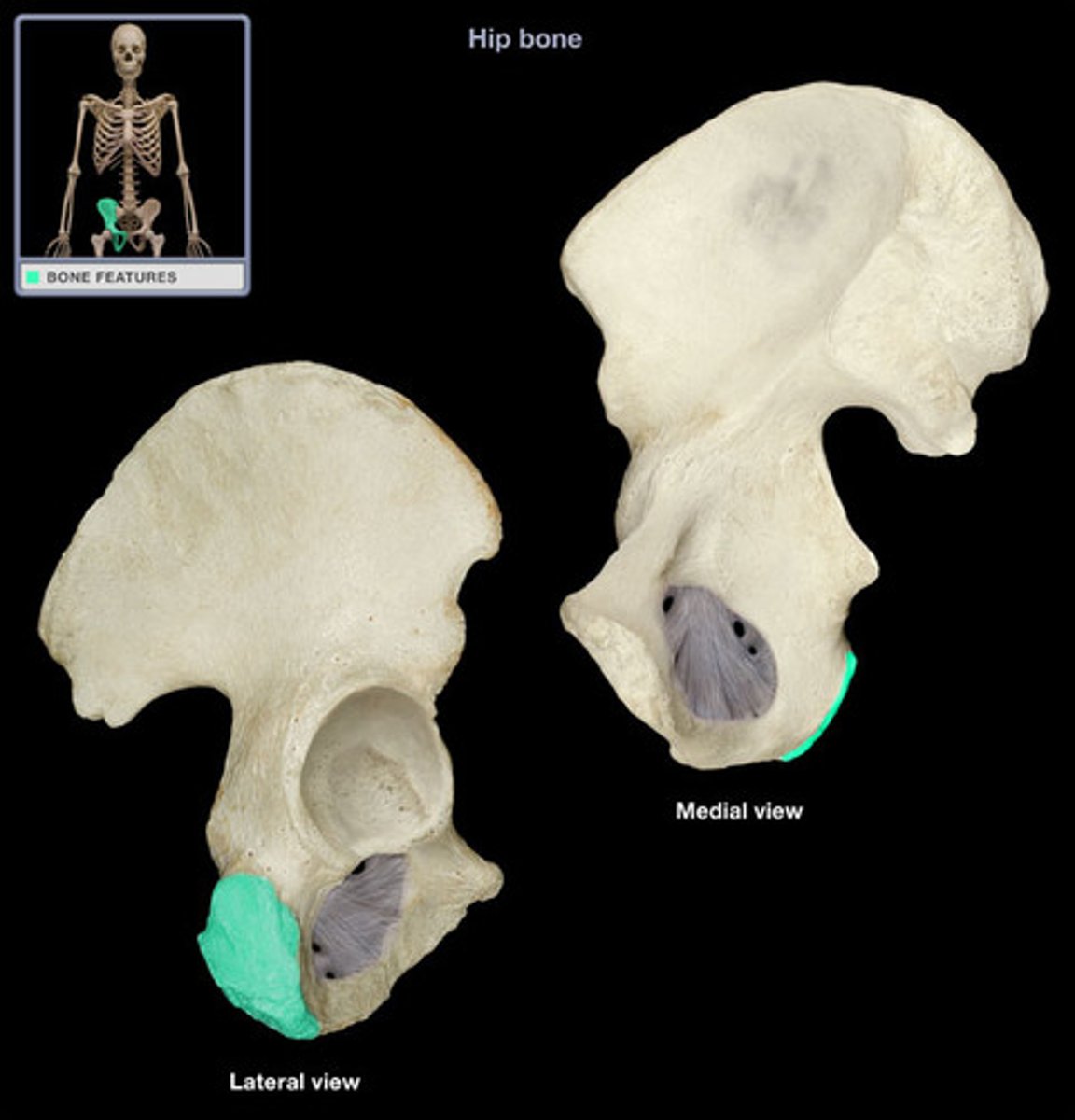
ischial tuberosity
- rough bone covering the rounded portion of the ischium
- provides attachment for the hamstring, adductor Magnus, and quadrates femurs muscles
pubis
- inferior, anterior part of the hip bone
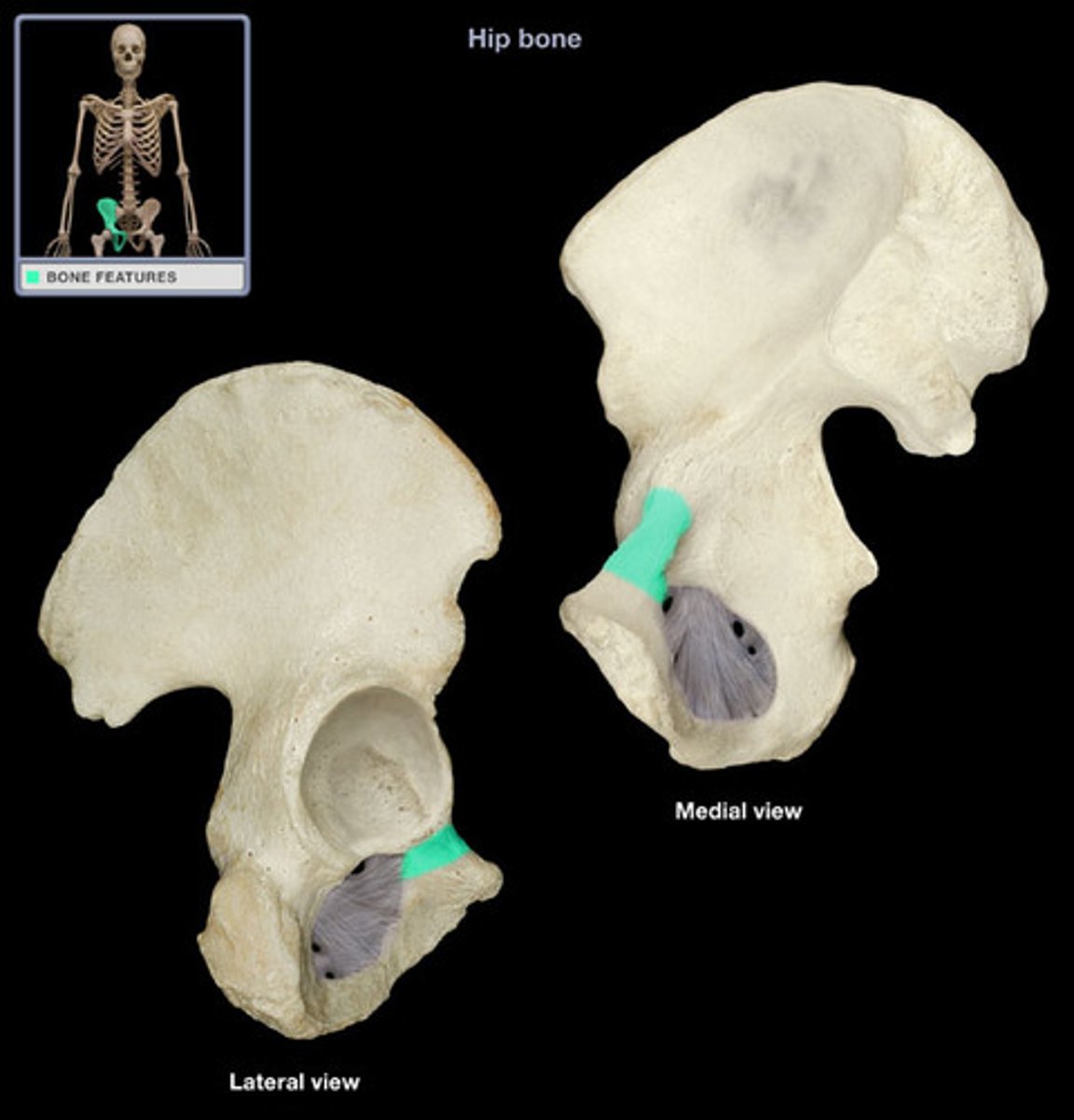
superior pubic ramus
- superior limb-iek bone
- superior border of the obturator foramen
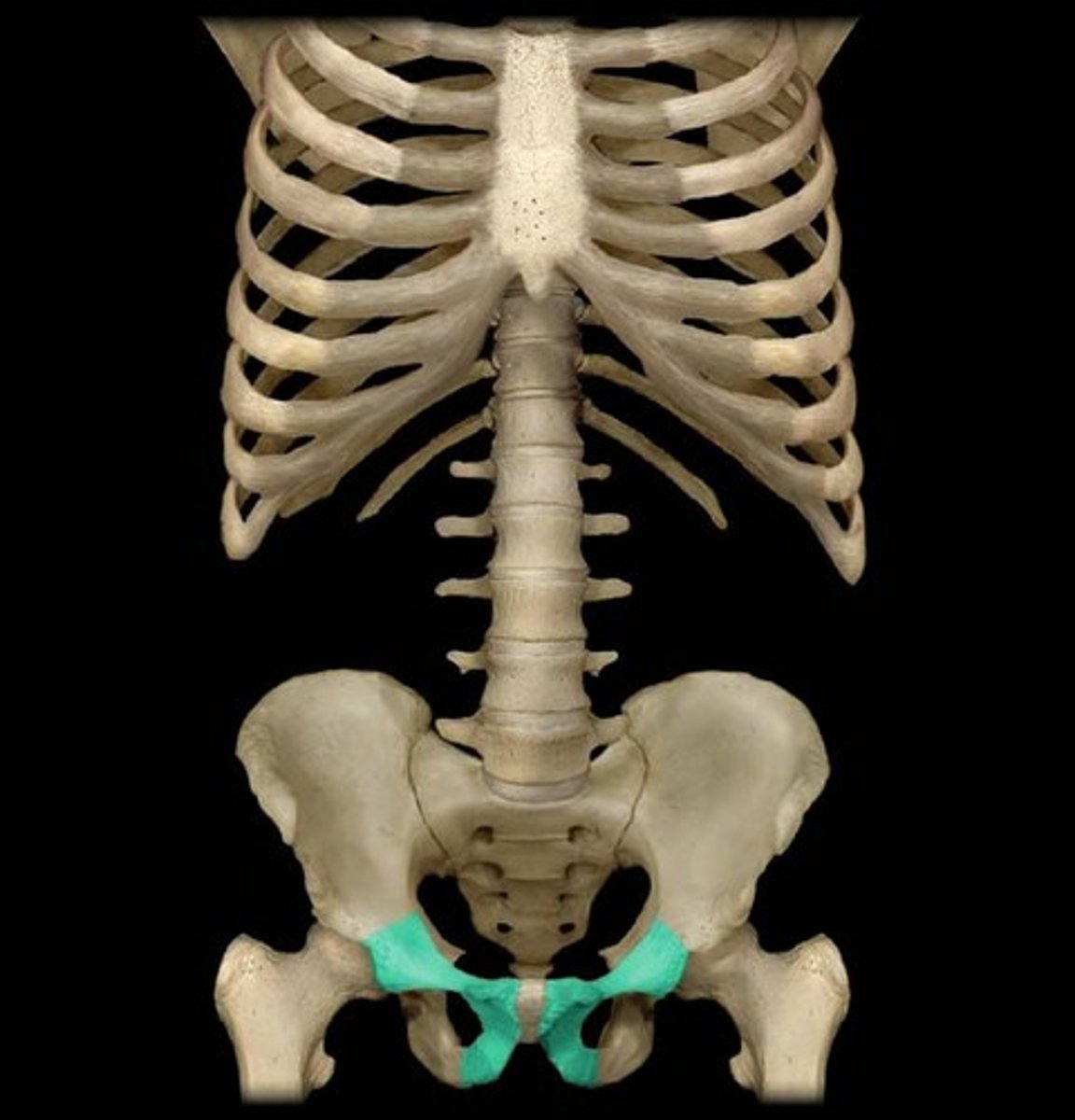
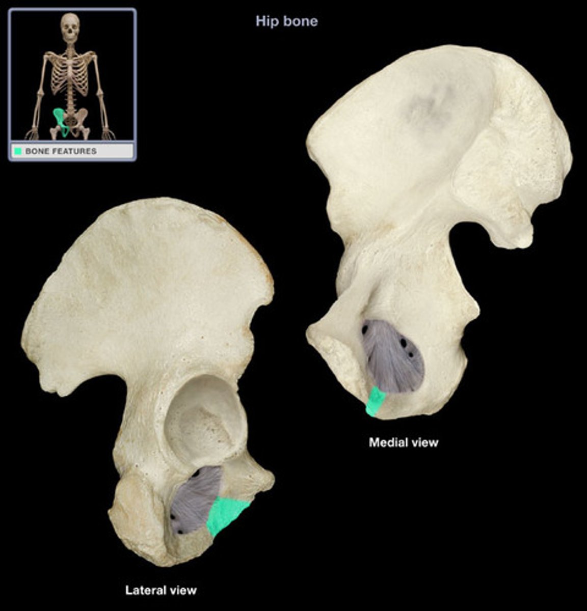
inferior pubic ramus
- inferior limb-like bone
- forms part of the inferior border of the obturator foramen
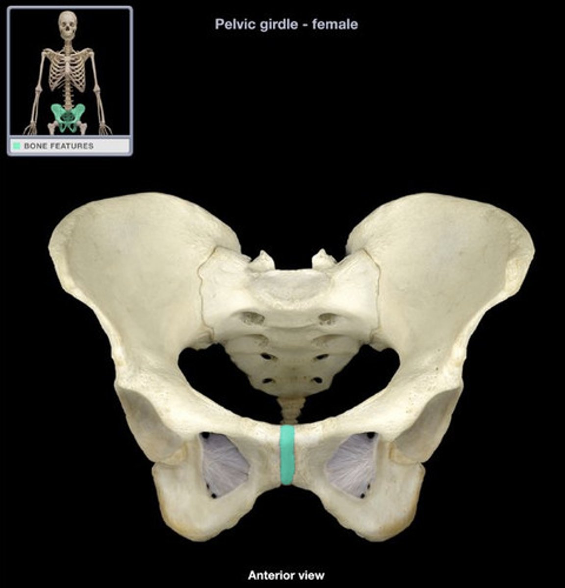
pubic symphysis
- anterior, midline site where the pubic bones meet
- fibrocartilage joint
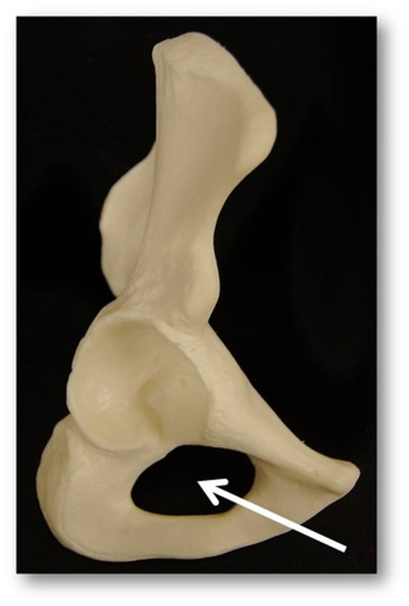
obturator foramen
- large space formed by the rami of the pubic and ischium
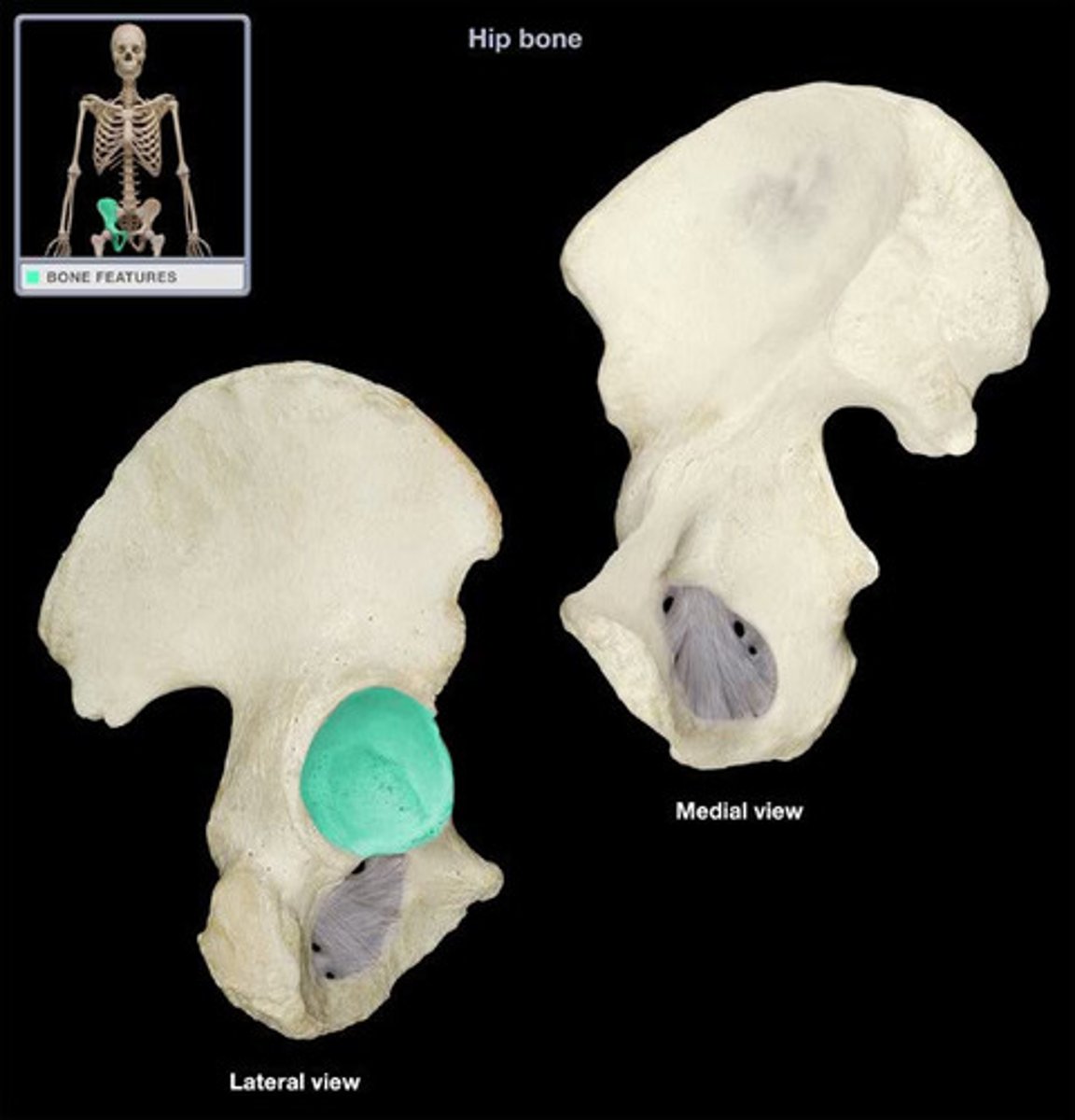
acetabulum
- hemispherical cavity on the lateral aspect of the hip bone
- "hip-socket"
- all three bones (ilium, ischium, and pubis) contribute to the acetabulum
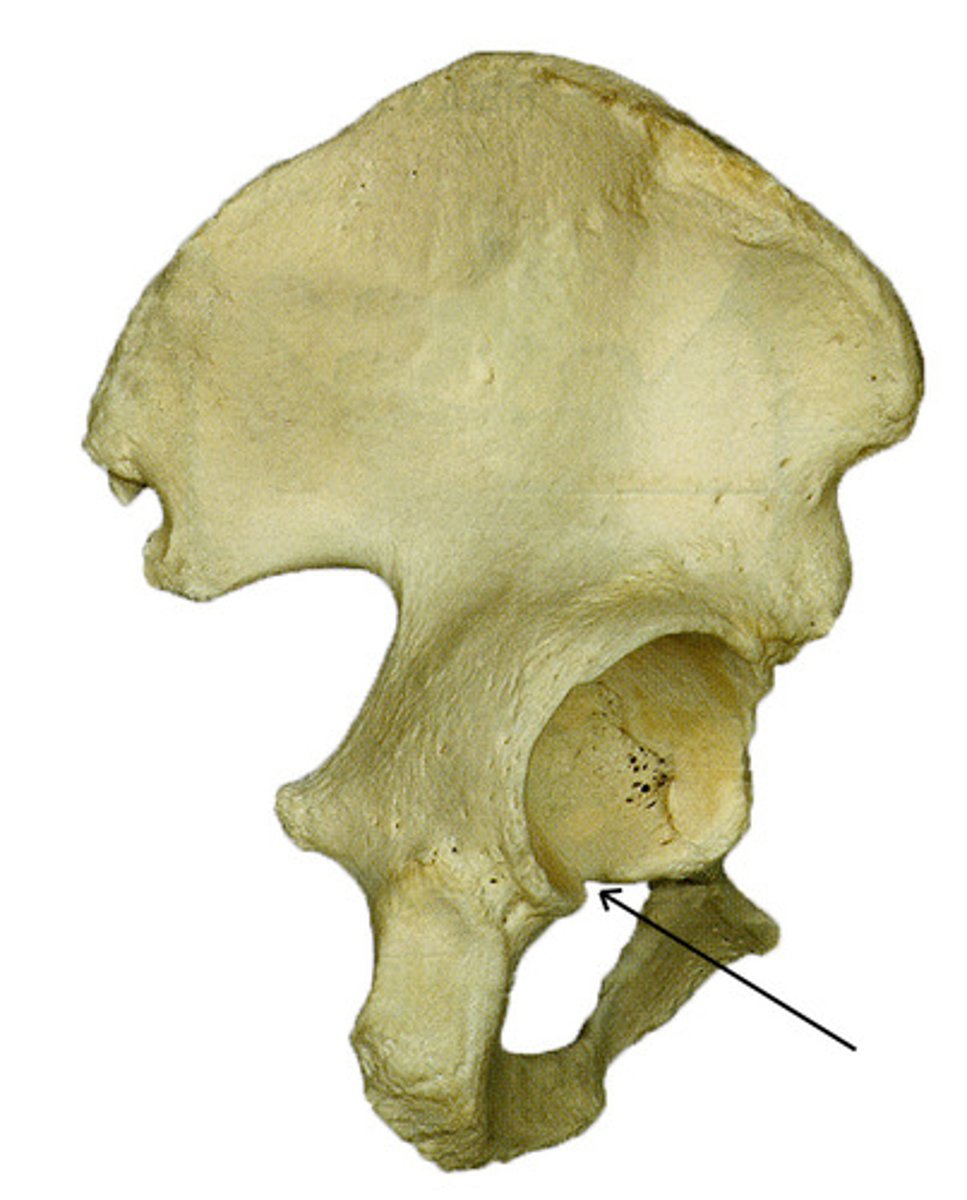
acetabular notch
- deep notch in the inferior part of the brim
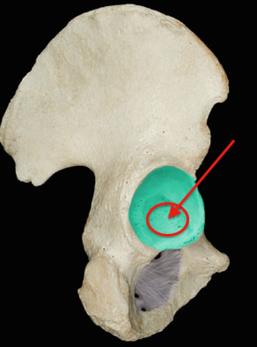
acetabular fossa
- circular depression located deep in the acetabulum
- site that does not articulate with the femur
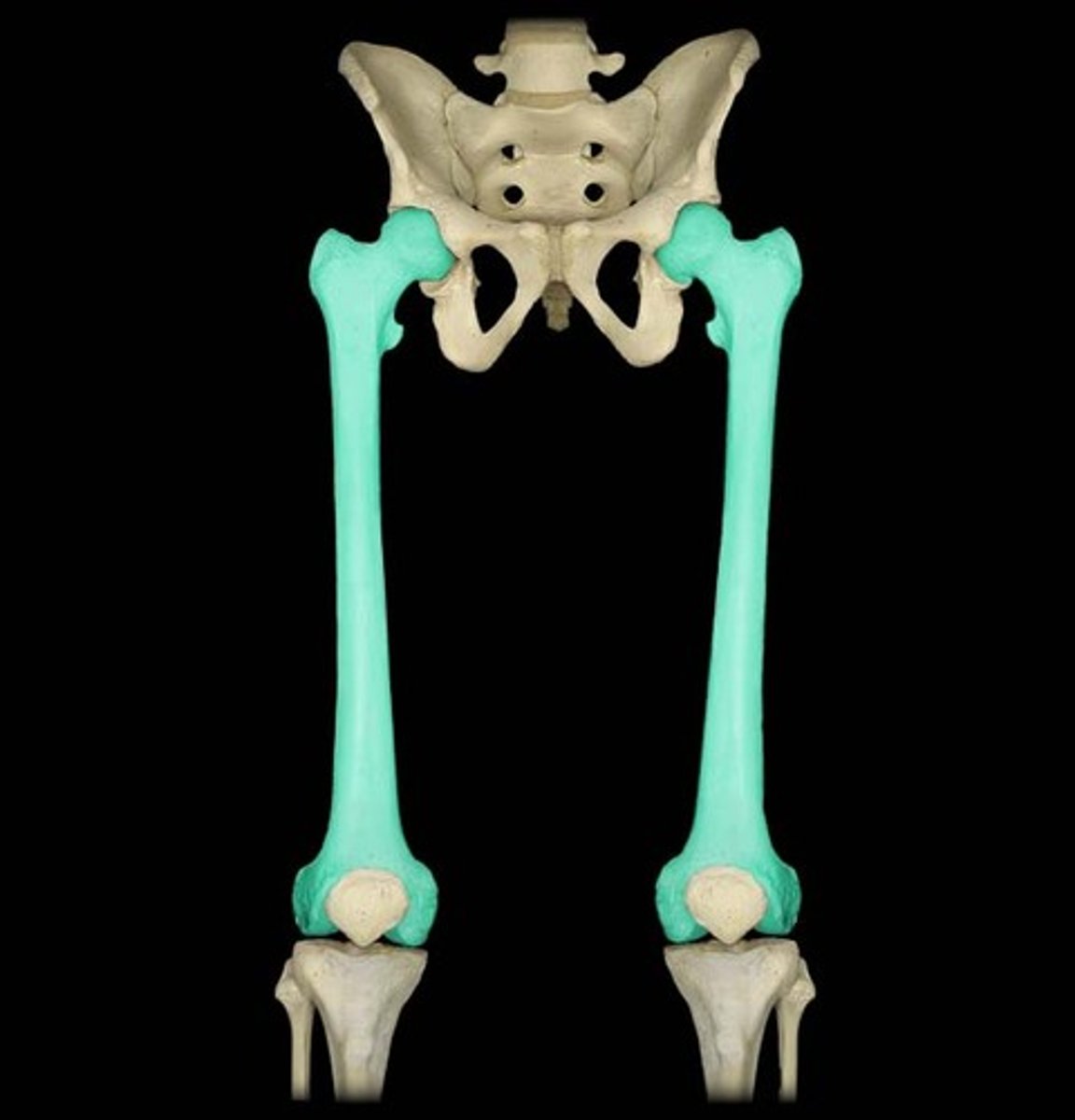
femur
- longest, strongest bone of the body
- "thigh bone"
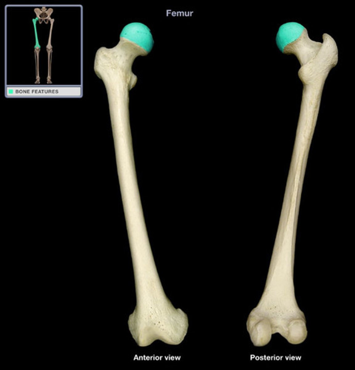
head of femur
- smooth, rounded, ball-lie proximal end
- articulates with the acetabulum to form the hip joint
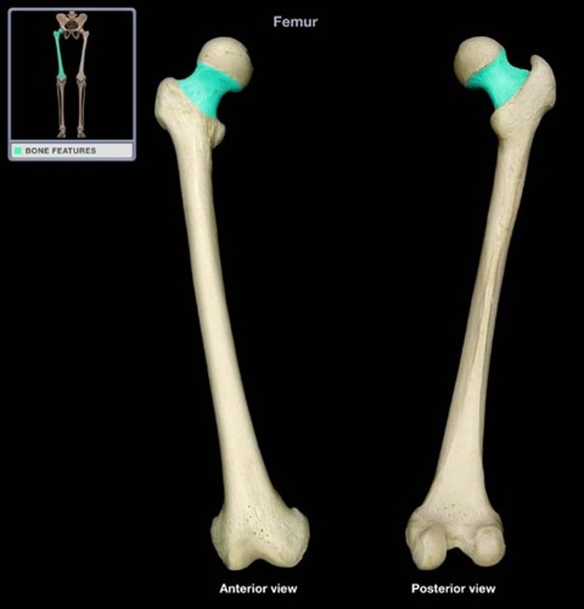
neck of femur
- oblique part between the head and shaft of the femur
- common site of fracture
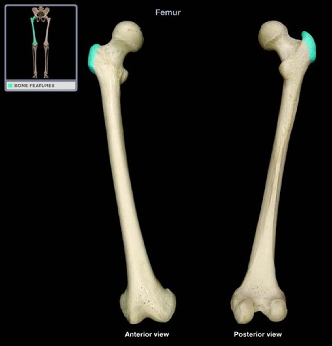
greater trochanter of femur
- large, lateral quadrangular prominence distal to the neck
- attachment site for gluteus maximus muscle
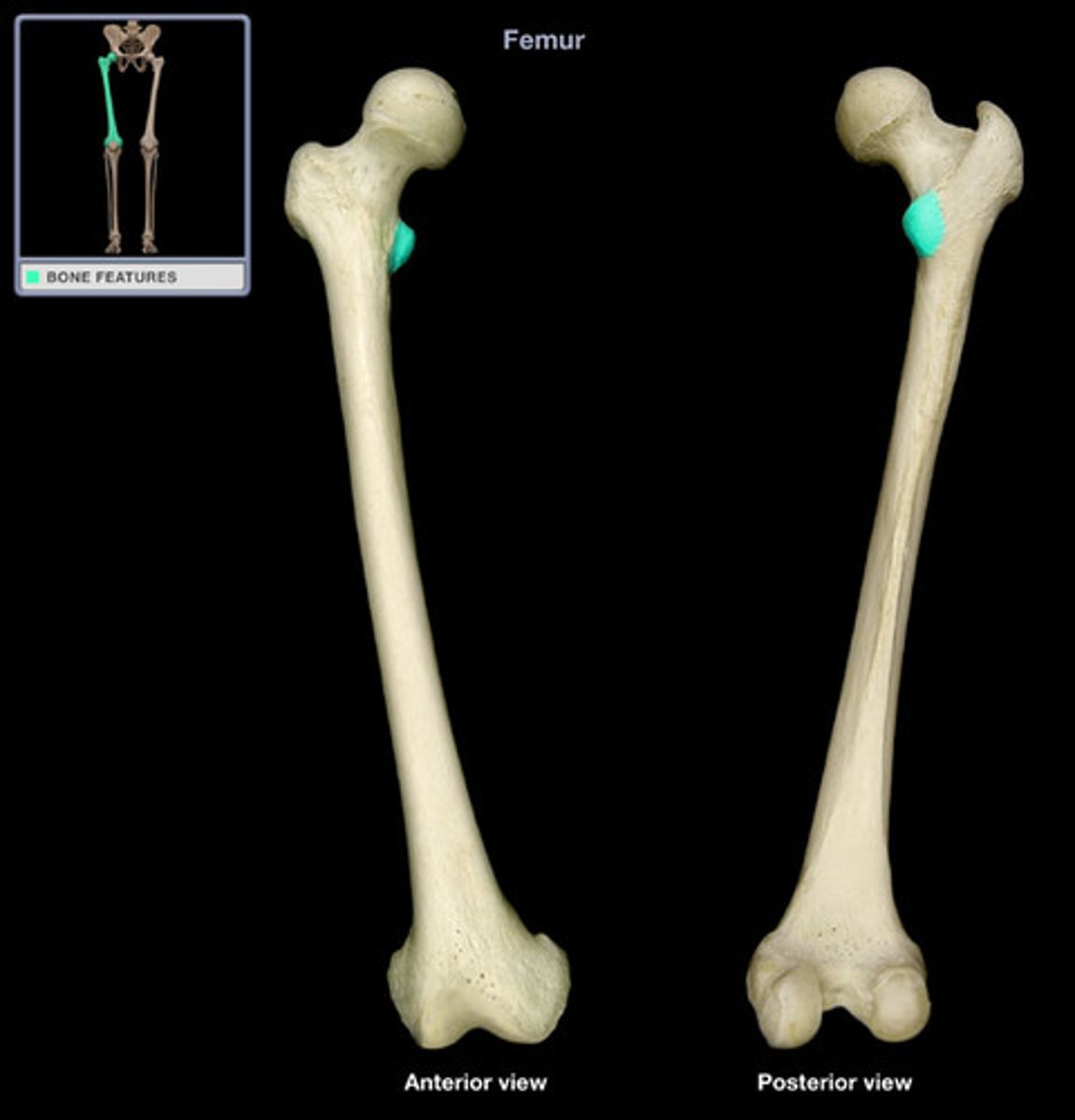
lesser trochanter of femur
- small, medial, and posterior prominence distal to the neck
- attachment for the iliopsoas muscle
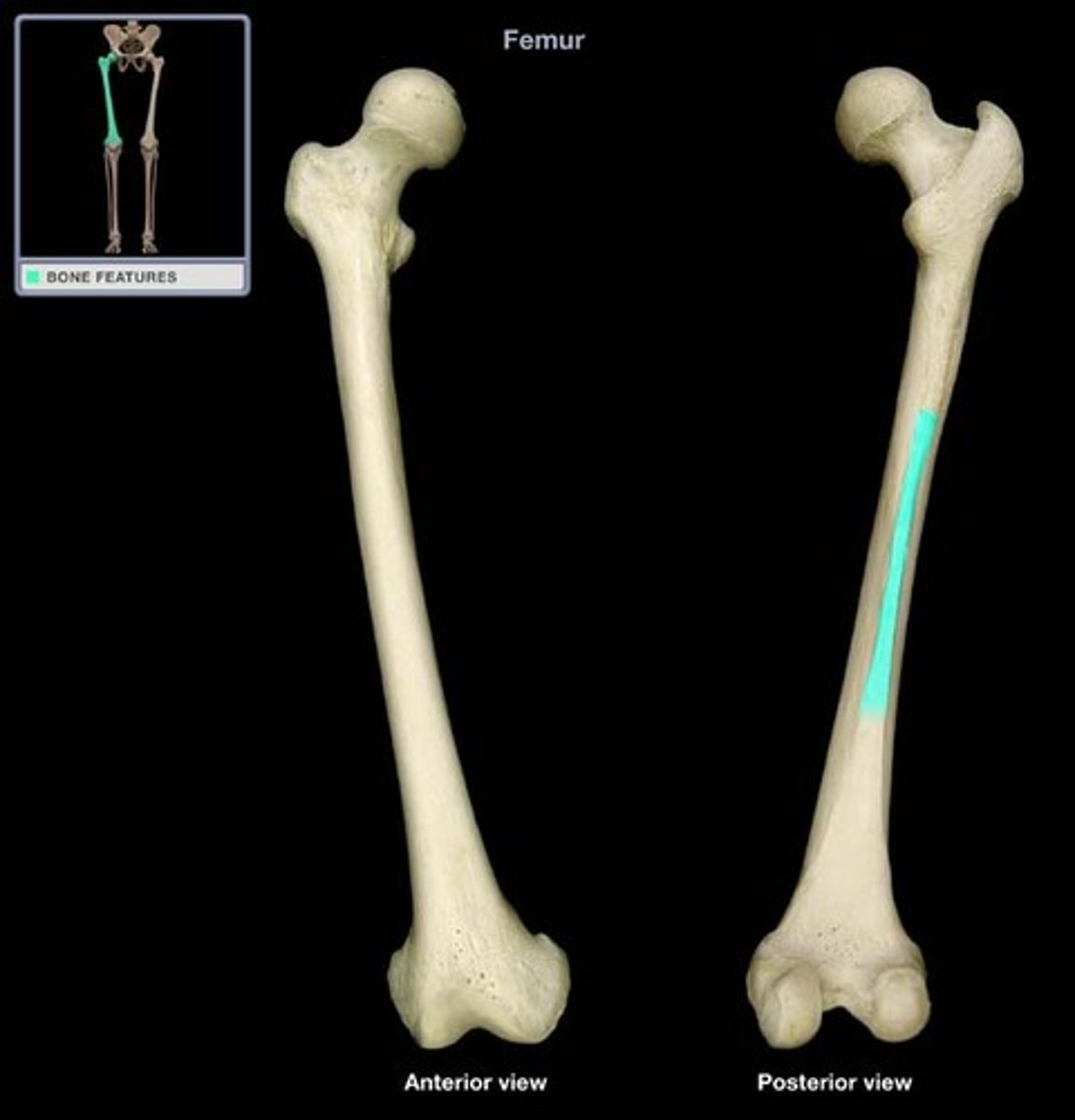
linea aspera
- prominent posterior ridge line on the posterior surface of the shaft
- attachment site for the biceps femoris (short head), adductor longus, adductor magnus, and vastus medialis muscles
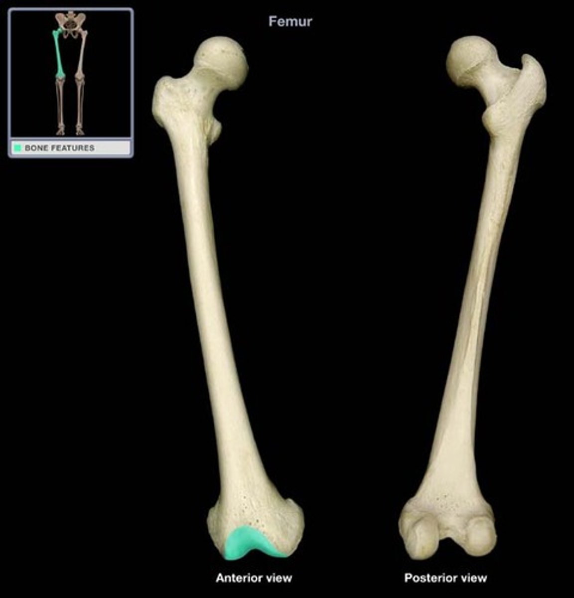
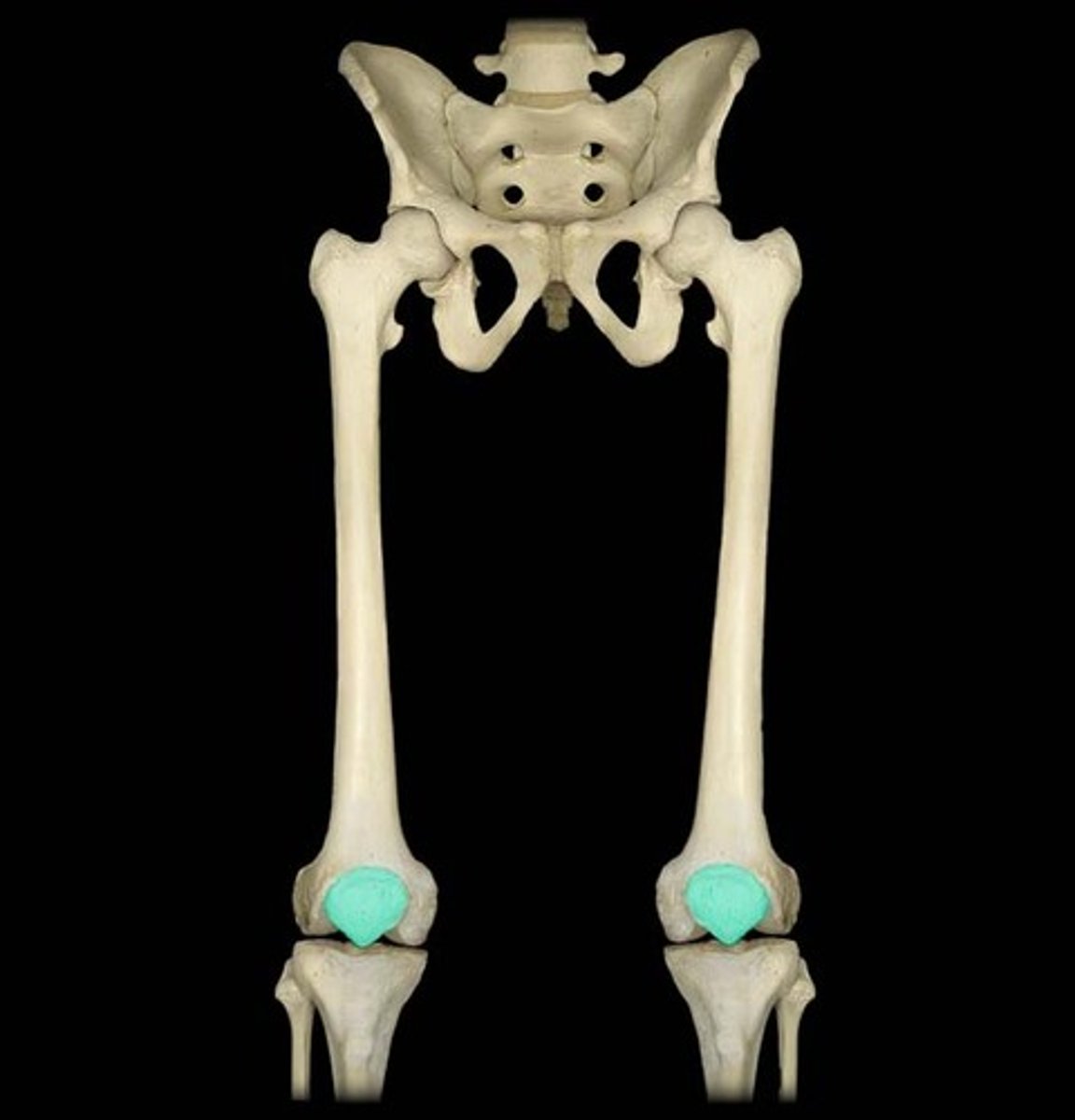
patellar surface
- smooth, anterior surface at the distal end
- articular surface for the patella
lateral epicondyle of femur
- prominent, lateral enlargement superior to the lateral condyle
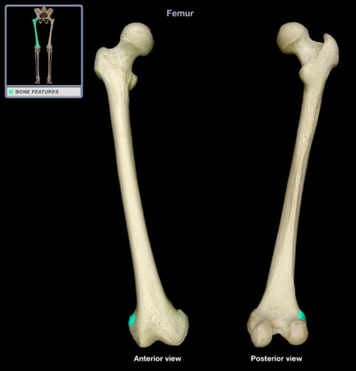
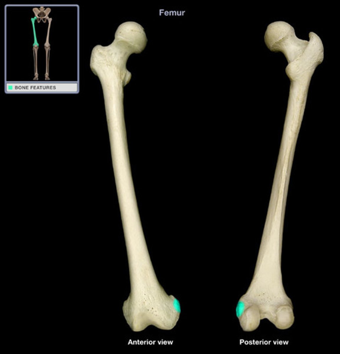
medial epicondyle of femur
- prominent, medial enlargement superor to the medial condyle
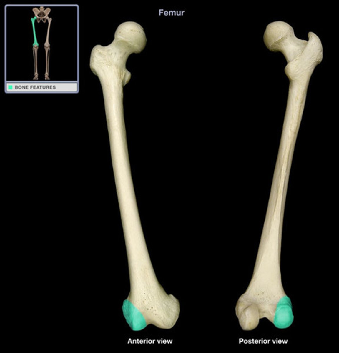
lateral condyle of femur
- smooth, lateral enlargement of the distal end
- articulates with the lateral condyle of the tibia
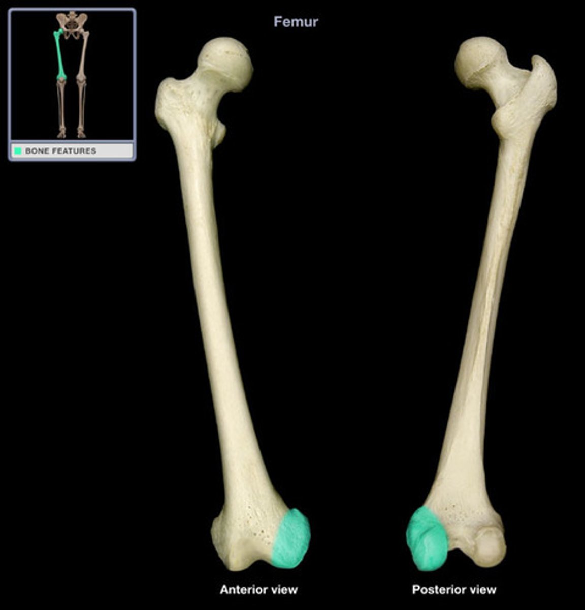
medial condyle of femur
- smooth, medial enlargement of the distal end
- articulates with the medial condyle of the tibia
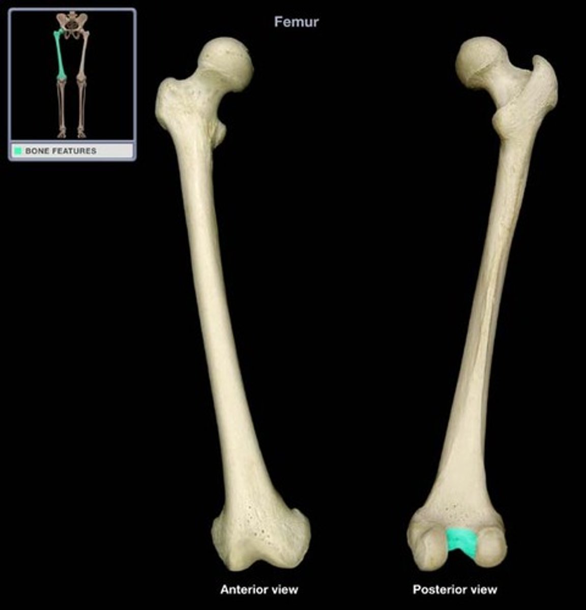
intercondylar fossa
- deep depression between the posterior parts of the medial and lateral condyles
- inter- means between
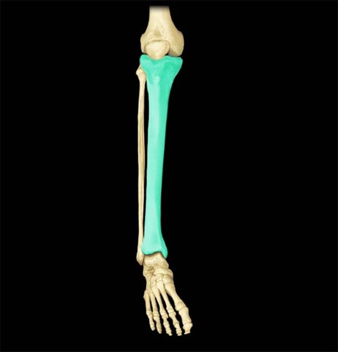
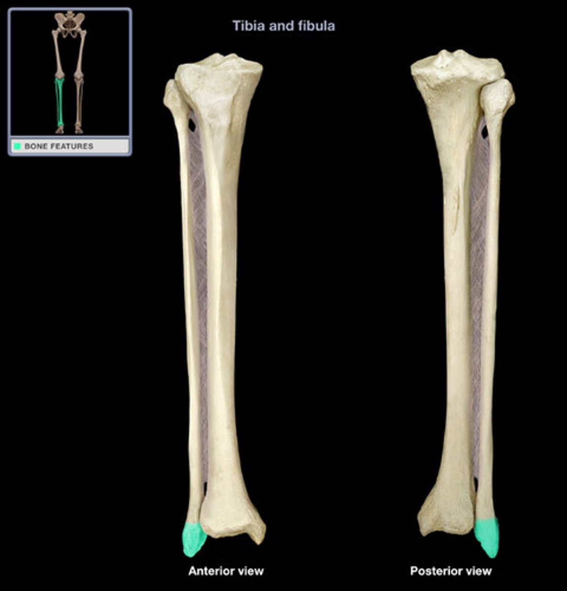
tibia
- thick, medial bone of the leg
- "shin bone"
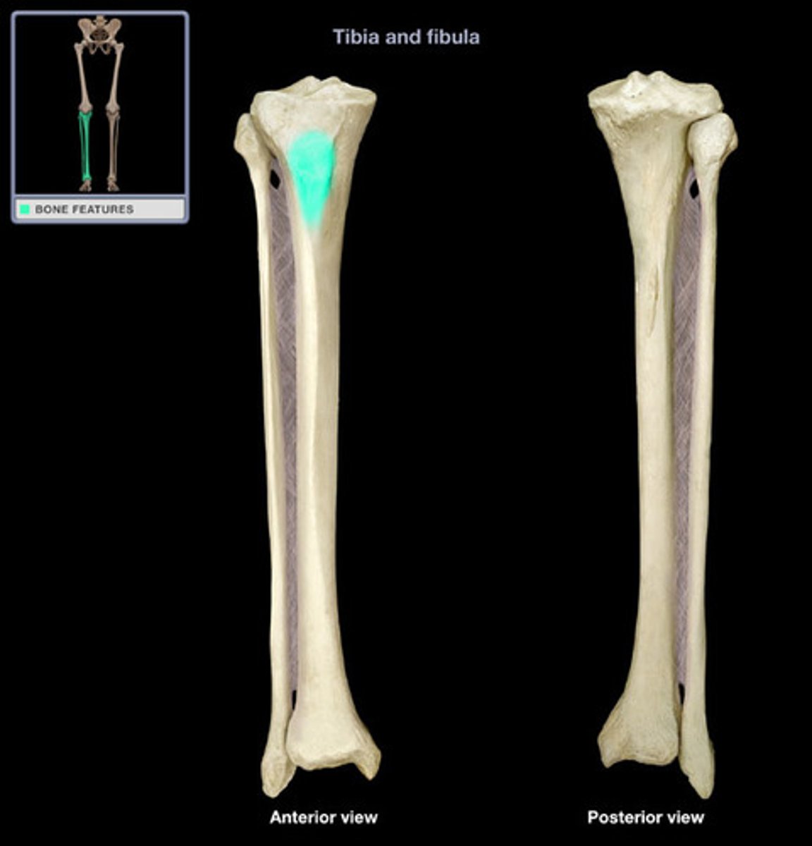
tibial tuberosity
- bump on anterior surface of the proximal end
- where the patellar ligament attaches
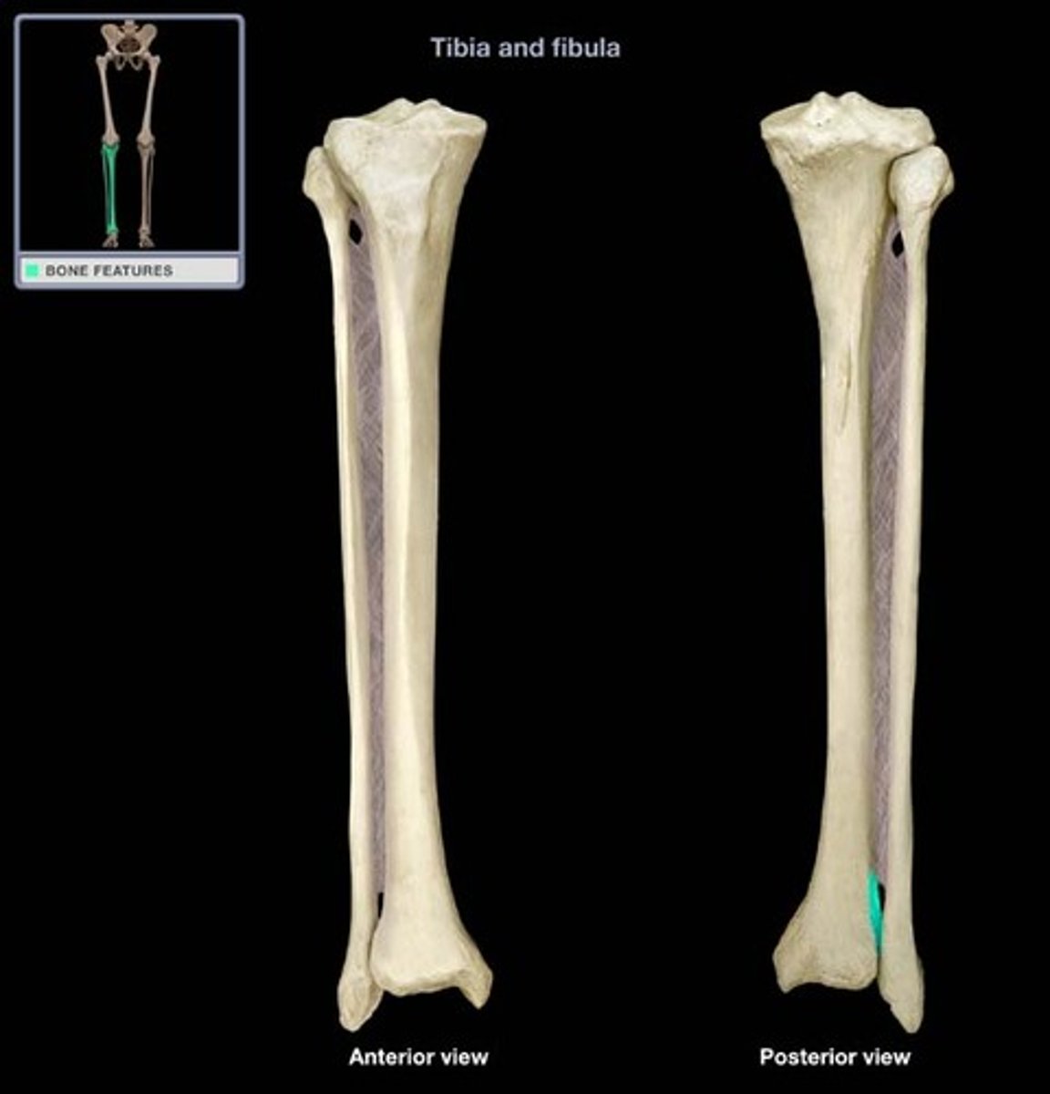
fibular notch
- groove on the distal, lateral surface of the tibia
- where the tibia and fibula articulate
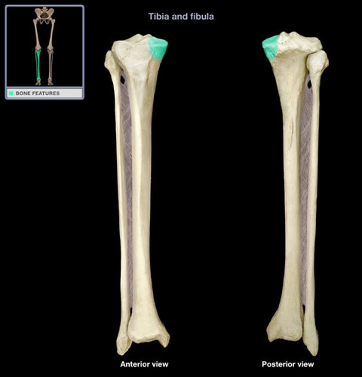
lateral condyle of tibia
- lateral protuberance at the proximal end
- articulates with the lateral condyle of the femur
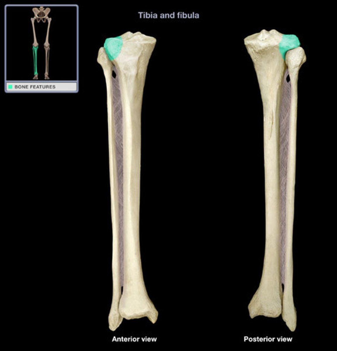
medial condyle of tibia
- medial protuberance at the proximal end
- articulates with the medial condyle of the femur
medial malleolus
- rounded, medial enlargement at the distal end
- forms the medial aspect of the ankle
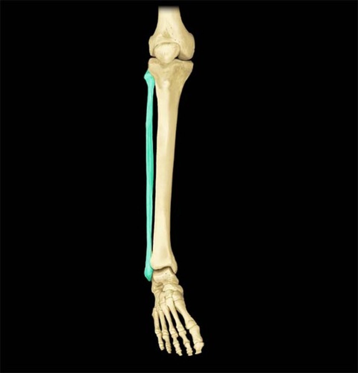
fibula
- thin, lateral bone of the leg
- "fibulA is lAterAl"
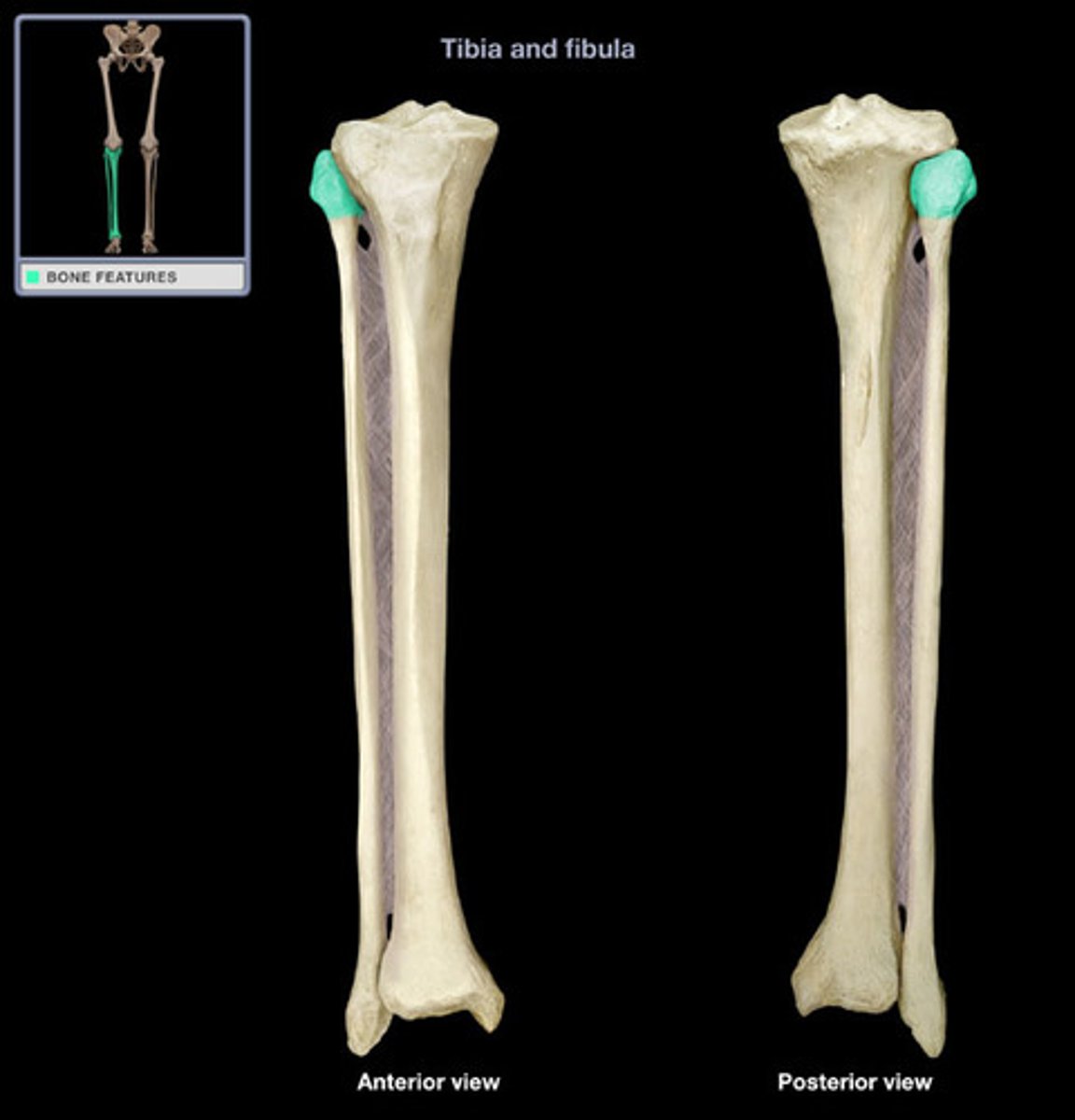
head of fibula
- rounded projection at the proximal end
- articulates with the fibular notch of the tibia
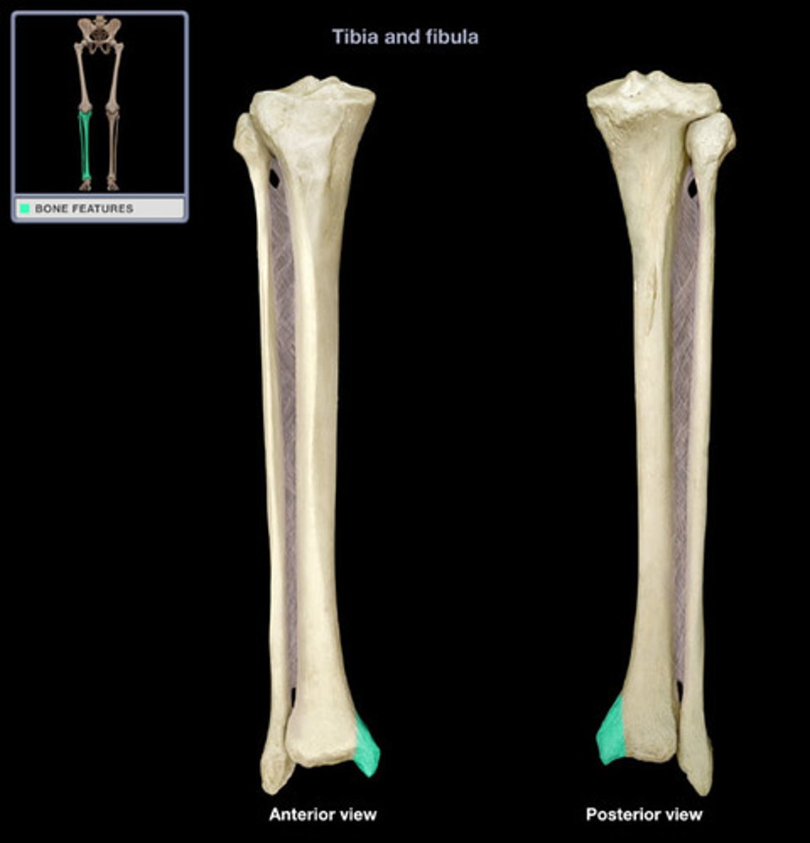
lateral malleolus
- rounded, lateral enlargement at the distal end
- forms the lateral aspect of the ankle
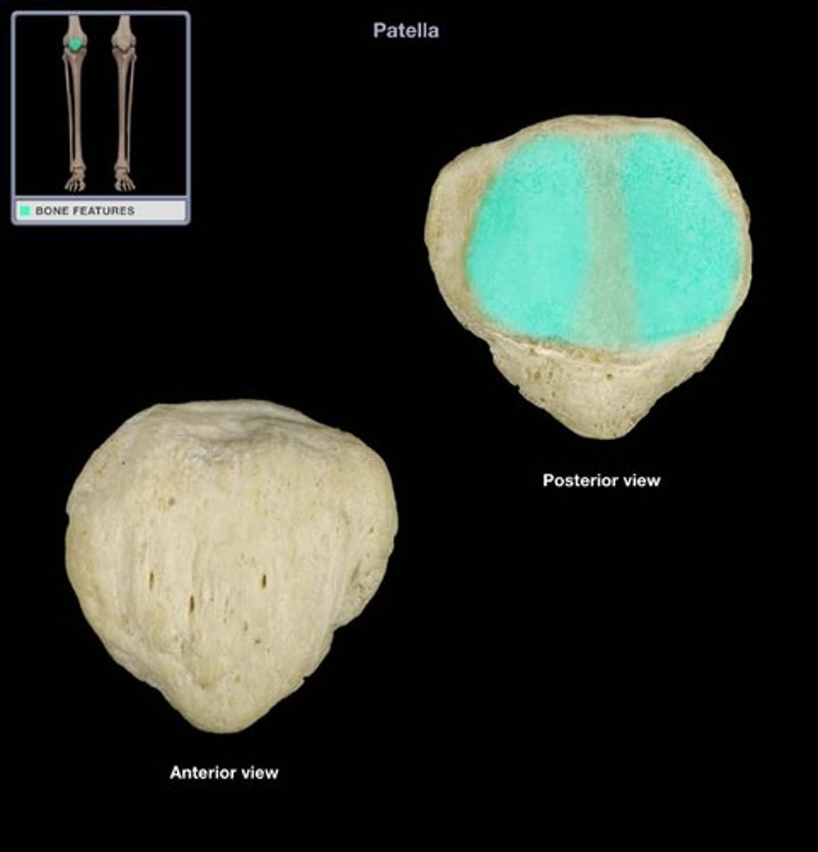
patella
- heart-shaped bone
- "knee cap"
apex of patella
- point at the distal end
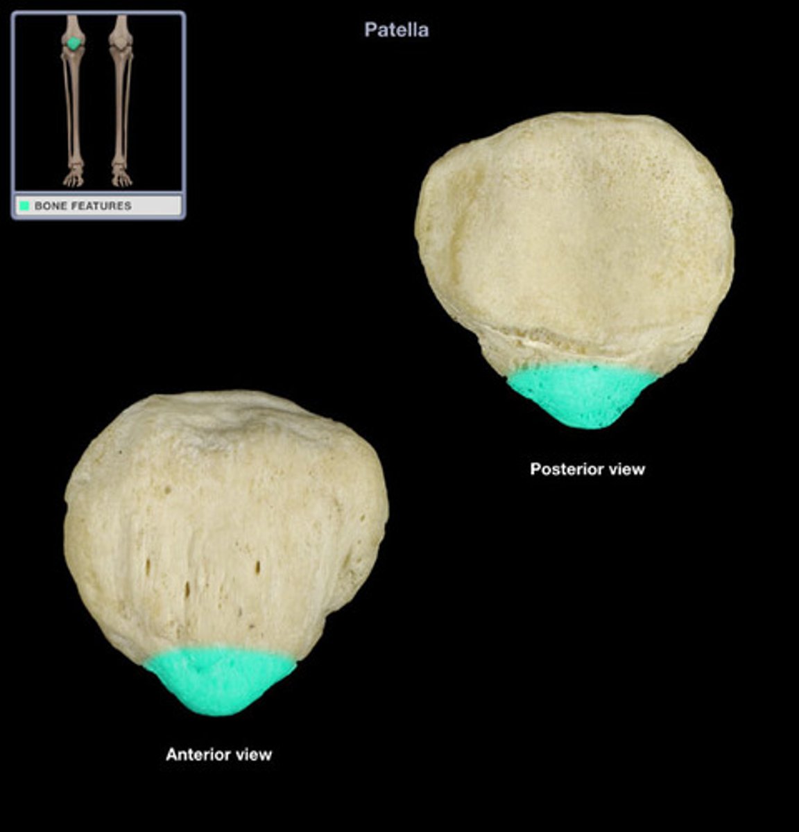
base of patella
- broad, flat surface at the proximal end
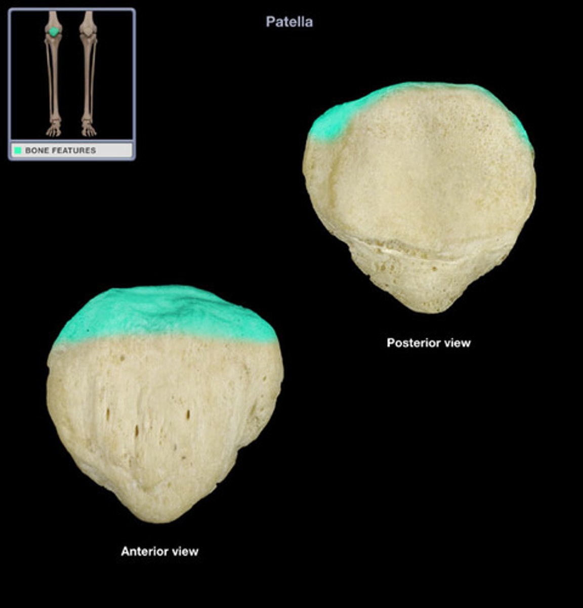
patellar articular facets
- two, smooth surfaces on the posterior side
- articulate with the patellar surface of the femur
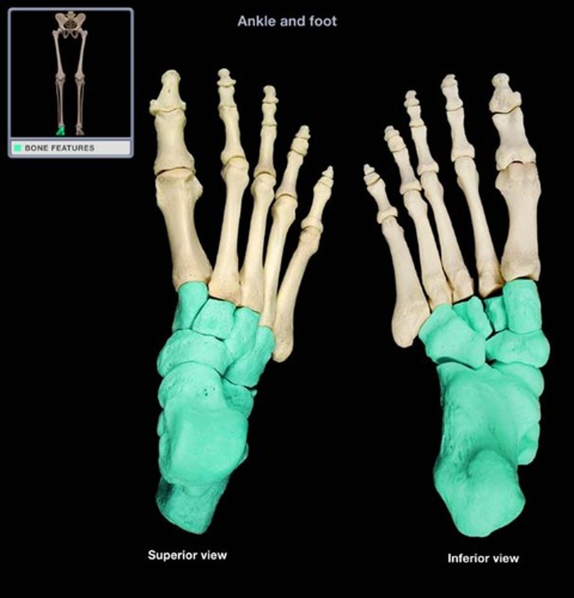
tarsal bones
- bones forming the ankle
- seven tarsal bones
talus
- most medial tarsal bone that articulates with the medial malleolus
- articulates with the tibia/fibula, navicular and calcaneus
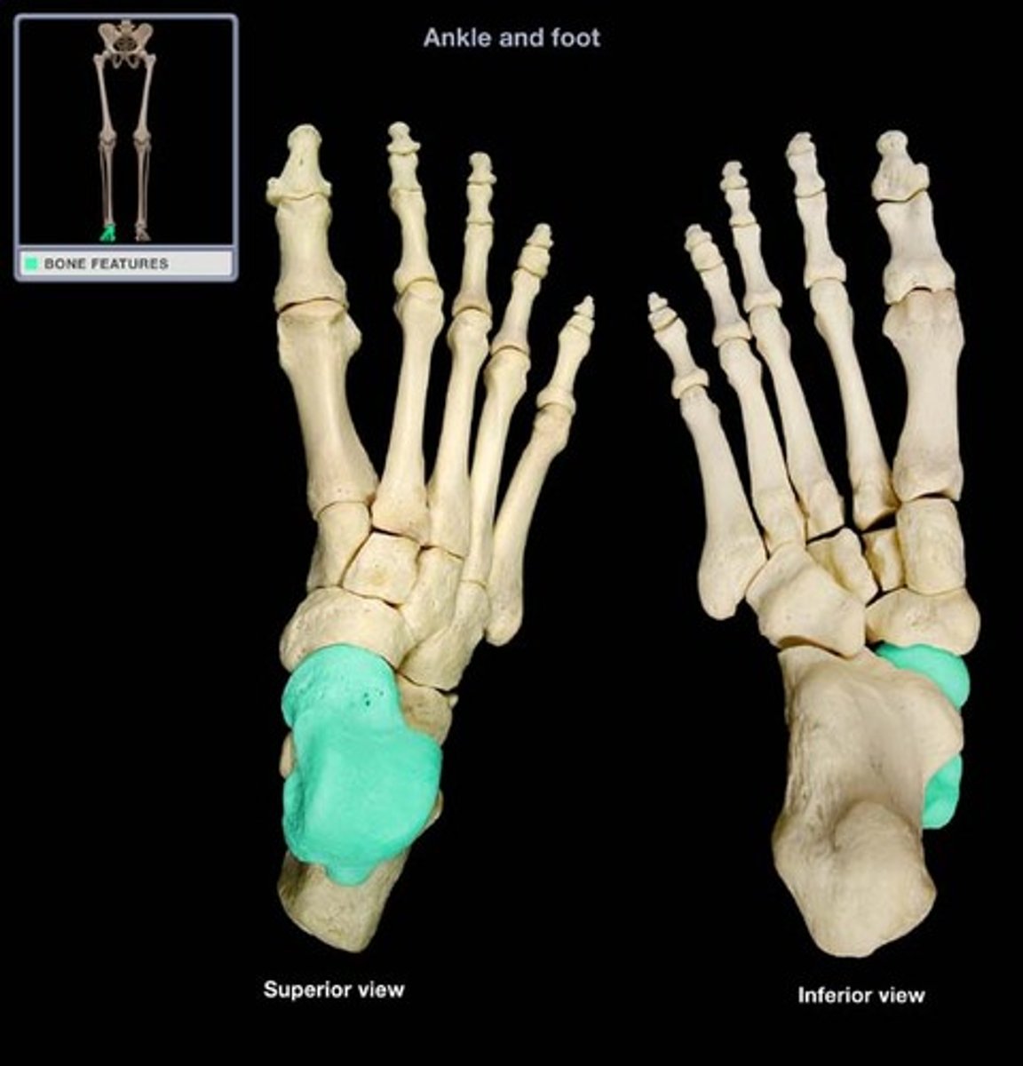
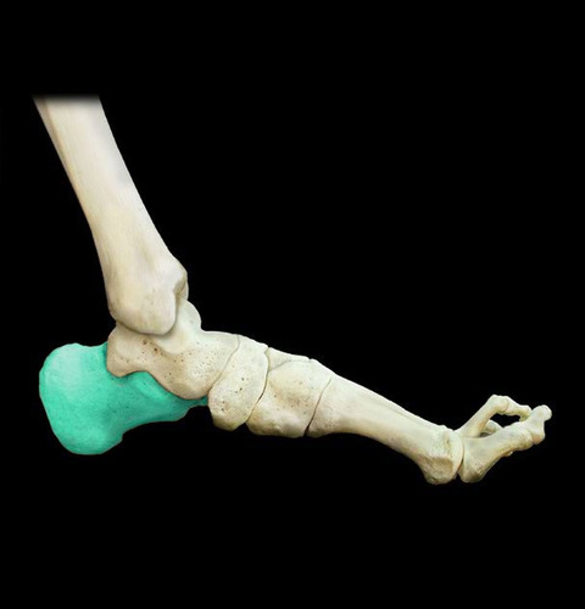
calcaneus
- largest, most posterior tarsal bone
- "heel bone"
- articulates with the talus and cuboid bone
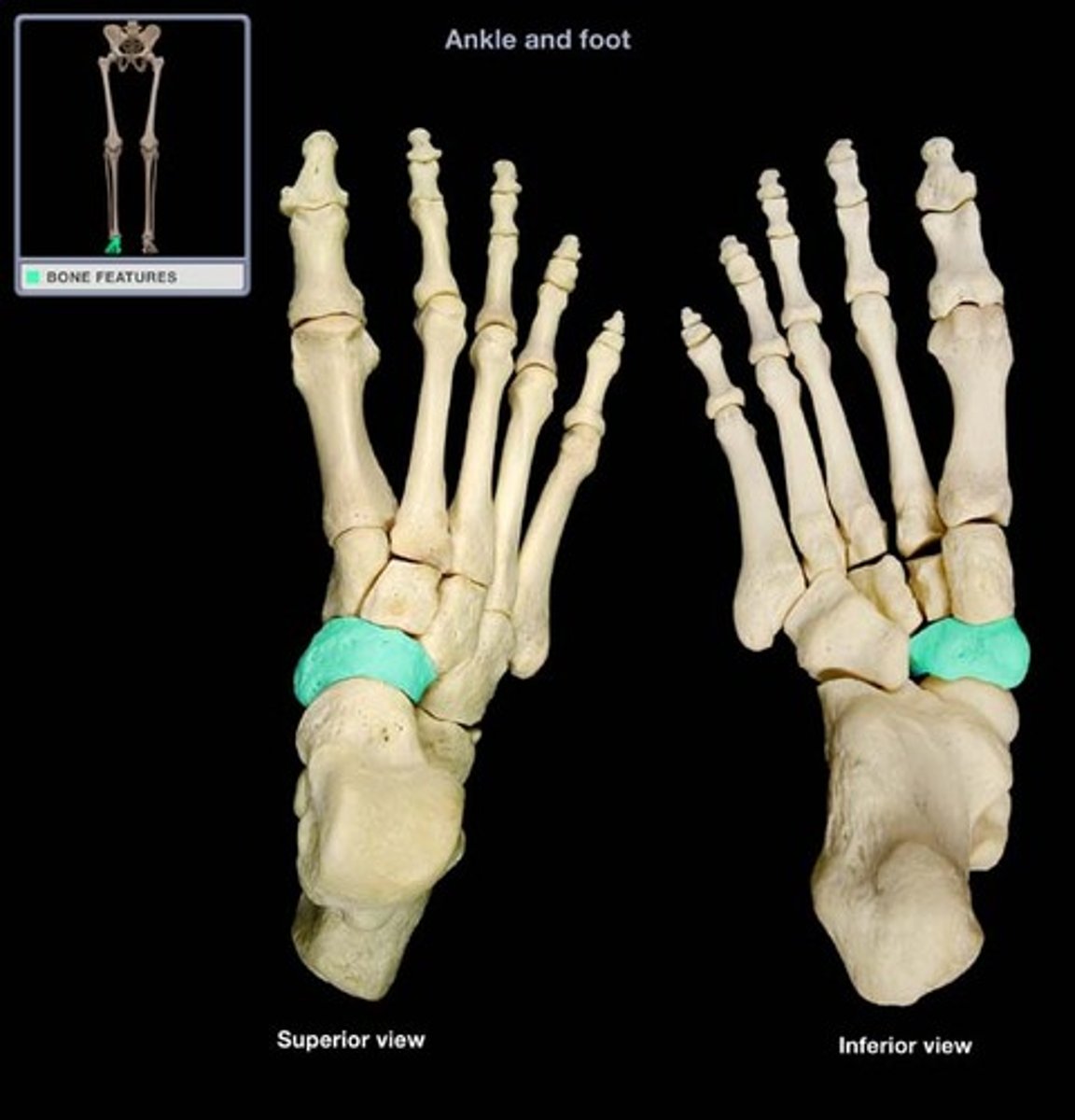
navicular
- nickel-shaped bone between the talus and cuneiform bones
- articulates with the talus and cuneiform bones
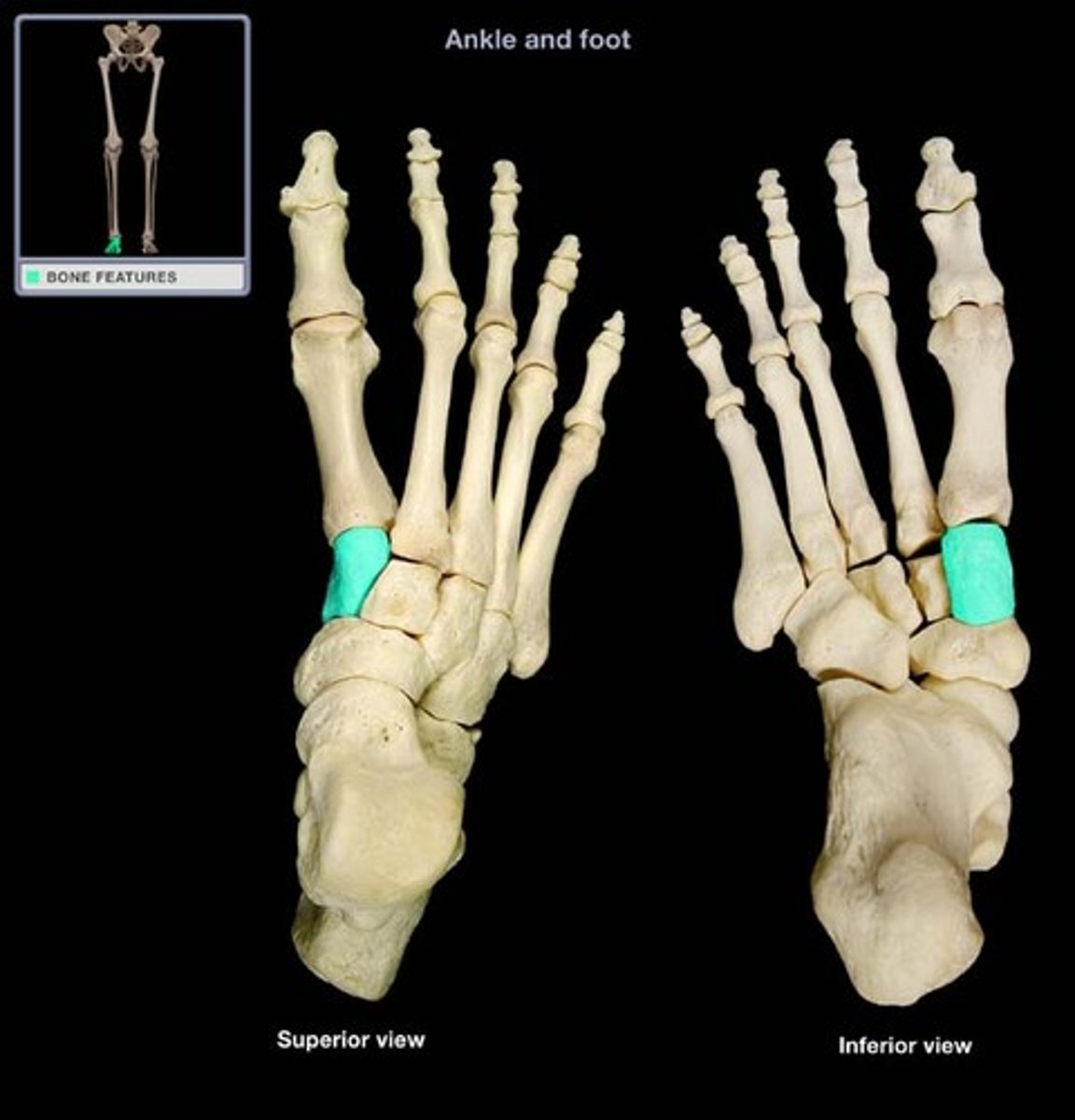
medial (first) cuneiform
- most medial and distal wedge-shaped tarsal bone
- articulates with the intermediate cuneiform, navicular and 1st and 2nd metatarsals
intermediate (second) cuneiform
- middle, wedge-shaped tarsal bone
- articulates with the lateral cuneiform, medial cuneiform, navicular, and 2nd metatarsal

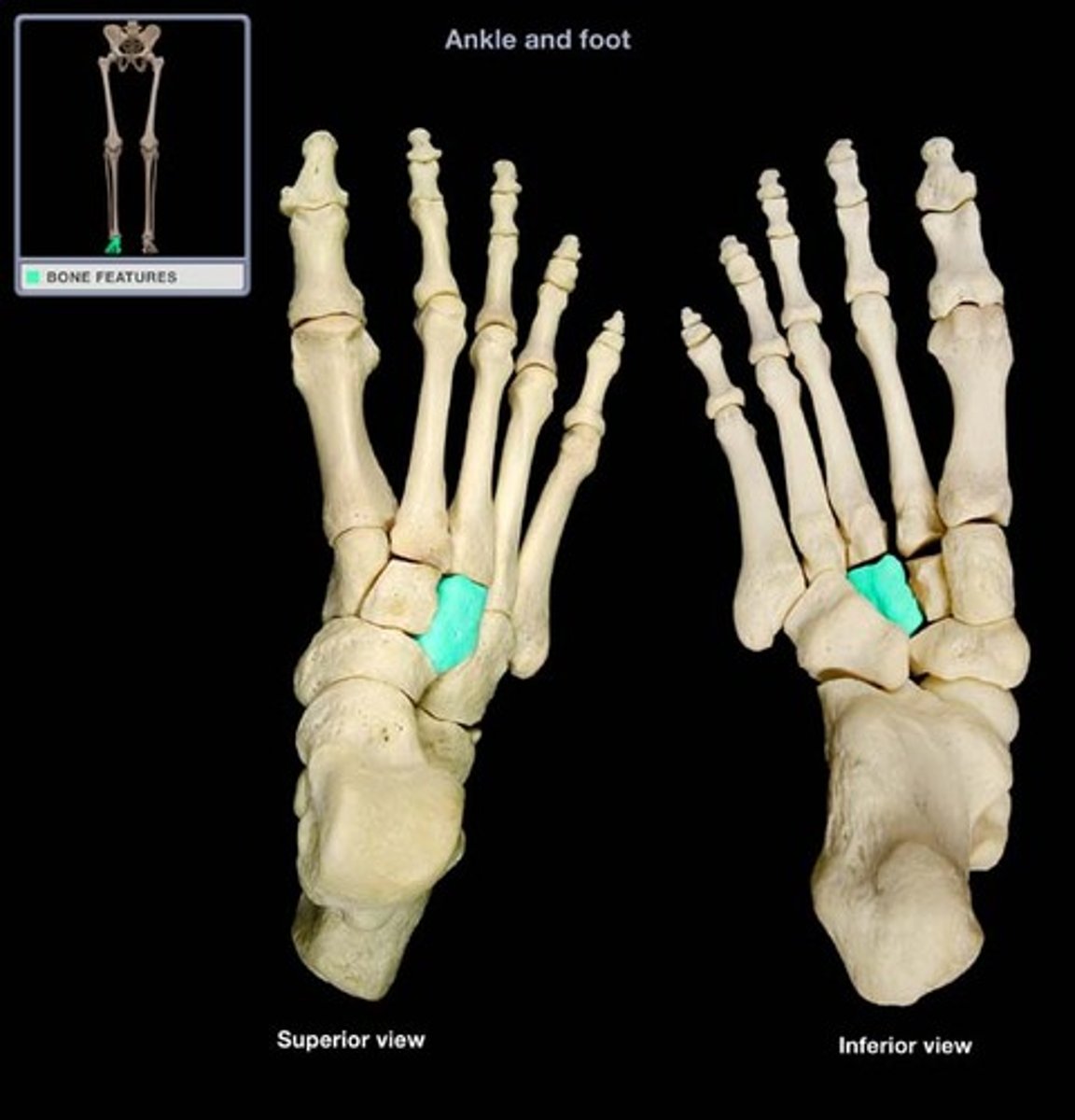
lateral (third) cuneiform
- lateral, wedge-shaped bone
- articulates with the cuboid, intermediate cuneiform, navicular, and 3rd metatarsal
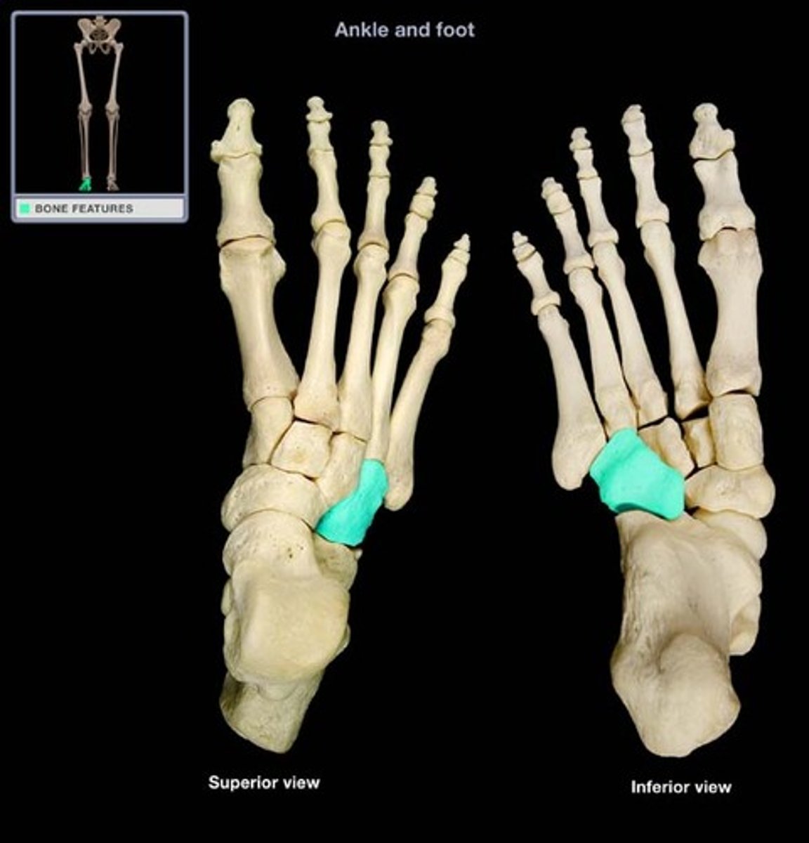
cuboid
- most lateral cube-shaped bone
- between the calcaneus and 4th and 5th metatarsals
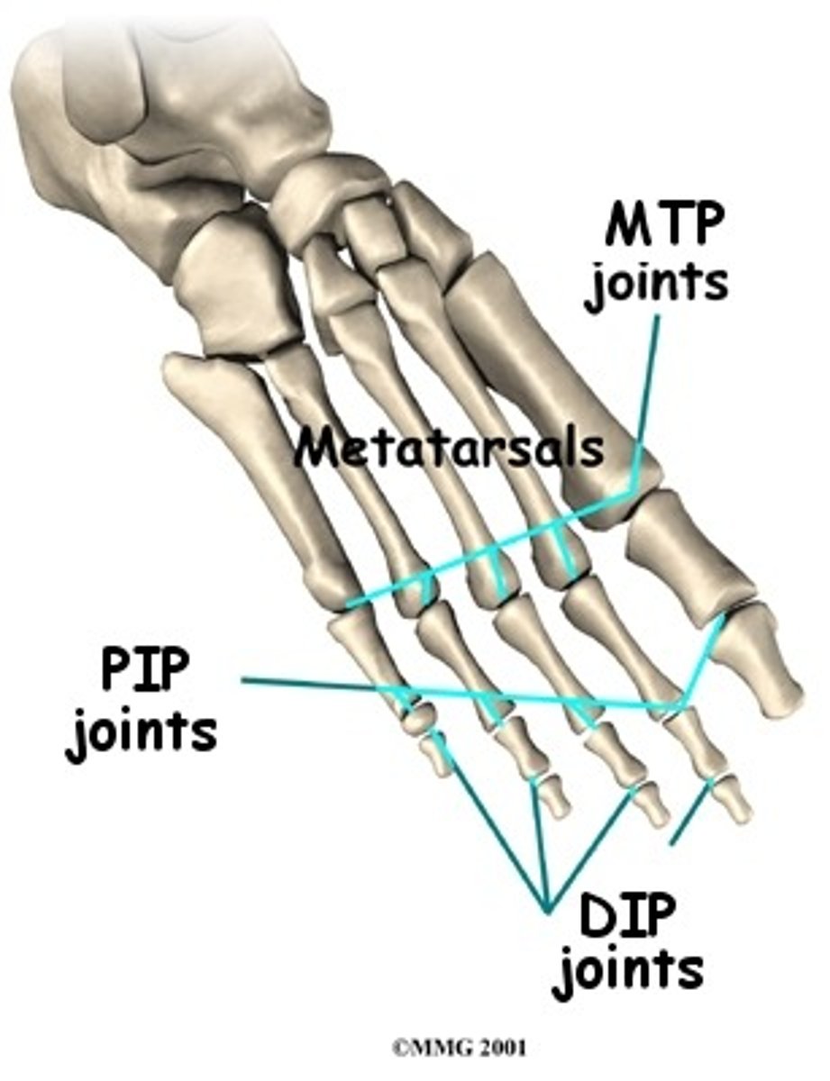
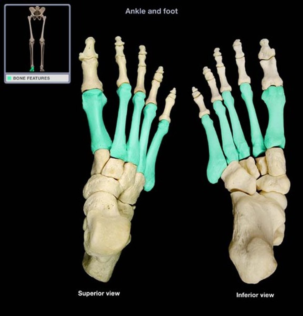
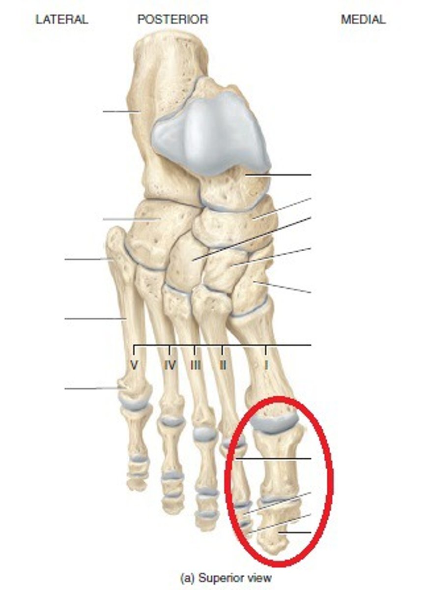
metatarsal bones
- bones forming a foot, between the tarsal bones and proximal phalanges
- numbered I-V from medial (big toe) to lateral (little toe)
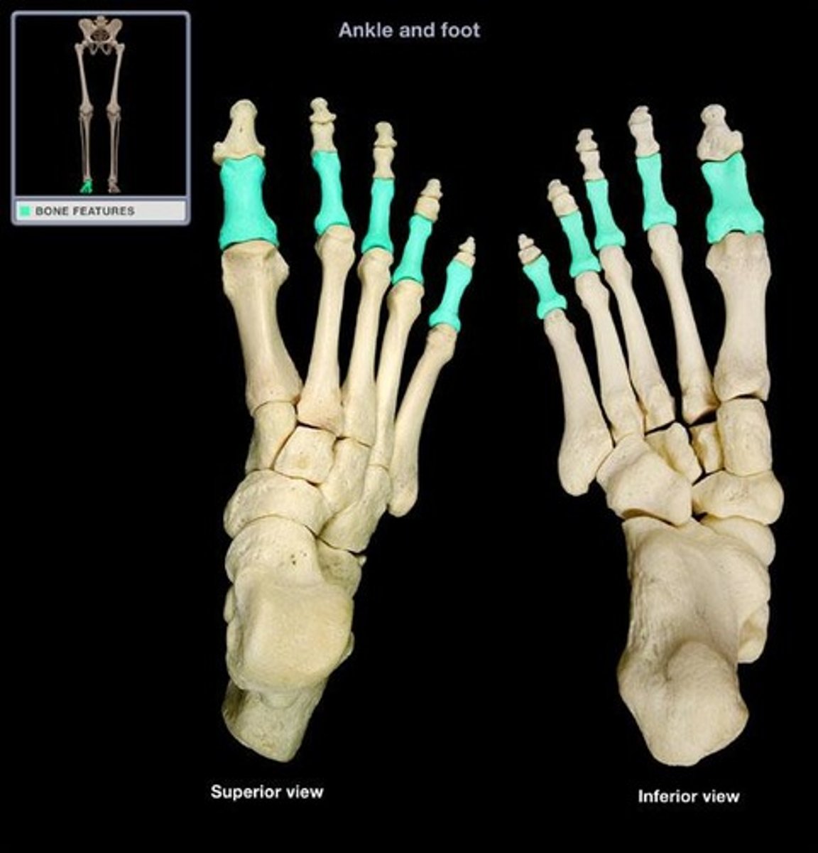
proximal phalanges
- articulates with the metacarpal bones and middle phalanges
- numbered I-V
- phalanges = plural
- phalanx = singular
middle phalanges
- articulates with the proximal and distal phalanges
- numbered II-V

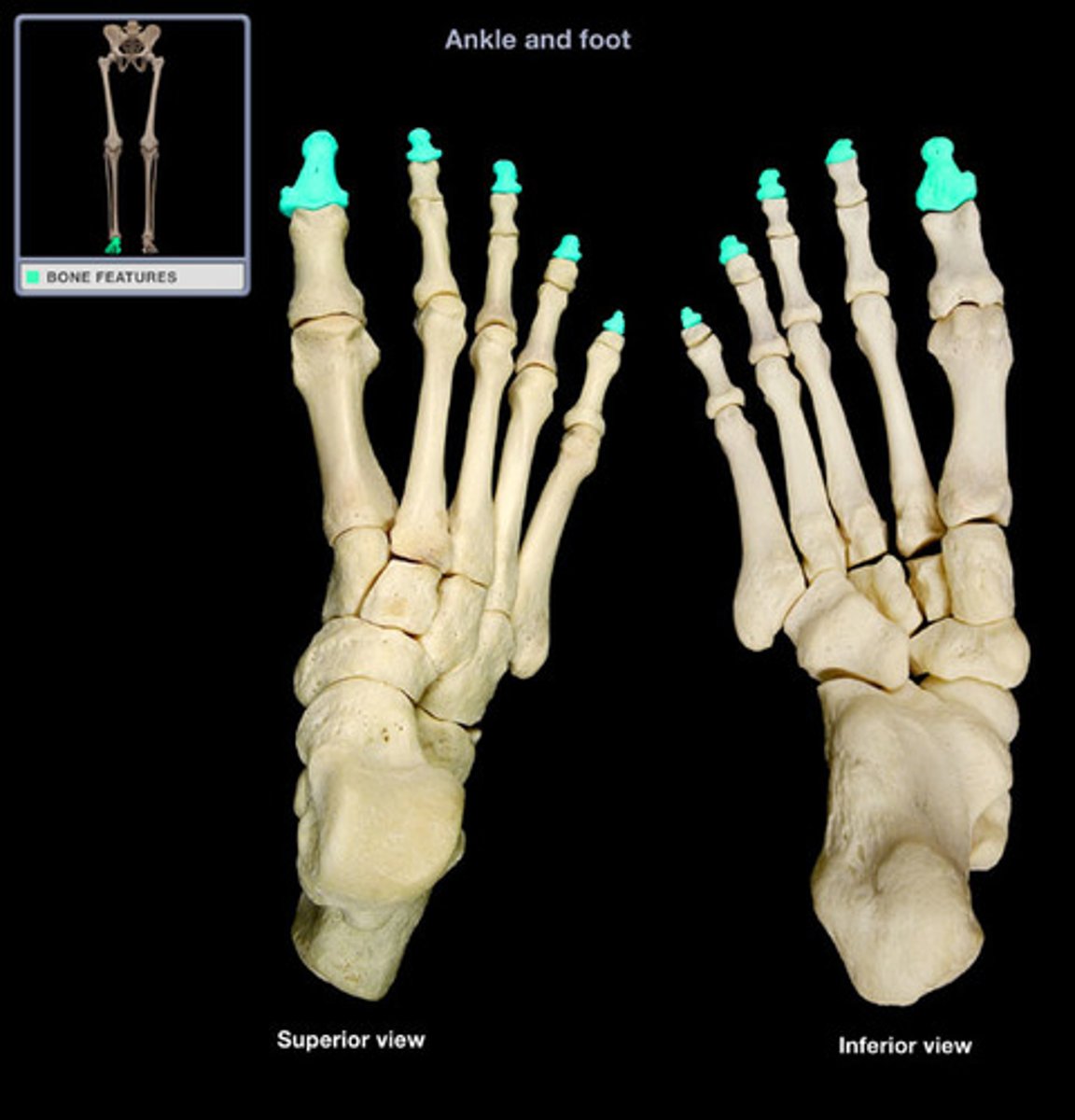
distal phalanges
- articulates with the middle phalanges
- numbered I-V
- "tips of toes"
hallux
- formed by the proximal and distal phalanx of digit 1
- "big toe"
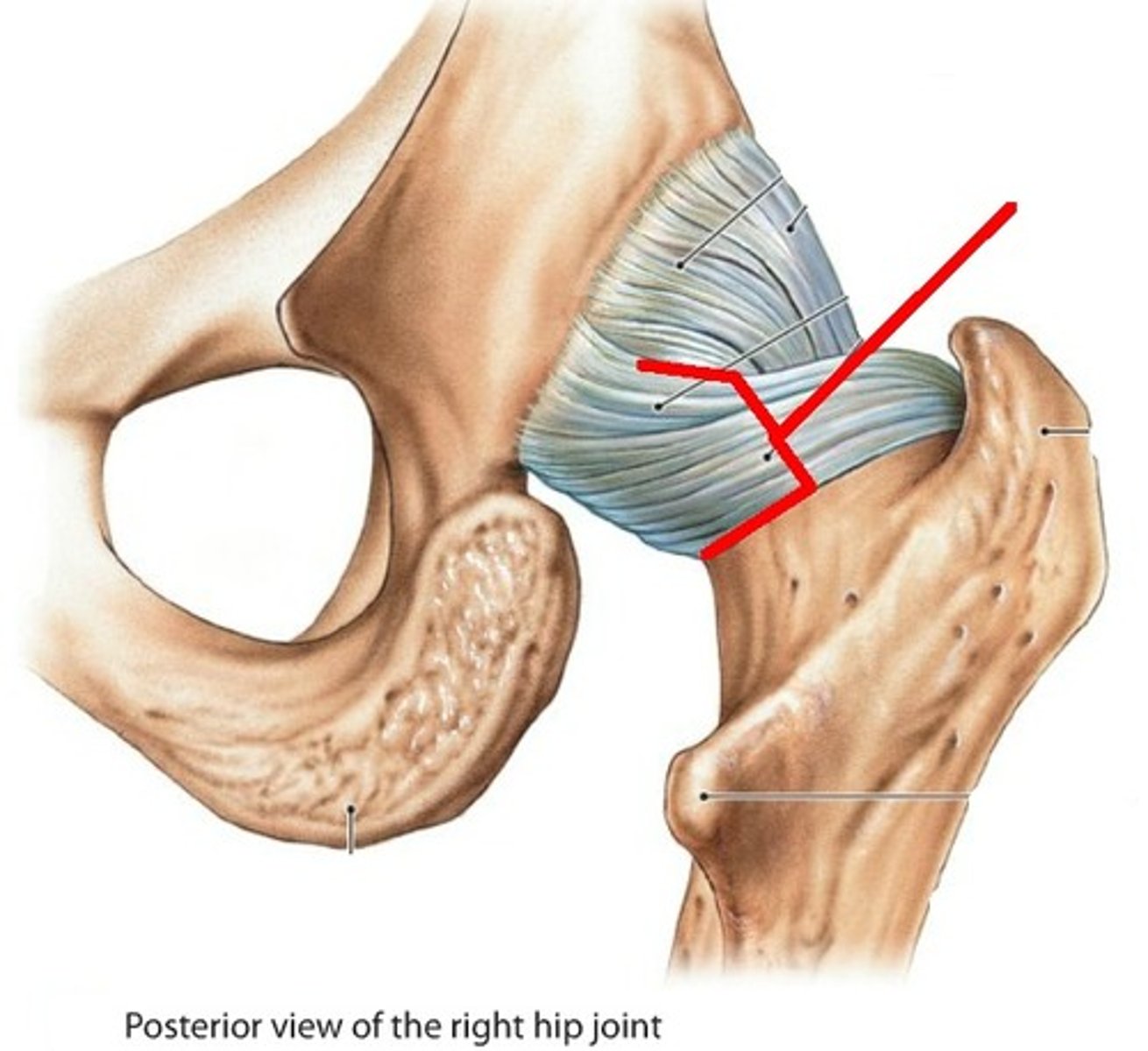
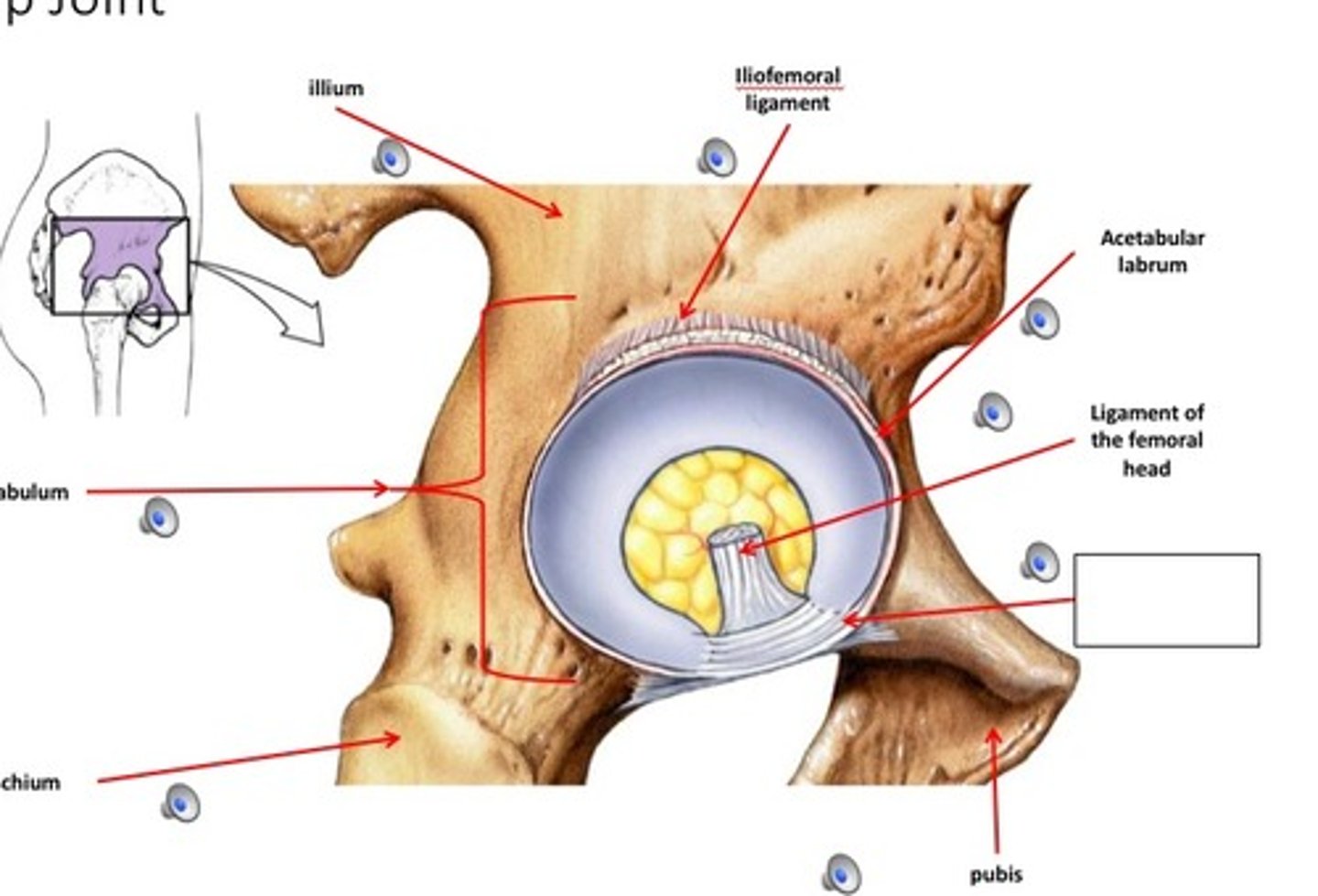
hip joint
- articulation of the head of the femur with the acetabulum of the coral bone
- ball-and-socket type of synovial joint
- permits flexion/extension, abduction/adduction, and internal/external rotation of the hip
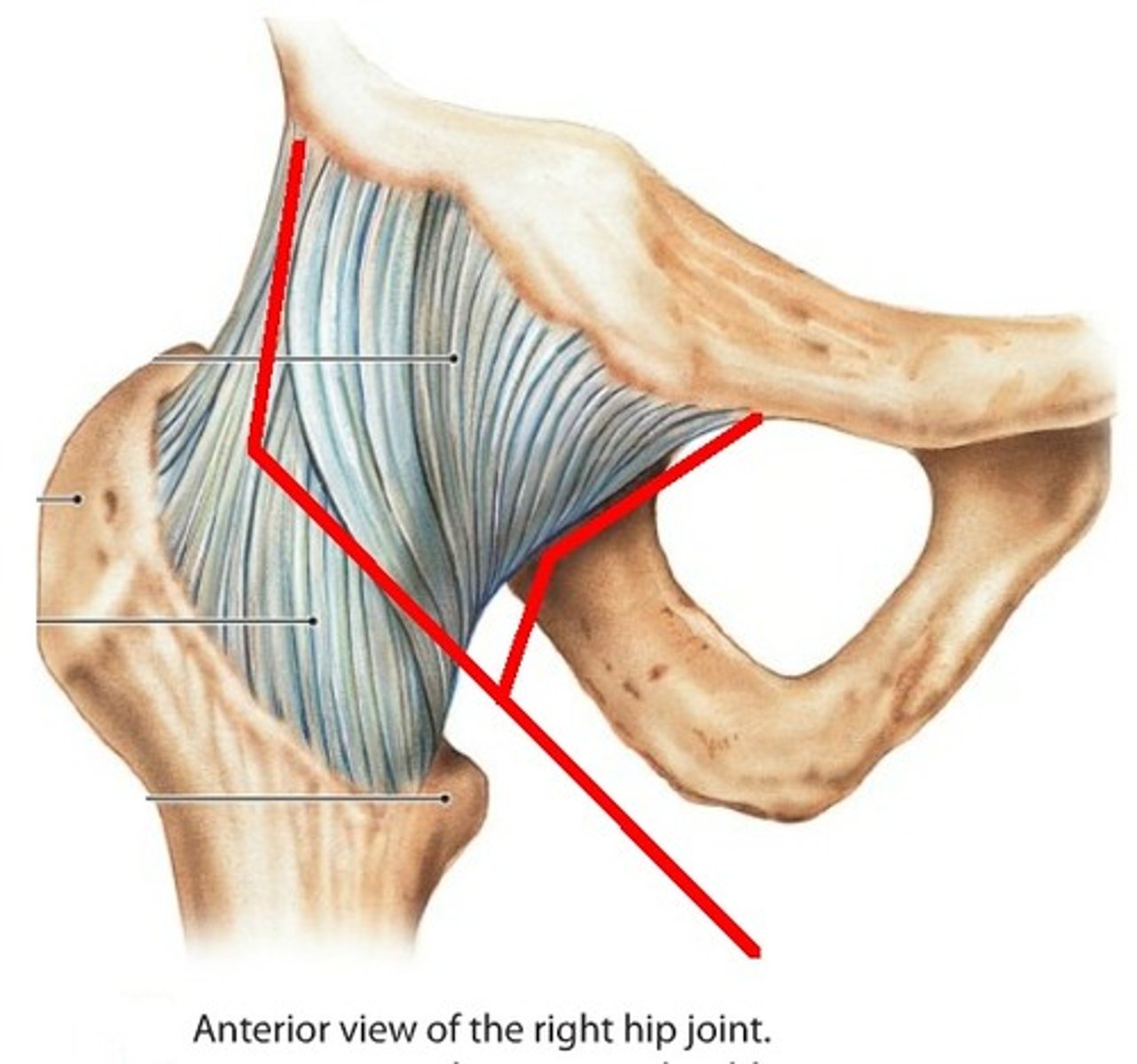
iliofemoral ligament
- ligament running from the ilium to the head of the femur
- Y-shaped ligament
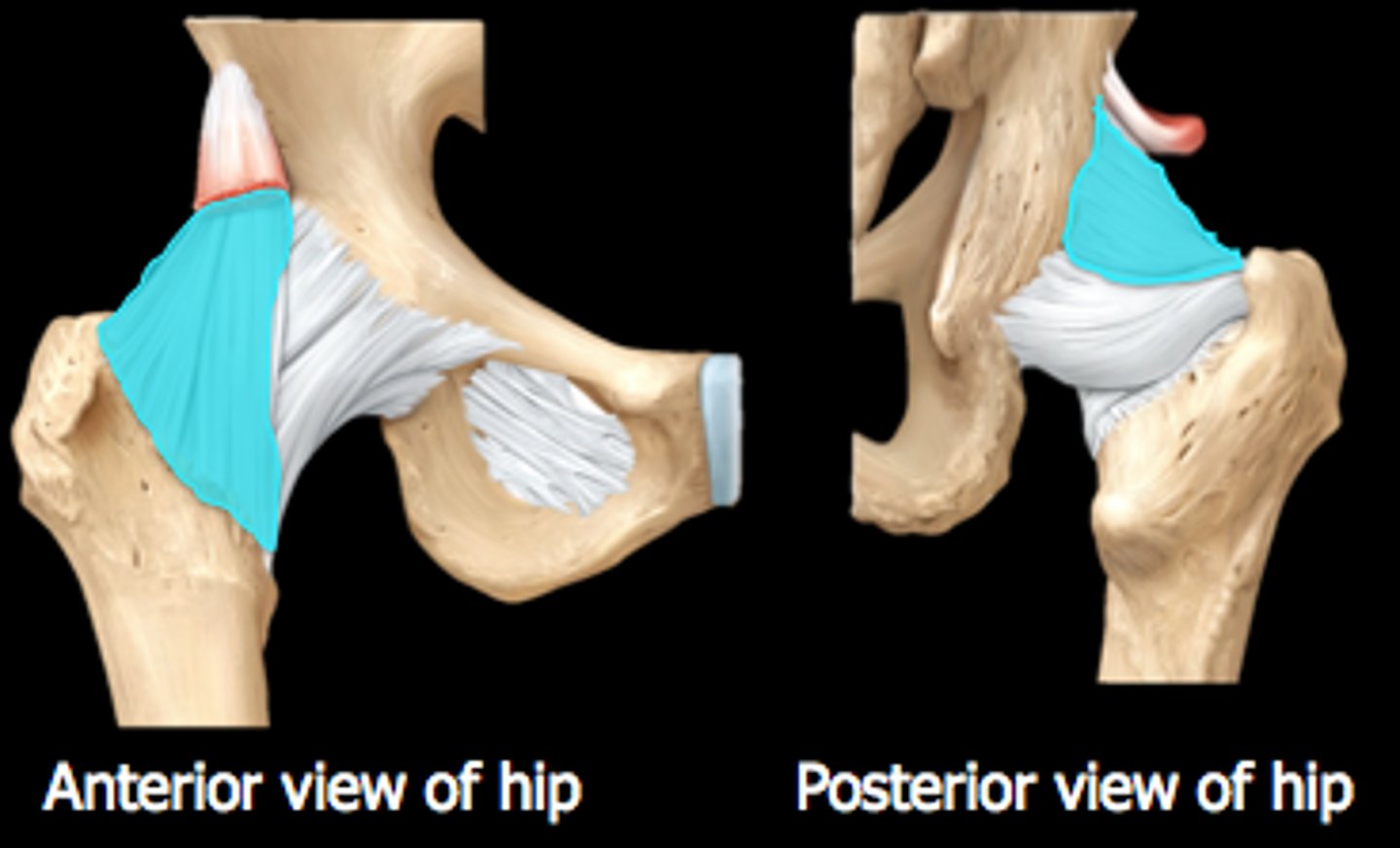
ischiofemoral ligament
- ligament running from the ischium to the head of the femur
pubofemoral ligament
- ligament running from the pubic to the head of the femur
- blends with the iliofemoral ligament
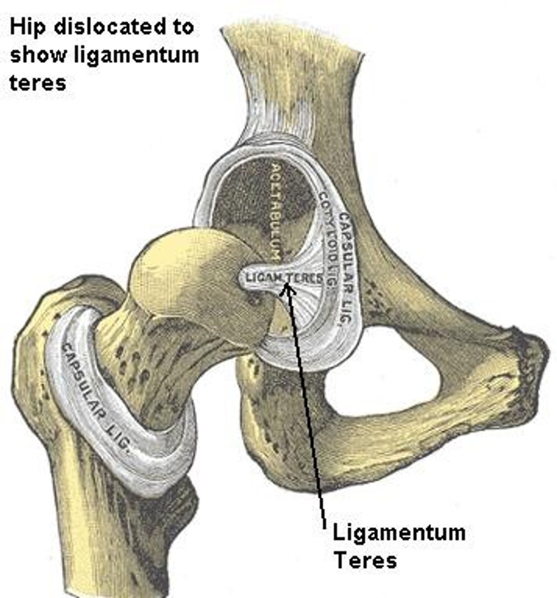
ligament of the femoral head
- ligament running from the head of the femur to the acetabular notch
acetabular labrum
- fibrocartilaginous ring lining the wall of the acetabulum
- increases surface area and decreases joint stress
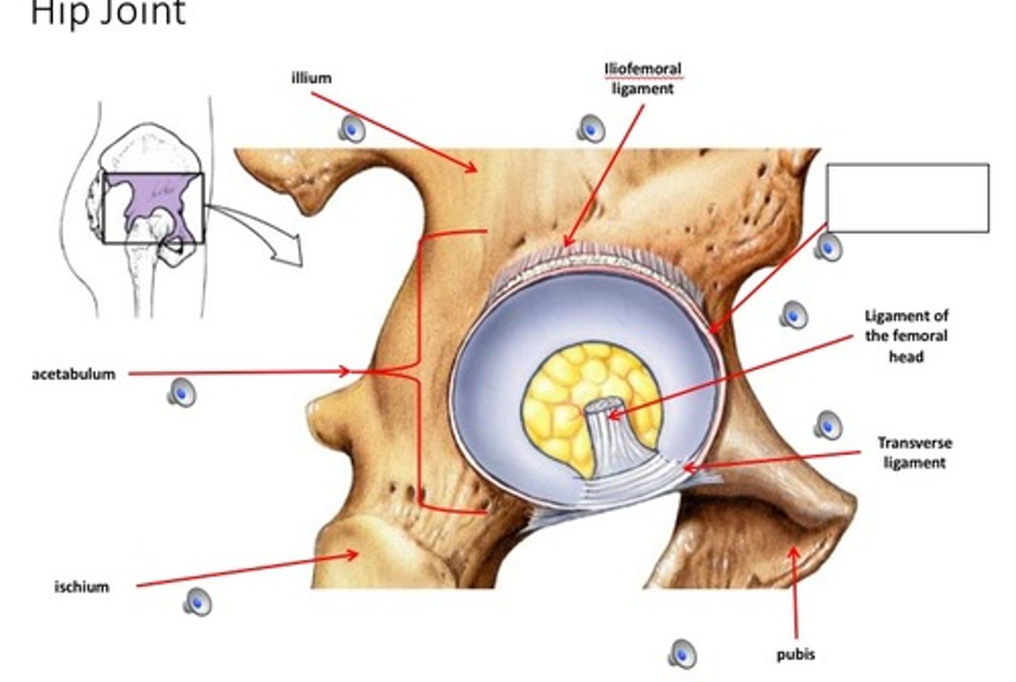
transverse ligament of the hip
- ligament running across the acetabular notch
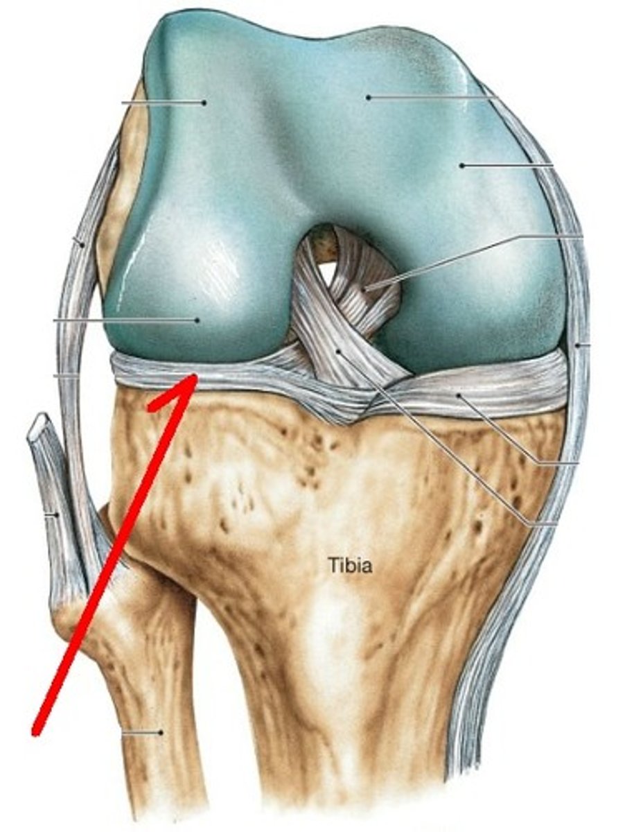
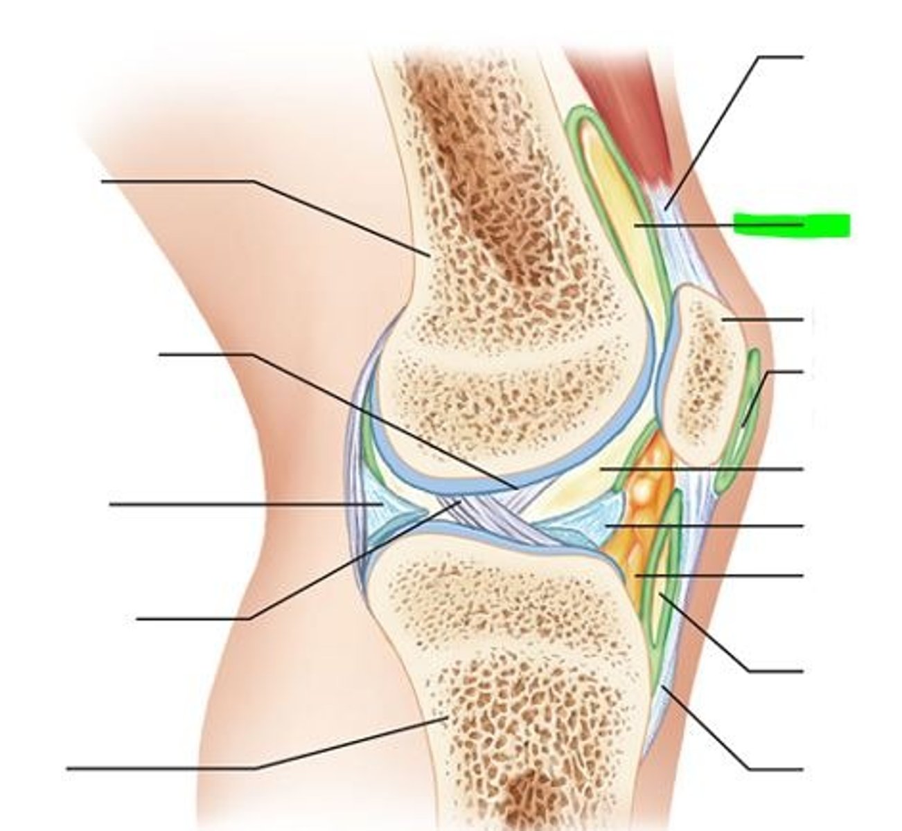
knee joint
- articulation of the medial/lateral femoral condyles with the medial/lateral tibial condyles
- hinge type of synovial joint
- permits flexion/extension of the knee
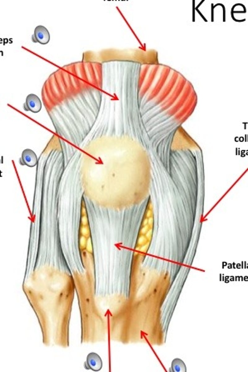
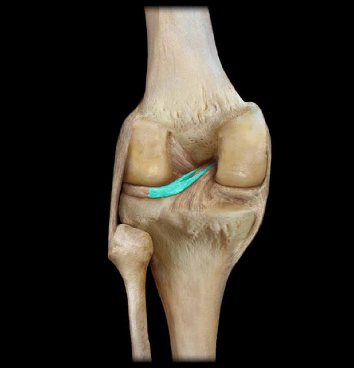
tibial (medial) collateral ligament
- ligament running from the medial epicondyle of the femur to the medial condyle of the tibia
- flat, band-like capsular ligament
- weaker Thant he fibular collateral ligament
fibular (lateral) collateral ligament
- ligament running from the lateral epicondyle of the femur to the head of the fibula
- cord-like extracapsular ligament

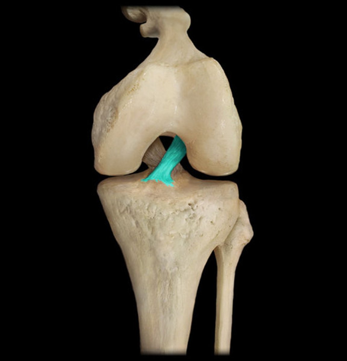
anterior cruciate ligament
- ligament running from the anterior aspect of the intercondylar area of the tibia to the medal aspect of the lateral femoral condyle
- prevents posterior displacement of the femur on the tibia and hyperextension of the knee joint
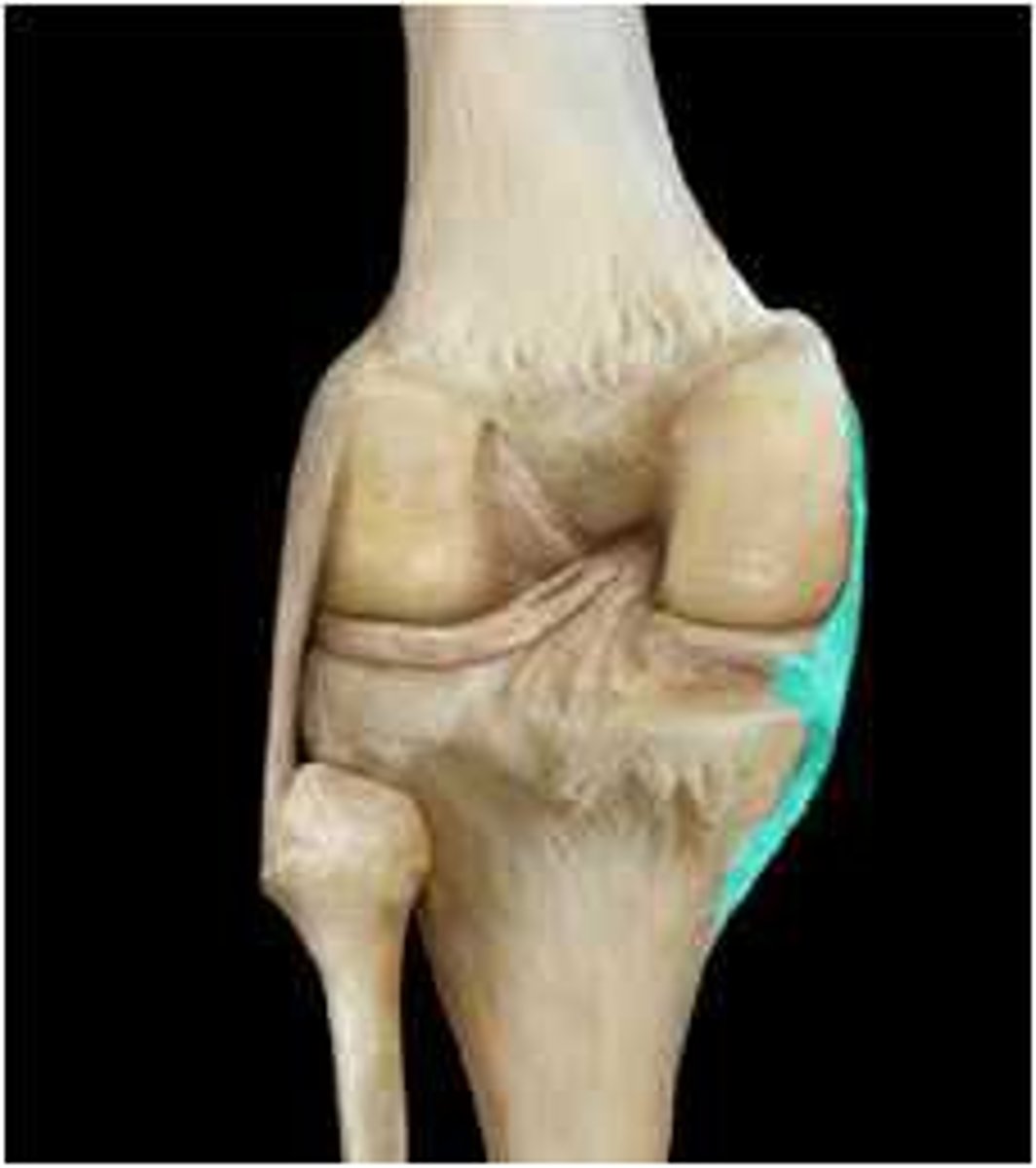
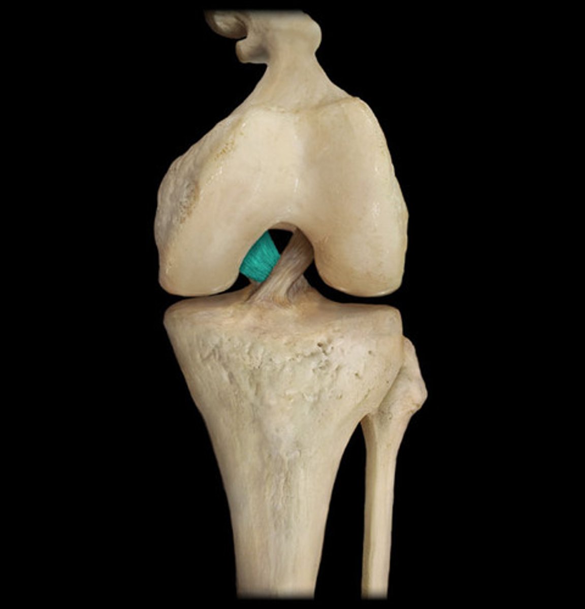
posterior cruciate ligament
- ligament running from the posterior aspect of the intercondylar area of the tibia to the lateral surface of the medial femoral condyle
- prevents anterior displacement of the femur on the tibia and hyperextension of the knee cap
medial meniscus
- semicircular cartilage on the superior surface of the medial tibial condyle
- commonly injured with the lateral force to the knee
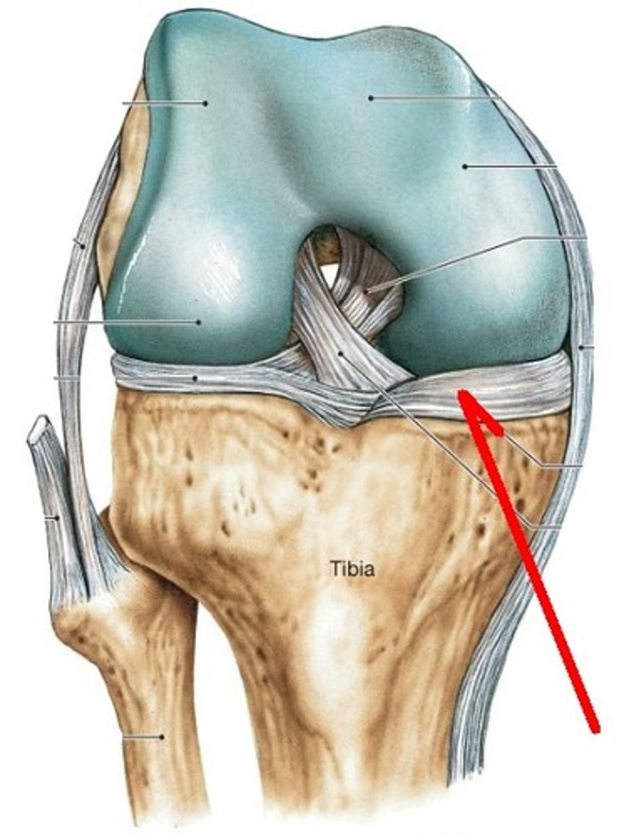
lateral meniscus
- semicircular cartilage on the superior surface of the lateral tibial condyle
- acts as a "shock absorber" for the knee joint
posterior meniscofemoral ligament
- ligament between the lateral meniscus and medial condyle of the femur
- fibrous band on the posterior knee
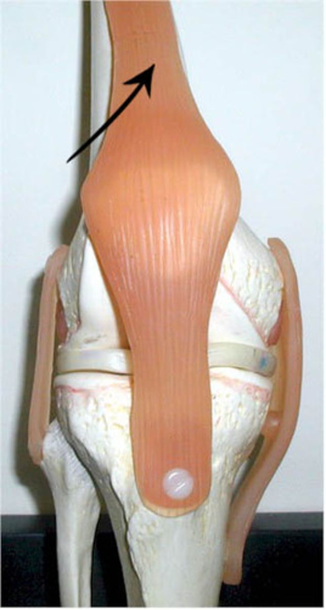
quadriceps tendon
- connecting the quadriceps muscles to the patella
- common insertion of the four quadriceps muscles
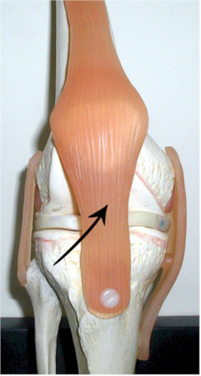
patellar ligament
- ligament between the patella and tibial tuberosity
- distal continuation of the quadriceps tendon
suprapatellar bursa
- large bursa between the anterior surface of the femur and the quadriceps tendon
- allows for movement of the quadriceps tendon
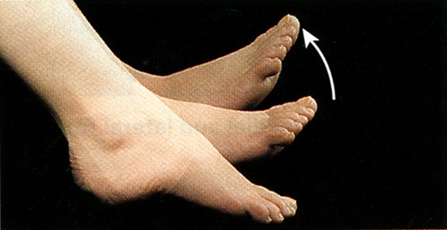
ankle joint
- articulation of the distal ends of the tibia and fibula with the superior surface of the talus
- hinge type of synovial joint
- permits plantarflexion/dorsoflexinon of the ankle
deltoid ligament
- fan-shaped ligament running from the medial malleolus of the tibia to the talus, navicular, and calcaneus
- broad, medial, extra capsular ligament of the ankle
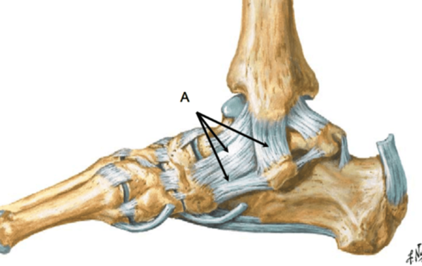
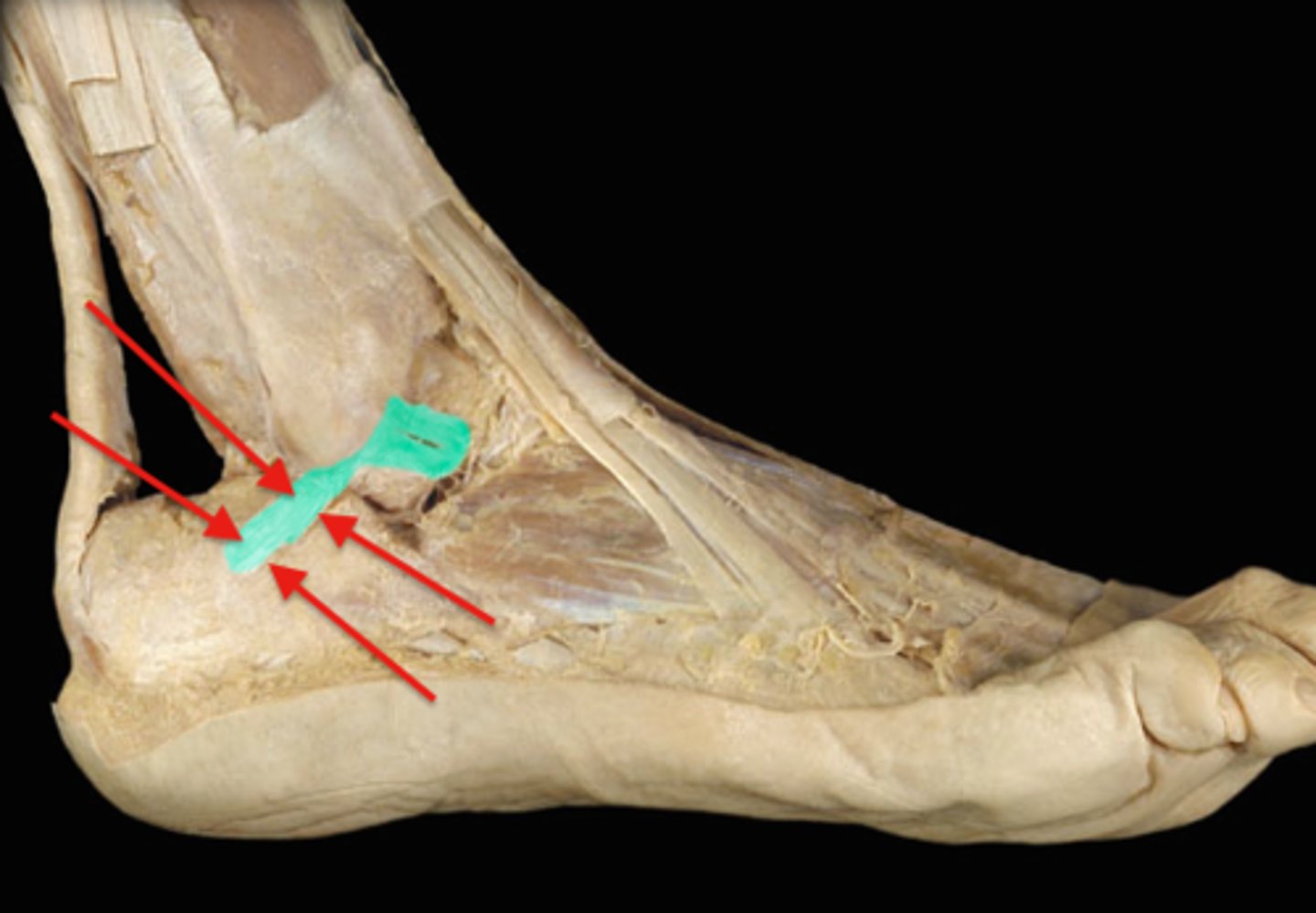
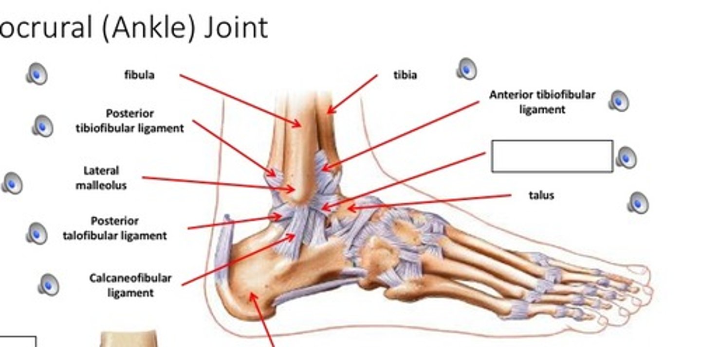
calcaneofibular ligament
- ligament running from the lateral surface of the calcaneus to the lateral malleolus of the fibula
- smaller, lateral extra capsular ligament of the ankle
- covered by the fibulas longs and braves tendons
posterior talofibular ligament
- ligament running from the posterior aspect of the talus to the fibula
- horizontal band between the posterior talus and fibula
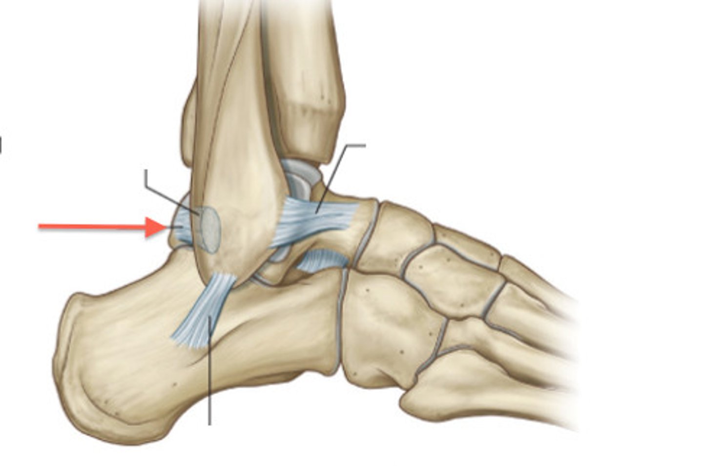
anterior talofibular ligament
- ligament running from the anterior aspect of the talus to the fibula
- horizontal band between the anterior talus and fibula
- most commonly injured ligament in a sprained ankle
posterior tibiofibular ligament
- ligament running between the posterior aspects of the tibia and fibula
- thick, triangular band extending obliquely between the posterior tibia and fibula
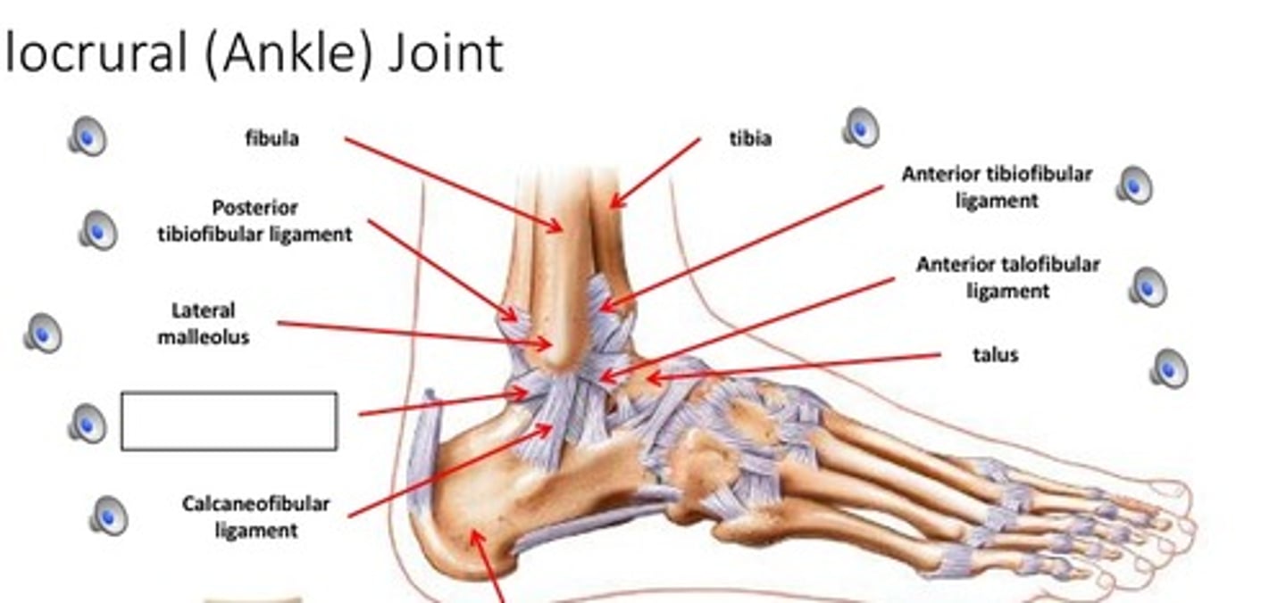
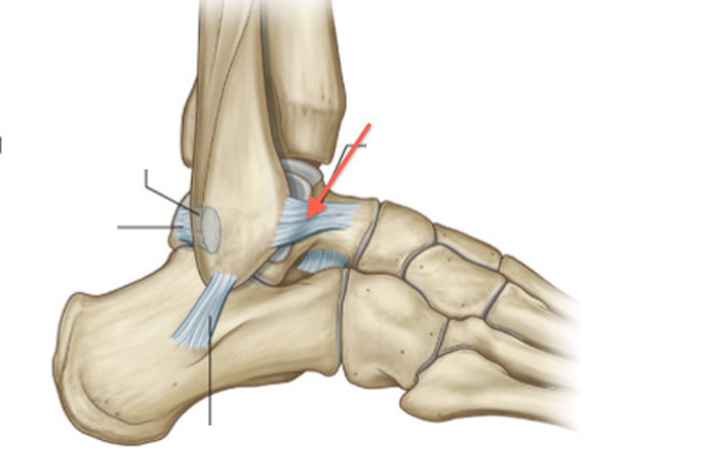
anterior tibiofibular ligament
- ligament running between the anterior aspects of the tibia and fibula
- thick, triangular band extending obliquely between the anterior tibia and fibula
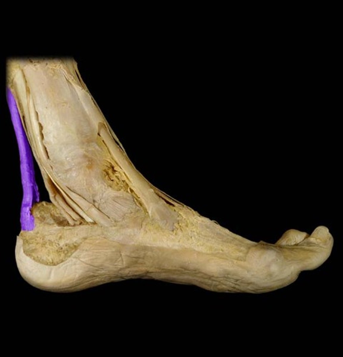
calcaneal tendon
- connecting the gastrocnemius and soles muscles to the calcaneus
- also known as the "achilles" tendon
- strongest tendon of the body
subtalar joint
- articulation of the inferior surface of the talus with the superior surface of the calcaneus
- plane type of synovial joint
- permits inversion/eversion of the foot
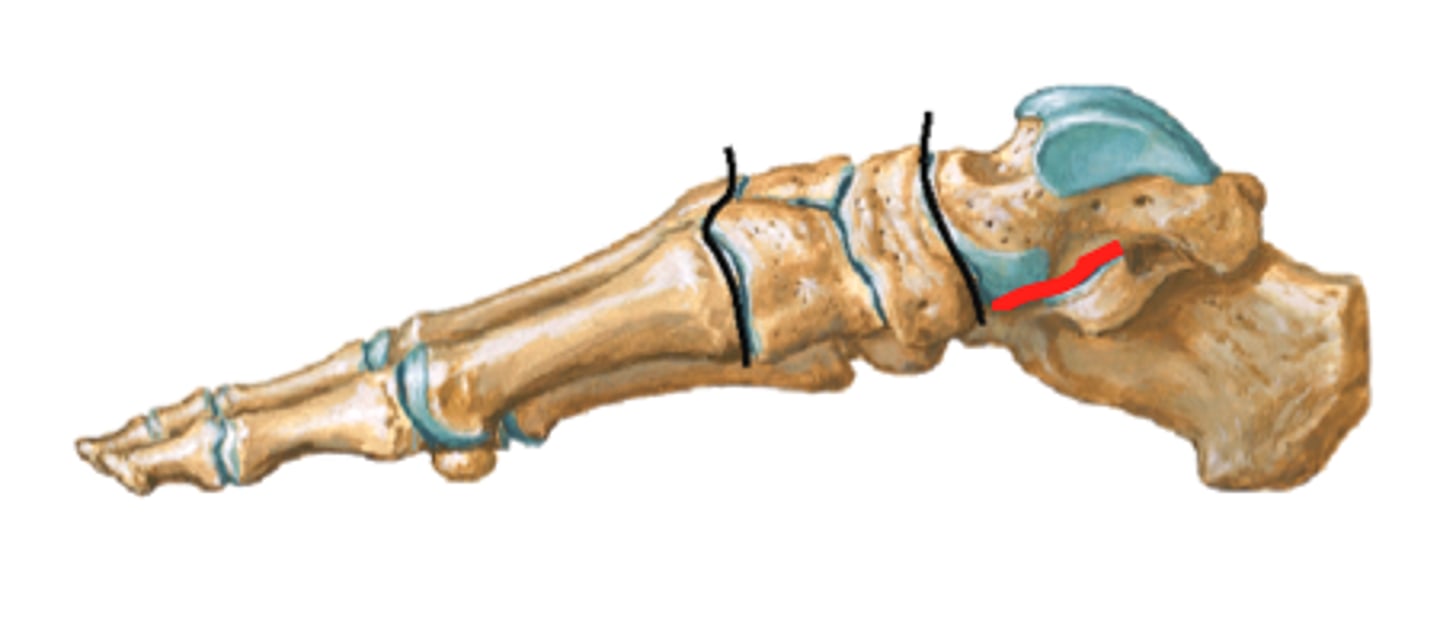
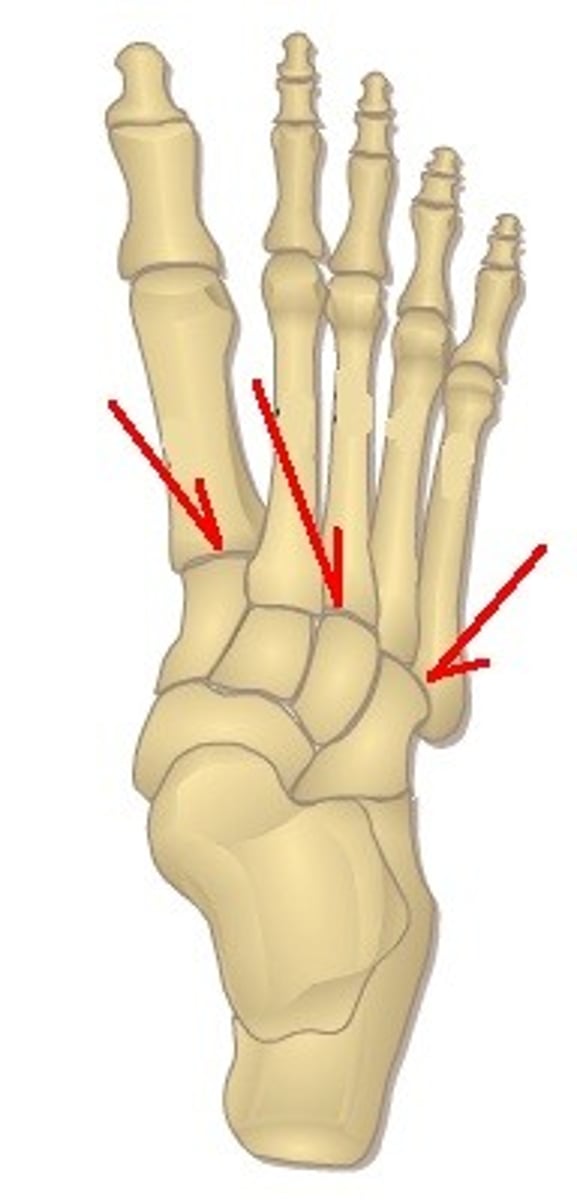
tarsometatarsal joints
- articulations of the tarsal bones with the metatarsals
- plane type of synovial joint
- permits gliding movements of the tarsal bones
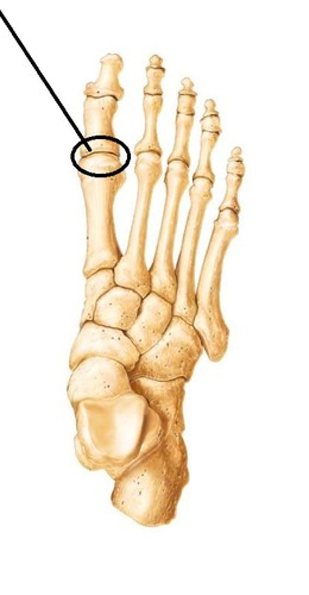
metatarsophalangeal joint
- articulation of the metatarsals with the proximal phalanges
- condyloid type of synovial joint
- permits flexion/extension and abduction/adduction of the digits
proximal interphalangeal joints of foot
- articulation of the proximal phalanges with the middle phalanges
- hinge type of synovial joint
- permits flexion/extension of the digits
- only present in digits 2-5

distal interphalangeal joints of foot
- articulation of the middle phalanges with the distal phalanges
- hinge type of synovial joint
- permits flexion/extension of the digits
- digit 1 only has an interphalangeal joint