Nervous System Development
1/35
There's no tags or description
Looks like no tags are added yet.
Name | Mastery | Learn | Test | Matching | Spaced |
|---|
No study sessions yet.
36 Terms
***RQ: what is produced during the process of neurulation?
neuroectoderm: gives rise to the brain, spinal cord, and PNS
primary neurulation: produces portion of neural tube that gives rise to brain and spinal cord through the lumbar levels
secondary neurulation: produces portion of neural tube that gives rise to the sacral and coccygeal levels of the spinal cord

***RQ: what are key events associated with the process of neurulation, and when (days of gestation) do those events occur?
key events:
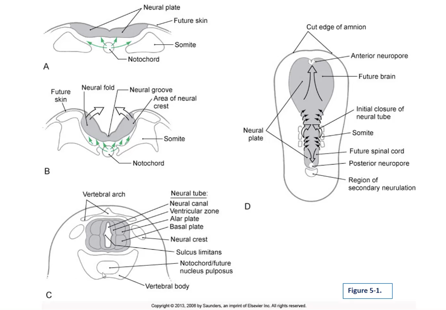
notochord: important function is induction (i.e., directing the ectoderm to form the neural plates) ~d16
neural groove: first indicator of neurulation; located on the posterior aspect of the embryo in the center of the neural plate
neural plate: gives rise to most of the nervous system
neural folds: thickening of neural plate at the lateral margins ~d18
neural tube: hollow structure that forms when the neural folds meet each other during in-folding at ~d20
the CNS develops from the neural tube
neural crest: arise from the lateral edge of neural plate; they detach and move lateral to the neural tube forming
the PNS develops from the neural crest
primary neurulation: produces portion of neural tube that gives rise to brain and spinal cord through the lumbar levels
secondary neurulation: produces portion of neural tube that gives rise to the sacral and coccygeal levels of the spinal cord- begins ~d20 and is completed by ~d42
lumen of the neural tube:
neuroblasts arise on luminal surface of tube and dividing cells cluster forming the ventricular zone
processes of these developing cells form the. marginal zone located lateral to the ventricular zone
cells in ventricular zone undergo their last division and migrate forming the intermediate zone located between the ventricular and marginal zones
anterior neuropore: rostral opening of neural tube closes at ~d24
posterior neuropore: caudal opening of neural tube closes at ~d26
***RQ: does spinal bifida result from failure of anterior or posterior neuropore closure?
posterior
***RQ: what are the 3 types of spina bifida?
spinal bifidas:
(1) spina bifida occulta
most mild form
small gap in 1 or more of the vertebra of the spine
posterior arches aren’t formed properly, but the spinal cord is still formed/retained intact
no signs or symptoms; also no neurological problems (goes unnoticed a lot!)
spina bifida cystica:
(2) meningocele
protective membranes around the spinal cord (meninges) will push out in the opening of the vertebra
membranes can be removed with surgery with little to no damage to the nerve pathways, as the spinal cord is still developed properly
(3) meningomyelocele
most severe
baby’s spinal canal remains open among several vertebrae in the lower/middle back region
both the membranes of the spinal cord and the neuro-tissue itself protrude
forms a sac on the back
nerves and tissues are exposed, making a baby vulnerable to infections
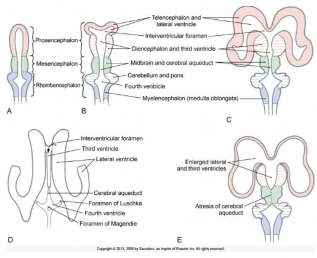
***RQ: what are the names of the primary brain vesicles?
prosencephalon (forebrain)
divides into 2 secondary brain vesicles
telencephalon
diencephalon
mesencephalon (midbrain)
remains as the midbrain
rhombencephalon (hindbrain)
divides into 2 secondary brain vesicles
metencephalon
myelencephalon
***RQ: what are the adult derivatives of the brain vesicles?
primary vesicles
prosencephalon (adult derivative: forebrain)
mesencephalon (adult derivative: midbrain)
rhombencephalon (adult derivative: hindbrain)
secondary vesicles
telencephalon: cerebral hemispheres and deep cortical structures
diencephalon: thalamus, hypothalamus, subthalamus, epithalamus
mesencephalon: midbrain
metencephalon: pons and cerebellum
myelencephalon: medulla
***RQ: which ventricles develops from the cavities of the telencephalon and diencephalon?
lateral ventricles: cavities of the telencephalic vesicles
third ventricles: cavity of the diencephalon
***RQ: do the alar plates give rise to the ventral or dorsal horns of the spinal cord?
dorsal
***RQ: do the basal plates give rise to sensory or motor cranial nerve nuclei?
motor
neurulation- neuroectoderm
gives rise to the brain, spinal cord, and PNS
neurulation- key events
***notochord: important function is ***induction (i.e., directing the ectoderm to form the neural plates) ~d16***
neural groove: first indicator of neurulation; located on the posterior aspect of the embryo in the center of the neural plate
neural plate: gives rise to most of the nervous system
***neural folds: thickening of neural plate at the lateral margins ~d18***
***neural tube: hollow structure that forms when the neural folds meet each other during in-folding at ~d20***
the CNS develops from the neural tube
***neural crest: arise from the lateral edge of neural plate; they detach and move lateral to the neural tube forming
***the PNS develops from the neural crest

neurulation- primary neurulation
produces portion of neural tube that gives rise to brain and spinal cord through the lumbar levels
neurulation- secondary neurulation
produces portion of neural tube that gives rise to the sacral and coccygeal levels of spinal cord- begins ~d20 and is completed by ~d42
lumen of the neural tube:
***neuroblasts arise on luminal surface of tube and dividing cells cluster forming the ventricular zone
processes of these developing cells form the marginal zone located lateral to the ventricular zone
cells in ventricular zone undergo their last division and migrate forming the intermediate zone located between the ventricular and marginal zones
neurulation- anterior neuropore
***rostral opening of neural tube closes at ~d24***
neurulation- posterior neuropore
***caudal opening of neural tube closes at ~d26***
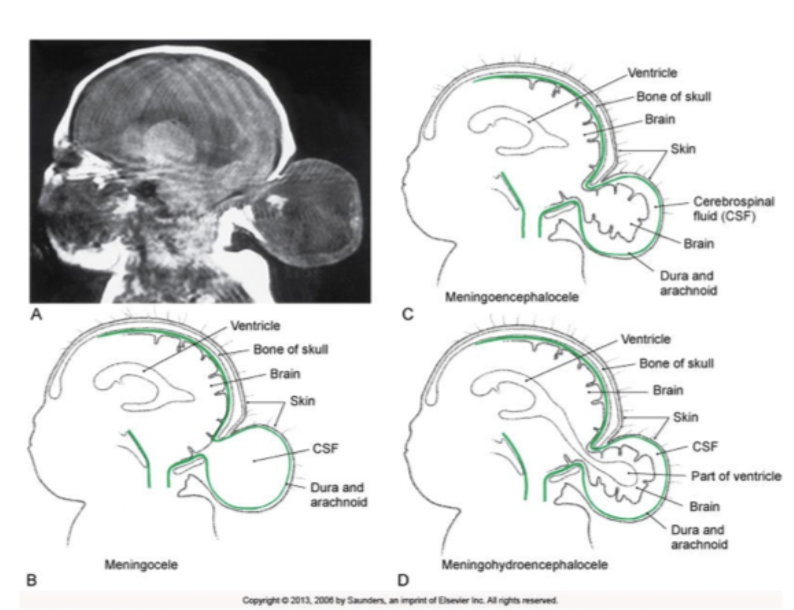
***dysraphic defects- failure of anterior neuropore closure
defective neurulation gives rise to congenital malformations collectively called dysraphic defects; failure of anterior neuropore closure (most failures happen here):
anencephaly
brain is not formed
surrounding meninges, protective coverings of the brain and skull are absent
facial deformities
neonatal death
encephalocele
opening is seen anywhere along the center of the skull (but usually in the occipital region)
requires surgery, where contents that have herniated have to be put back in and the defect has to be closed up
meningocele
least severe
cystic structure will produce in the defect of the cranium, containing the meninges
meningoencephalocele
cystic structure contains meninges and brain tissue
meningohydroencephalocele
most severe
cystic structure contains meninges, brain tissues, and part of the ventricular system
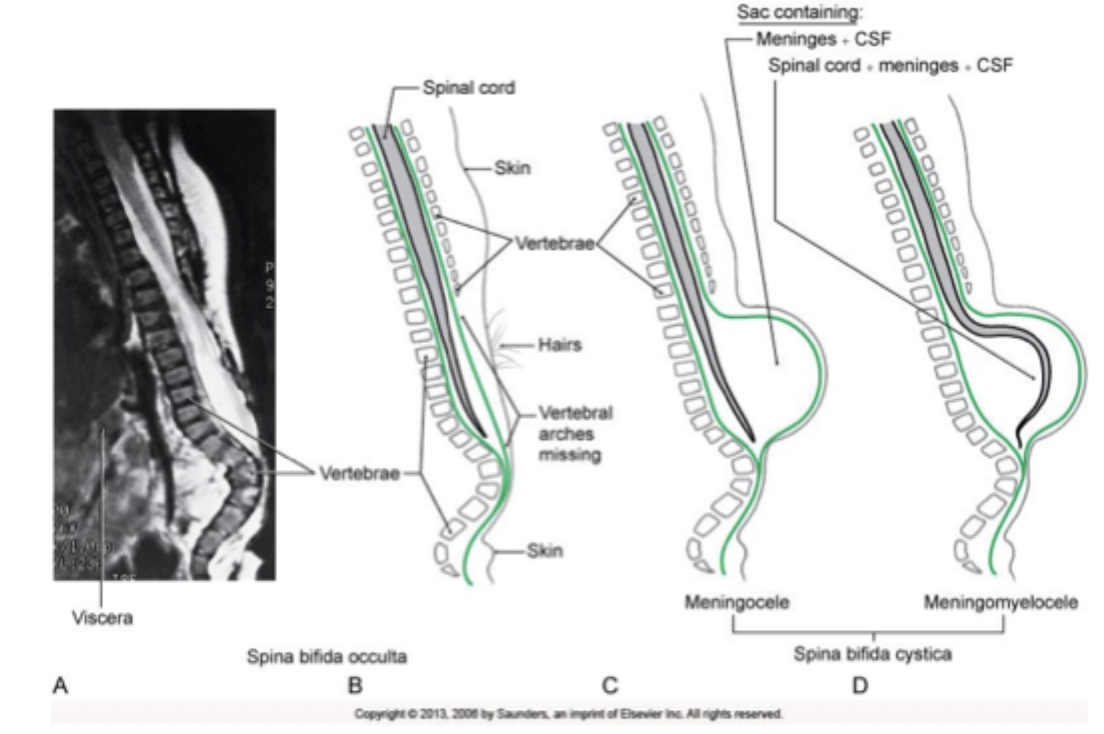
***dysraphic defects- failure of posterior neuropore closure
defective neurulation gives rise to congenital malformations collectively called dysraphic defects; failure of posterior neuropore closure: an opening in the vertebral column, usually in the lumbar/sacral regions
spinal bifidas:
(1) spina bifida occulta
most mild form
small gap in 1 or more of the vertebra of the spine
posterior arches aren’t formed properly, but the spinal cord is still formed/retained intact
no signs or symptoms; also no neurological problems (goes unnoticed a lot!)
spina bifida cystica:
(2) meningocele
protective membranes around the spinal cord (meninges) will push out in the opening of the vertebra
membranes can be removed with surgery with little to no damage to the nerve pathways, as the spinal cord is still developed properly
(3) meningomyelocele
most severe
baby’s spinal canal remains open among several vertebrae in the lower/middle back region
both the membranes of the spinal cord and the neuro-tissue itself protrude
forms a sac on the back
nerves and tissues are exposed, making a baby vulnerable to infections
***dysraphic defects- abnormal secondary neurulation
tethered cord syndrome
caused by tissue attachments that limit the movement of the spinal cord within the spinal column
prevents the spinal cord from moving along with the growing vertebral column in an appropriate way
causes abnormal stretching of the spinal cord
progressive course
may go undiagnosed until adulthood, due to a delay in presentation

***brain vesicles- primary brain vesicles
prosencephalon (adult derivative: forebrain)
divides into 2 secondary brain vesicles
telencephalon
diencephalon
mesencephalon (adult derivative: midbrain)
remains as the midbrain
rhombencephalon (adult derivative: hindbrain)
divides into 2 secondary brain vesicles
metencephalon
myelencephalon
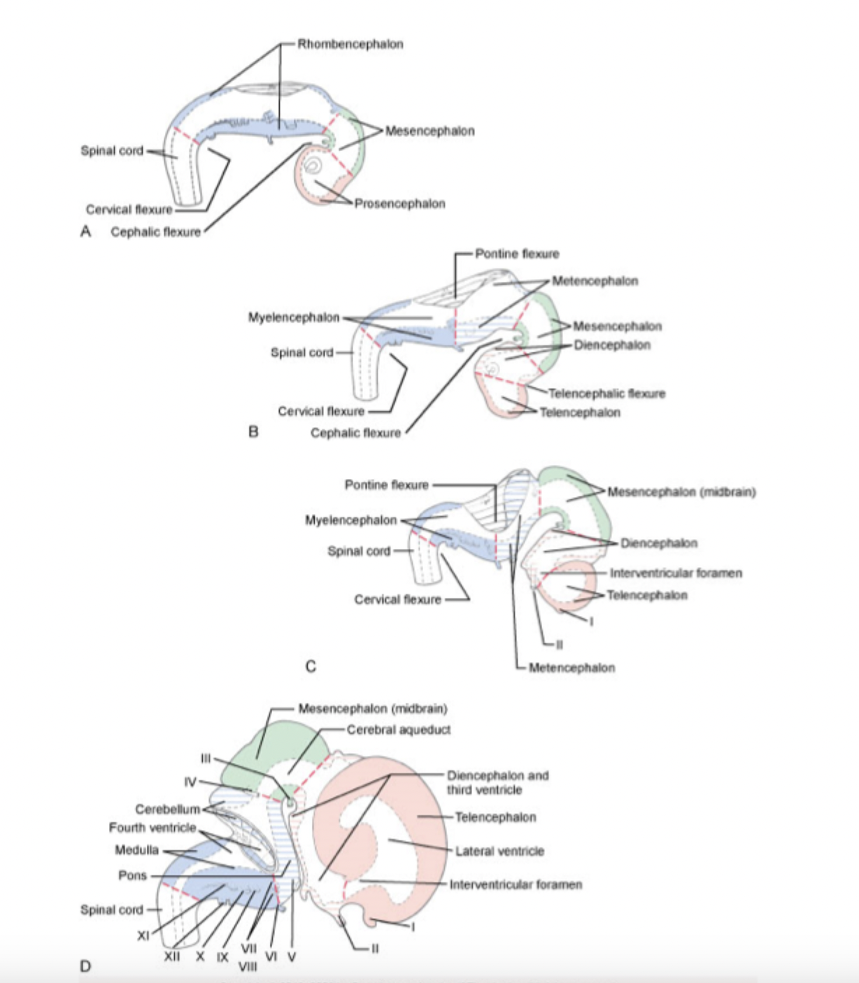
***brain vesicles- secondary brain vesicles
telencephalon: cerebral hemispheres and deep cortical structures
diencephalon: thalamus, hypothalamus, subthalamus, epithalamus
mesencephalon: remains as the midbrain
metencephalon: pons and cerebellum
myelencephalon: medulla

development of ventricles
ventricles develop from the cavities of the developing brain vesicles
lateral ventricles: cavities of the telencephalic vesicles
third ventricle: cavity of the diencephalon
fourth ventricle: cavity of the rhombencephalon
cerebral aqueduct: cavity of the mesencephalon- connects the 3rd and 4th ventricles
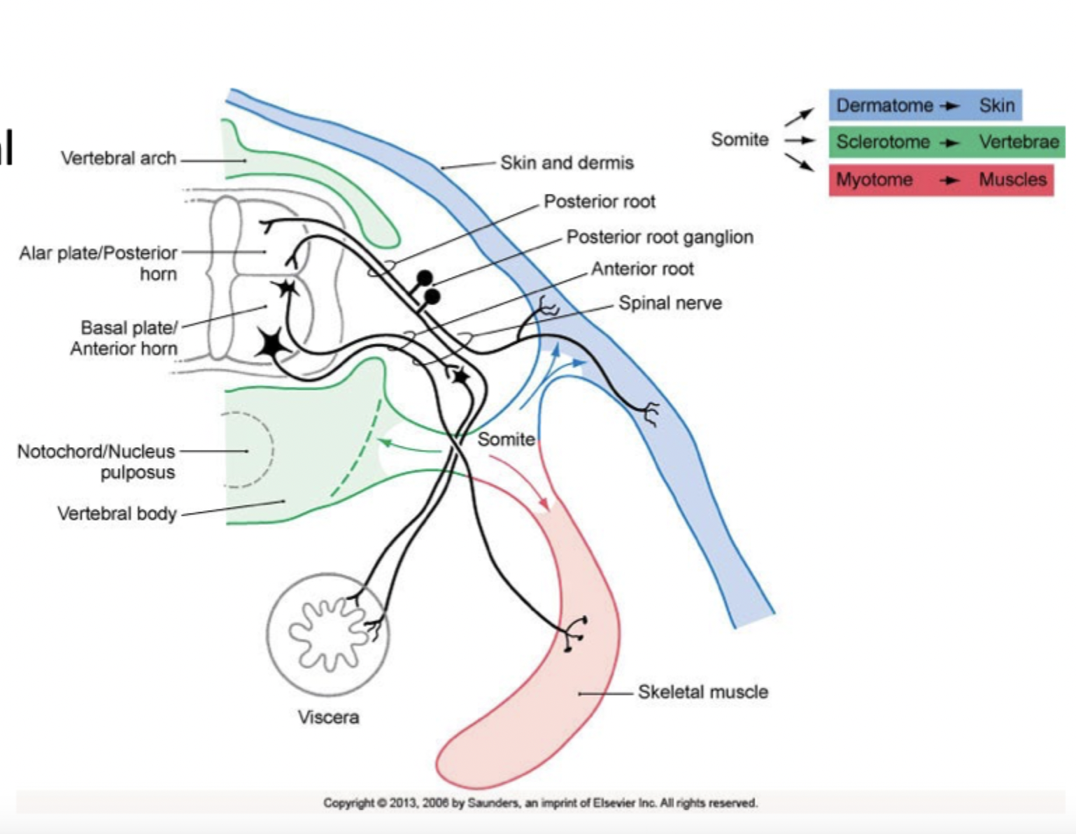
development of the spinal cord
***spinal cord: develops from caudal aspects of the neural tube
***neural canal becomes central canal of spinal cord
***neuroblast proliferating in ventricular layer migrate to form the intermediate zone and these zones fuse to form 4 longitudinal plates:
***paired basal plates: anteriorly located- becomes the anterior (ventral) horn
***paired alar plates: posteriorly located- becomes the posterior (dorsal) horn
plates on each side are separated by the sulcus limitans
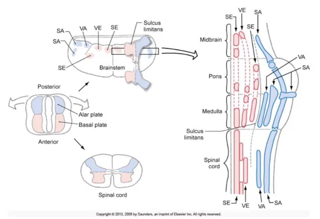
development of brainstem CN nuclei
***brainstem
myelencephalon (medulla)
part of metencephalon (pons)
mesencephalon (midbrain)
***rotation of the basal and alar plates dorsolateral resulting in a lateral to medial orientation of the alar and basal plates in the brainstem
basal plate: positioned medially in the brainstem- gives rise to motor cranial nerve nuclei
alar plate: positioned laterally in the brainstem- gives rise to sensory cranial nerve nuclei
***RQ: the notochord will induce the primitive ectoderm to form which of the following structures?
neural plate
on ~ d16 of gestation the notochord (formed by cells in the mesoderm layer of the embryo) will induce the overlying cells in the ectoderm to form the neural plate
the neural plate will thicken at its margins on ~d18 of gestation forming the neural folds
the neural crests form from lateral portions of the neural folds that break off when the folds form the neural tube
***RQ: true/false- when the neural folds contact each other during in-folding on day 20 of gestation, this is the beginning of the formation of the neural tube
true
at day 20 the neural folds will contact each other to begin the formation of the neural tube
***RQ: the neural tube is considered fully formed with the closing of the anterior neuropore on day________ and the closing of the posterior neuropore on day_______________
18; 20
24; 26
20; 42
24; 26
the neural tube is considered fully formed with the closure of the anterior neuropore which takes place on ~d24, and the closure of the posterior neuropore which takes place on ~d26
18; 20
key event taking place on ~d18 is the formation of the neural folds
key event taking place on ~d20 is the beginning of the formation of the neural tube with the initial contact of the neural folds
20; 42
days 20 - 42 mark the period of secondary neurulation
***RQ: which of the following cell layers (zones) on the lumen of the neural tube will merge together to form the basal and alar plates?
ventricular and intermediate zones
along the lumen of the neural tube, cells will proliferate forming the ventricular zone (these cells will become the neurons, glial cells, and ependymal cells)
the processes of the developing cells will produce a marginal layer
cells in the ventricular zone will migrate to what is called the intermediate zone
the ventricular and intermediate zones fuse to form 4 longitudinal plates:
2 basal plates (located anteriorly giving rise to motor neurons)
2 alar plates (located posteriorly - giving rise to sensory neurons)
***RQ: true/false- the process of primary neurulation gives rise to the portion of the neural tube that develops the brain and the spinal cord through the lumbar levels, and the process of secondary neurulation gives rise to the remainder of the spinal cord.
true
primary neurulation will give rise to the portion of the neural tube that will develop into the brain and the spinal cord through the lumbar levels
the remainder of the spinal cord develops during secondary neurulation.
***RQ: true/false- failure of the anterior neuropore to close can result in spina bifida
false
failure of the anterior neuropore to close results in either anencephaly or an encephalocele
***RQ: which of the following is considered the most severe form of an encephalocele?
meningohydroencephalocele
encephaloceles are defects in the cranium that invovle various degress of hernination of intracranial contents
meningohydroencephaloce is considered the most severe form
it is a cystic structure that contains meninges, brain tissue & part of the ventricular system
***RQ: what are considered primary brain vesicles? Select all that apply.
prosencephalon (forebrain)
mesencephalon (midbrain)
rhombencephalon (hindbrain)
***RQ: the rhombencephalon will divide to become the__________and ___________.
metencephalon; myelencephalon
the prosencephalon will divide creating the telencephalon and the diencephalon
mesencephalon doesn't divide and remains the mesencephalon
***RQ: which brain vesicle gives rise to the cerebral cortex?
mesencephalon
telencephalon
diencephalon
metencephalon
myelencephalon
telencephalon
become the cerebral cortex, subcortical white matter, olfactory bulb, basal nuclei, amygdala, and the hippocampus
mesencephalon
stays the midbrain
diencephalon
becomes the thalamus, subthalamus, hypothalamus, and the epithalamus
metencephalon
becomes the pons and cerebellum
myelencephalon
becomes the medulla
***RQ: the cavity of which of the following brain vesicle becomes the cerebral aqueduct?
telencephalon
diencephalon
rhombencephalon
mesencephalon
mesencephalon
cavity of the mesencephalon (midbrain) becomes the cerebral aqueduct (which is the communication between the 3rd and 4th ventricles)
telencephalon
cavity of the telencephalon becomes the lateral ventricles (there are 2)
diencephalon
cavity of the diencephalon becomes the 3rd ventricle
rhombencephalon
cavity of the rhombencephalon becomest the forth ventricle
foramen of Monro
interventricular foramen
foramina of Luschka
lateral apertures